
Abstract
Objective:
Our aim of the study was to investigate the expression level and methylation status of the secreted frizzled-related protein 2 in esophageal squamous cell carcinoma and to evaluate the clinical utility of the marker.
Material and Methods:
We first used Immunohistochemistry (ICH) to explore the expression level of secreted frizzled-related protein 2 protein in esophageal squamous cell carcinoma tissues and adjacent normal tissues and then used methylation-specific polymerase chain reaction and bisulfite sequencing polymerase chain reaction to detect methylation status of secreted frizzled-related protein 2.
Results:
Secreted frizzled-related protein 2 expression was notably reduced in patients with esophageal squamous cell carcinoma, whereas methylation of secreted frizzled-related protein 2 was increased in the majority of esophageal squamous cell carcinoma specimens.
Conclusion:
Sum up, we have demonstrated the abnormal DNA hypermethylation, causing reduced or absent gene expression. Methylation testing of secreted frizzled-related protein 2 using epigenetic marker may be a significative screening method for patients with esophageal squamous cell carcinoma.
Keywords
Introduction
Esophageal cancer is considered to be a common malignant tumor worldwide, over 80% of which occurred in developed regions. 1 -3 In China, the incidence and mortality of esophageal cancer are about 2 times higher than that in the world. 4 The incidence of esophageal cancer in Xinjiang has reached to 155.9/100 000. The incidence rates far exceed those in other countries. 5 -7 Histological types of esophageal cancer include esophageal squamous cell carcinoma (ESCC) and esophageal adenocarcinoma (EAC). Esophageal squamous cell carcinoma remains the main predominant histological type. 8,9 The incidence of ESCC has obvious regional differences, among which Xinjiang has the higher incidence than other regions. 10 The residents in Xinjiang have nutritional deficiencies, intake of pickled vegetables, nitrosamine-rich or mycotoxin-contaminated foods, and low socioeconomic status, all of this reasons are likely to contribute to ESCC. 11 Patients with esophageal squamous cell carcinoma have poor survival since patients usually diagnosed at the advanced stage. 12 Meanwhile, our knowledge of potential molecular mechanisms of DNA methylation biomarkers remains unclear. 13 To our knowledge, epigenetic gene silencing constitutes an alternative or complementary mechanism to mutational events in tumorigenesis. Screening, therefore, is the key for detecting patients before they are advanced. Thus, it is critical to identify the underlying epigenetically silenced cancer-related genes as new prognostic and therapeutic targets in ESCC diagnosis and treatment.
As we all know, Wnt signaling pathway plays a very essential role in the occurrence and development of various tumors. 14,15 Esophageal squamous cell carcinoma can develop through aberrant Wnt signaling which influences gene expression levels and methylation status, finally leading to carcinoma initiation and progression. 16 -20 Wnt signaling pathway is partially regulated by its family members including secreted frizzled-related proteins (SFRPs). The SFRPs constitute a family of extracellular Wnt signaling antagonists, which have 5 members (SFRP1-5). Secreted frizzled-related proteins have been found to inhibit several tumor activation by downregulating the growth rate of tumor cells. Several studies have shown that SFRPs could inhibit activation of canonical Wnt signaling. 21 A previous study confirmed SFRPs as tumor suppressors, such as colorectal cancer, 22 gastric cancer, 23 and oral squamous cellcarcinoma. 24 Suggesting that SFRP2 is a tumor suppressor. But the underlying regulatory tumor growth mechanism of SFRP2 remains unclear. Here, we aimed to assess the effects of SFRP2 antagonism on tumor growth and to investigate the contribution of the expression levels and methylation status of SFRP2 in patients with ESCC to identify the gene with gradually altering expression can be potentially regulated by DNA methylation. We want to find an useful screening marker for patients with ESCC.
Materials and Methods
Tissue Samples
Whole gene expression profiling was performed on 90 human ESCC and 90 histologically normal adjacent tissues; they were obtained from surgically removed tumors in the First Affiliated Hospital of Xinjiang Medical University. Written informed consent was obtained from all patients, and the study was approved by the ethics committee of the First Affiliated Hospital of Xinjiang Medical University (20180223-08), between 2007 and 2018. We have collected all the clinical data of the patients who involved in the study. The average age of the patients was 64.72 years; the youngest patient was only 43 years old and the oldest patient was 84 years old at the time of surgery. Their mean age ± standard deviation was 64.72 ± 13.71. In this study, 90 cases of ESCC tissues were examined at the Department of Pathology, First Affiliated Hospital of Xinjiang Medical University. All samples were classified by American Joint Committee on Cancer (AJCC) staging, involving tumor size (T), lymph node involvement (N), and distant metastasis (M). The 90 patients were classified according to AJCC as follows: pathological stage T0, 10 (11%); pathological stage T1, 29 (32%); pathological stage T2, 27(30%); pathological stage T3, 24 (27%). The degree of differentiation for ESCC was classified as high, middle, and low grades. Lymph node metastasis was defined as negative and positive. No restrictions were placed in terms of age, sex, or disease stage. The patient did not receive any therapy prior surgery operation, such as chemotherapy or radiation.
Immunohistochemistry
The identification of SFRP2 protein level can be achieved using immunohistochemical (IHC) staining analysis. Four micrometer sections were cut, which were baked at 65°C for 120 minutes, then deparaffinized in xylene for 30 minutes and dehydrated in graded ethanol for 5 minutes. The sections were sterilized with 1% sodium citrate buffer under high pressure and then cooled to room temperature. The sections were autoclaved in 1% sodium citrate buffer (pH 6.0), cooled at room temperature. In order to block endogenous peroxidase activity, it is necessary to culture in 3% H2O2 for 15 minutes after cooling and rinsing in distilled water. Samples were preincubated with a protein blocking solution for 10 minutes and the sections were incubated at 4°C overnight in anti-SFRP2 rabbit polyclonal antibody at a dilution of 1:100. After washing 3 times in phosphate-buffered saline, the slides were incubated with biotinylated secondary antibody for 90 minutes at 37°C.
The percentage and intensity of positively stained SFRP2 in each sample was assessed by 2 pathologists who had no knowledge of the patients’ characteristics on optical microscopes. Briefly, every core was scored by the staining depth (0 for negative staining, +1 for faint yellow, +2 for brown madder, and +3 for dark brown). The score of the results was 0 to 4, according to the percentage and degree of positive cells. Then the images were scored according to the percentage of positive cells by the given intensity value in the total tumor cells, score 4: ≥76%; score 3: 51% to 75%; score 2: 11% to 50%; score 1: 1% to 10%. The score of the same slide was summed to produce a final score (Final score = staining depth score × the number of positive cells score): ≥ score 5: strong positive (+++); score 4: middle positive (++); score 3: weakly positive (+); < score 3: negative (−).
DNA Extraction, Methylation-Specific Polymerase Chain Reaction
DNA was isolated from formalin-fixed, paraffin-embedded cancer tissues and their adjacent noncancer specimens using the QIAamp DNA formalin-fixed, paraffin-embedded kit (Qiagen, Hilden, Germany), following the manufacturer’s instructions. The extracted DNA was quantitatively detected by ultraviolet spectroscopy and stored below −20°C for reserve. Stored DNA was modified with the Qiagen Epitect Fast Bisulfite Conversion kit (Qiagen), according to the manufacturer’s instructions. The bisulfite-modified DNA was subjected to polymerase chain reaction (PCR) in a blinded manner using primer pairs designed to amplify specifically the methylated or unmethylated alleles of respective genes. All the primers were synthesized by Shanghai Biosune Biotechnology Company in China. The primer sequences used are shown in Table 1. The PCR protocol according to the manufacturer’s instructions was 95°C for 5 minutes, 35 cycles of 95°C for 30 seconds, 60°C for 30 seconds, 72°C for 40 seconds, and a final extension at 72°C for 5 minutes. The PCR products were then electrophoresed on a 2.5% agarose gel and visualized under ultraviolet illumination (ChemiDoc XRS; Bio-Rad, Hercules, California).
List of Primer Sequences.
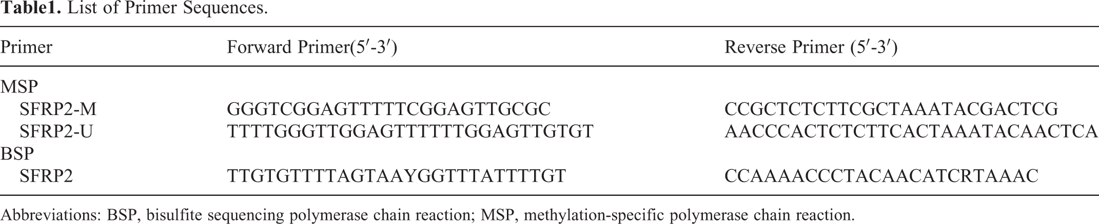
Abbreviations: BSP, bisulfite sequencing polymerase chain reaction; MSP, methylation-specific polymerase chain reaction.
Bisulfite Sequencing PCR
Ninety samples were selected and methylation status was detected by methylation-specific PCR (MSP). Methylation status of the gene was further analyzed on 10 samples which were proved to be methylated or unmethylated by MSP and were verified by bisulfite sequencing PCR (BSP). The primers used to detect methylation of SFRP2 CpG islands were designed by PyroMark Assay Design software (Version 2.0) in order to discriminate between methylated and nonmethylated sequences. The primer sequences used are shown in Table 1. The size of the PCR product is 269 bp. The PCR amplification was carried out with the following thermocycling conditions: 95°C for 2 minutes, 36 cycles × (95°C × 15 seconds, 56°C × 15 seconds, 72°C × 15 seconds), then 72°C for 7 minutes. In the end of PCR amplification, PCR products were subjected to DNA sequenced with 3730 measuring sequence analyzer (ABI, Foster City). Each experiment was performed in triplicate to validate the results.
Follow-Up and Survival Analysis
We followed up the patients. The duration from surgery to death is defined as the overall survival period of the patient. The follow-up deadline was April 30, 2018. Based on the follow-up data, we use the Kaplan-Meier method to calculate survival curves for them. The relations of SFRP2 expression and methylation status with age, sex, and lymph node metastasis were analyzed. When P is less than .05, the difference has statistical significance.
Statistical Analysis
The researchers used the SPSS version 19.0 software package for all data statistics. The continuous variables were expressed as means ± standard error of the mean. χ2 test or Fisher exact method was applied in order to determine the statistical significance of the correlations between SFRP2 expression and the different clinicopathological parameters, and meanwhile, to assess the association between the methylation gene and the different clinicopathological parameters using the same method. The patients were routinely followed up clinically. All P values were 2-sided and the significance level was P < .05.
Results
Silencing of SFRP2 in ESCC Tissues
In our study, we found that SFRP2 decreases expression in ESCC samples compared to paired normal samples (31/90, 34.44% vs 70/90, 77.78%; Figure 1). The difference was significant (P < .01). This decreasing expression was validated using IHC staining in ESCC samples and normal samples. By doing so, we found that SFRP2 expression levels were 2.26-fold upregulated in normal samples relative to all ESCC samples. The statistical analysis suggested that there was no association between the expression status of SFRP2 with age, gender, nation, tumor location, tumor size, AJCC stage, infiltration degree, and lymph node metastasis in ESCC. The results are listed in Table 2. Based on these results, we set out to assess whether DNA methylation was involved in the downregulation.

The SFRP2 protein expression in ESCC tissues and normal controls. A, The SFRP2-positive expression in adjacent normal tissues; (B) The SFRP2-negative expression in adjacent normal tissues; (C) The SFRP2-positive expression in ESCC tissues; (D) The SFRP2-negative expression in ESCC tissues. ESCC indicates esophageal squamous cell carcinoma; SFRP2, secreted frizzled-related protein 2.
Correlation Between the SFRP2 Methylation Gene Promoter Methylation Status, SFRP2 Protein Expression Level, and Clinicopathological Parameters in ESCC.
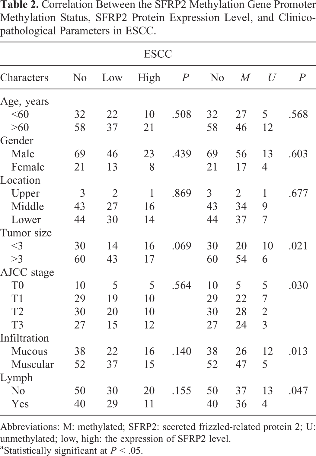
Abbreviations: M: methylated; SFRP2: secreted frizzled-related protein 2; U: unmethylated; low, high: the expression of SFRP2 level.
a Statistically significant at P < .05.
Silence of SFRP2 Expression via Hypermethylation of SFRP2
To research whether the DNA methylation status of SFRP2 gene in formalin-fixed, paraffin-embedded cancer tissues had diagnostic value for ESCC and involved in the downregulation, we investigate the frequency of DNA methylation of the gene by MSP analysis in 90 patients with ESCC. The SFRP2 promoter showed hypermethylation in 73 (81.11%) tumor samples. However, the SFRP2 promoter methylation was performed in only 16 (17.78%) corresponding normal tumor-adjacent samples. The frequency of SFRP2 promoter methylation in ESCC tissues was significantly higher than that in the adjacent tissues (χ2 = 4.39; P = .046). The difference was significant. Furthermore, we also studied the relationship between the methylation status of SFRP2 and the clinicopathological parameters of patients. The analysis results are shown in Table 2. Statistical analysis indicated that methylation of the SFRP2 gene was significantly related to tumor size, AJCC stage, lymph node metastasis, and infiltration degree. However, there was no statistical correlation between the SFRP2 promoter methylation status and age, gender, nation, and tumor location. The agarose gel electrophoresis results of the SFRP2 gene using MSP are shown in Figure 2. Notably, using BSP analysis, all CpG islands in the promoter region of SFRP2 gene have been extensively methylated, whereas only limited methylation was found in paired normal epithelial tissues (Figures 3 and 4).

Representative results showing the SFRP2 promoter methylation status identified by MSP. Control indicates blank control group; MSP, methylation-specific polymerase chain reaction; M, methylated; N, corresponding normal tumor-adjacent tissues; SFRP2, secreted frizzled-related protein 2; T, ESCC tissues; U, unmethylated.

The BSP histogram result of ESCC and corresponding normal tumor-adjacent tissues. The figure comes from 3730 measuring sequence analyzer. ESCC indicates esophageal squamous cell carcinoma; BSP, bisulfite sequencing polymerase chain reaction.

Bisulfite sequencing of the SFRP2 CpG island in ESCC and corresponding normal tumor-adjacent tissues.ˆ: unmethylated;•: methylated CpG sites. A, ESCC tissues. B, Corresponding normal tumor-adjacent tissues. ESCC indicates esophageal squamous cell carcinoma; SFRP2, secreted frizzled-related protein 2.
Analysis of SFRP2 Methylation Status and the Correlation With SFRP2 Expression
In our study, all the 73 (81.11%) cases with SFRP2 promoter methylation-positive ESCC tissues showed almost all IHC results were negative. The similar results were observed in the corresponding normal tissues. Interestingly, of the 16 cases with SFRP2 promoter methylation-negative tissues, 14 cases showed positive immunoreactivity for SFRP2 (14/16, 87.50%). There was a significant correlation between SFRP2 promoter hypermethylation and SFRP2 protein expression results (χ2 = 25.153, P < .01). DNA methylation may lead to downregulation or deletion of protein expression, which can be confirmed by IHC, MSP, and BSP. The SFRP2 expression level of all data sets was potentially regulated by DNA methylation.
Prognostic Significance of SFRP2 Protein Expression and SFRP2 Promoter Methylation Status
Ninety patients were followed up for 1 to 72 months, and the survival dates were analyzed. The overall survival rate of patients with ESCC with SFRP2-positive expression was higher than the patients with SFRP2-negative expression, but not statistically significant (P > .05; Figure 5A). The 5-year survival rate was 10%. Overall survival curves based on SFRP2 unmethylation and methylation status were constructed by the Kaplan-Meier method. The results showed that the patients with methylation of SFRP2 had a poorer prognosis than those with unmethylation of SFRP2, but has no statistical significance (P > .05; Figure 5B). Furthermore, there were no significant differences between lymph node metastatic group and nonmetastatic group (χ2 = 1.379, P = .240; Figure 5C). The invasion depth reached the muscularis and mucosa layers (χ2 = 1.522, P = .217; Figure 5D), male patients and female patients (χ2 = 0.084, P = .772), between ≥60 years old patients and <60 years old patients (χ2 = 1.613, P = .204; figures are not shown).

The relationship between the SFRP2 and survival curve. A, The overall survival rate of patients with ESCC with SFRP2 expression; (B) the overall survival rate of patients with ESCC with SFRP2 methylation status; (C) the overall survival rate of patients with ESCC with lymph node metastatic; (D) the overall survival rate of patients with ESCC with ethnic differences. ESCC indicates esophageal squamous cell carcinoma; SFRP2, secreted frizzled-related protein 2.
Discussion
In China, ESCC is regarded as a very prevalent malignant tumor threatening human health with a very poor outcome, 25,26 particularly in Xinjiang, northwest China. 5,6,9 About 1 million new cases are diagnosed worldwide, and over 50% of these patients will die due to this disease. Although the incidence of EAC is rapidly increasing in Western countries, ESCC still remains the main biological type in China. Advanced esophageal cancer is one of the most lethal malignant tumors in the world. It has the biological characteristics of strong invasiveness and poor survival rate, and its research is not deep enough. 27 Therefore, it is necessary to study the key role of biological markers in the occurrence and development of ESCC.
Acknowledged, Wnt signaling pathway plays an essential role in a variety of biological processes. 14,15 A delicate control of Wnt signaling is crucial for the proper maintenance of the organism, while aberrant Wnt signaling may lead to developmental defects and disease initiation and progression. 16 -18 The SFRPs are a group of negative modulators of the Wnt signaling pathway, of which 5 members (SFRP1-5) have been identified to date. Secreted frizzled-related proteins 2 may become a new underlying biology marker in the diagnosis and treatment of ESCC.
In a majority of studies, promoter methylation of abnormal SFRP2 gene has been described. Recent Hao’s study has demonstrated that the expression of SFRP2 was silent in 7 esophageal cancer cell lines with different differentiation, but relatively high in one normal esophageal epithelial cell line. 28 Huang et al 26 divided the patients into 3 groups: CRC group, adenoma group, and normal control group. Feces and serum were collected and SFRP2 methylation status was studied. The sensitivity of SFRP2 methylation in fecal DNA was significantly higher than that in serum DNA. The SFRP2 methylation rates were detected in fecal samples in patients with adenoma (46%) and colorectal cancer (84%), respectively. The SFRP2 methylation rates were detected in serum samples in patients with adenoma (6%) and colorectal cancer (67%), respectively. The silencing expression of proteins is correlated with methylation of promoter region, which has been confirmed by Tang et al. 29 They suggest that for patients with ESCC, the main reason for SFRP2 protein inactivation is methylation of promoter region.
However, the biological mechanism of SFRP2 methylation regulation has not been well investigated in the development of esophageal carcinogenesis. To investigate the role of SFRP2 in esophageal carcinogenesis, we detected SFRP2 expression by IHC in ESCC tissues and corresponding paired normal tissues. The results of our laboratory indicate that SFRP2 loses expression in ESCC tissues. To more clearly define the regulation of SFRP2, we generated functional methylation studies to SFRP2. In other words, we want to assess whether DNA methylation was involved in the downregulation. Our results indicate that there was a significant correlation between SFRP2 promoter hypermethylation and SFRP2 protein expression results (χ2 = 25.153, P < .01). We will further validate the effect of SFRP2 methylation on the expression of SFRP2 using MSP and BSP methods. We used MSP to detect the DNA methylation of SFRP2 in FFTF samples of tumor and nontumor from 90 patients with ESCC. Among them, 10 patients were selected using BSP detection to further verify.
In our analysis, we confirmed that compared to ESCC, the expression of SFRP2 in normal tissues was 2.26 times higher than that in tumors. Our results confirmed that the study of SFRP2 promoter showed that the incidence of CpG methylation in ESCC tissues was significantly higher than that in adjacent nontumor tissues, which indicated that abnormal methylation in SFRP2 promoter region was not a cell line-specific event but a common phenomenon in the development of ESCC. Furthermore, MSP analysis data indicate that the promoter region methylation may serve as a regulation element of SFRP2 expression.
Many studies have shown that SFRP2 is an antioncogene and may play an important role in ESCC. It is suggested that the detection of methylation of a biological marker in tissues is helpful for early detection of tumors, evaluation of metastasis and prognosis, and guidance of clinical treatment. In ESCC tissues, we detected a significant increase in methylation of SFRP2, suggesting that SFRP2 may be a marker for early detection of ESCC. Our research has elucidated the role of SFRP2 in ESCC and confirmed that SFRP2 may be a promising new biological target for ESCC. We included fewer samples in this study. We only studied ESCC tumor tissues and adjacent normal tissues and did not include patients with precancerous lesions. In this study, we also neglected the effect of race on SFRP2 expression and methylation status. Hence, further studies with larger amounts of tissues from patients with ESCC will be needed to validate SFRP2 as biomarkers for population-based screening of ESCC.
Conclusion
Secreted frizzled-related protein 2 stimulates antagonism, inhibits tumor growth, and provides evidence that SFRP2 is a tumor suppressor rather than a tumor promoter. In summary, we have demonstrated that SFRP2 expression level was potentially regulated by DNA methylation. The test of SFRP2 methylation status may be a promising screening method for ESCC. Furthermore, its methylation status might be a predictive epigenetic marker of ESCC and remodeling on the expression by demethylation can provide a potential viable therapeutic strategy. Hence, further studies on the epigenetic regulation of SFRP2 expression are necessary, and the regulation of SFRP2 expression by epigenetic drugs may have great promise for cancer prevention and therapy. Limitations of this study include its small sample size and we need a molecular mechanistic study of SFRP2 downstream genes.
Footnotes
Authors’ Note
Qian Liu and Ya-Xing Zhou are co-first authors. Written informed consent was obtained from all patients and the study was approved by the ethics committee of the First Affiliated Hospital of Xinjiang Medical University (20180223-08).
Declaration of Conflicting Interests
The author(s) declared no potential conflicts of interest with respect to the research, authorship, and/or publication of this article.
Funding
The author(s) declared the following potential conflicts of interest with respect to the research, authorship, and/or publication of this article: This study was supported by the grant from “The National Natural Science Foundation of China” (No. 81860422 and State Key Laboratory of Pathogenesis, Prevention and Treatment of High Incidence Diseases in Central Asia Fund (SKL-HIDCA-2017-8).