
Abstract
The safety of blood transfusions faces significant challenges due to the presence of leukocytes, which can trigger severe immune responses and facilitate virus transmission. Existing leukocyte reduction filters, predominantly composed of melt-blown nonwovens, encounter difficulties in completely eliminating leukocytes for specific patient populations such as organ transplant recipients. This research introduces electrospun membranes composed of PBS and CS/PBS nanofiber membranes as a novel filtration material. Through the optimization of electrospinning parameters (including voltage, receiving distance, and solution volume), membranes with adjustable pore sizes ranging from 1.3 to 2.1 μm and substantial specific surface areas were developed. Blood filtration trials revealed that nanofiber membranes with an optimal pore size range of 1.9–2.1 μm achieved complete leukocyte removal while preserving a red blood cell (RBC) recovery rate of 87.72%–90.65%. In comparison to conventional filters, these electrospun nanofiber membranes present a promising strategy for highly efficient leukocyte depletion, catering to the demands of critical transfusion scenarios.
Introduction
Blood transfusion is a widely utilized clinical intervention. Nonetheless, unprocessed whole blood harbors a substantial leukocyte population, leading to various adverse effects upon transfusion to recipients, including non-hemolytic febrile reactions, transfusion-related acute lung injury, and platelet transfusion refractoriness. 1 This is mainly due to the leukocyte antibody produced by the allogeneic immune reaction between the imported donor leukocyte and the recipient. 2 Furthermore, viruses spread through blood transfusions, such as hepatitis B virus, hepatitis C virus, human immunodeficiency virus and other cytotropic virus, also exist on white blood cell (WBC). 3 Thus, reducing the leukocyte content in transfused blood is crucial for enhancing the safety of blood transfusions.4,5
Currently, filtration is the most economical and practical method for removing white blood cells.6,7 This method separates leukocytes from other blood cells by leveraging differences in cell diameter, deformability during passage through filter media, and the material’s selective adsorption affinity. 8 Blood filters have evolved over time. The initial filters, developed in the 1960s–1970s, employed surface or depth filtration with pore sizes around 170–260 μm. Second-generation filters, introduced in the late 1970s, utilized consolidated fibrous mesh with mesh openings of 20–40 μm in a columnar design. In the 1990s, third-generation leukoreduction filters were introduced, employing nonwoven fabrics as the filter medium. This method is now widely used globally in blood banking and clinical settings. 4
The third-generation leukoreduction filters on the market are made of melt blown nonwovens9,10 which have fine fiber, small effective pore size, low price and stable chemical properties.11,12 The author studied the nonwovens used in leukocyte filter on the market, and found that its fiber diameter is 2-3 µm, the average pore size is about 8 µm, and the pore size distribution is uneven. After the blood is filtered by leukocyte filter on the market, the number of WBC decreases from 109/L to 105∼106/L, and RBC recovery rate is about 90%. 13 This greatly reduces the occurrence of side effects of blood transfusion, but there is still a high risk for some special patients such as pregnant women and organ transplant patients. For example, in organ transplant patients, leukocytes from blood transfusion may induce graft-versus-host disease, and leukocyte depletion is theoretically an effective method to prevent this complication. 14 Hence, enhancing the efficacy of leukocyte filtration and minimizing leukocyte content is imperative for specific patient populations. Nevertheless, the inherent constraints of the melt-blown process pose challenges in decreasing fiber fineness and pore size of melt-blown nonwovens, thereby hindering further enhancement of leukocyte filtration efficiency.
The emergence of electrospinning has enabled a more convenient approach to nanofiber production. Electrospun nanofibers, in contrast to meltblown microfibers, demonstrate narrower diameters, smaller and more evenly dispersed pores, increased specific surface area, and a comparably disordered fiber alignment.15,16 These characteristics make them a promising candidate for next-generation blood filtration membranes. 17 Guo et al. 13 fabricated PBT nanofibrous membranes via electrospinning and integrated them with PBT melt-blown nonwovens to develop a novel filter. SEM analysis revealed significantly reduced average fiber diameter and pore size in the composite material, enhancing filtration efficiency and adsorptive capacity. Leukocyte filtration experiments further confirmed that incorporating the electrospun PBT membrane reduced leukocyte counts from 105 to 104/L. However, this led to a longer filtration time. Mayuri P. V et al. 18 prepared poly (ethylene-vinyl alcohol) electrospun membranes and stacked multiple layers to fabricate a leukocyte removal filter. Whole blood filtration experiments demonstrated that this filter achieved a 100% leukocyte removal rate while capturing 8% of red blood cells and 91% of platelets. However, the length of filtration through the developed filter was high when compared to that of marketed filter. Gian Vincent Dizon 19 introduced the formation of a composite cellulose acetate membrane for the removal of blood cells from blood-derived solutions. These membranes showed significant potential for facilitating the separation of cells from plasma, which could be applied to the detection of biomarkers in plasma.
Throughout history, the evolution of white blood cell filtration materials has demonstrated a progression towards finer fibers, reduced pore sizes, and enhanced filtration efficacy. While electrospun nanofiber materials offer advantages in filtration applications, optimizing the pore size of these membranes for blood filtration presents a challenge. The goal is to maximize white blood cell removal, enhance red blood cell recovery, and reduce filtration duration. This study utilizes electrospinning technology to fabricate polybutylene succinate (PBS) and chitosan/polybutylene succinate (CS/PBS) fiber membranes with varying pore sizes by manipulating spinning parameters. The research aims to determine the ideal pore size for achieving superior filtration efficiency, ultimately improving blood filtration for specific patient populations.
Experimental
Materials
Chitosan powder (CS, Viscosity 200–400 mPas) was purchased from Shanghai Aladdin Biochemical Technology Co., Ltd. Polybutylene succinate powder (PBS, Mw ≈ 1000000) and Hexafluoroisopropanol (HFIP, AR, 99.5%) were purchased from Chengdu MICXY REAGENT Co. LTD. Whole blood samples from humans (provided by Bethune International Peace Hospital).
Instruments
Electrospinning device (HZ-10, Yunfan Instrument Co., Ltd), Optical contact angle measuring instrument (DataPhysics Instruments GmbH, OCA15EC), PMI Porometer (PMI, CFP-1100AX ), Scanning electron microscope (Hitachi, S-4800-I), Electronic universal strength machine (Shenzhen Sanqi Zongheng Technology Co., Ltd, UTM5305), Pressure steam sterilizer (Jiangyin Binjiang Medical Equipment Co., Ltd, YX280), Digital Display Constant Temperature Magnetic (Shanghai Sile Instrument Co., Ltd, HJ-4A), Incubator (Wenzhou Darong Textile Instrument Co., Ltd, Y(B)802G), Electronic balance (Sartorius AG, BS223S), Blood cell counting plate (Beijing Solaibao Technology Co., Ltd).
Preparation of electrospun PBS and CS/PBS membrane
Polybutylene succinate (PBS) is a novel polyester developed after the 1990s, characterized by recyclability, excellent biocompatibility, and non-toxicity towards cells, rendering it versatile in biomedicine. 20 Chitosan (CS), a biocompatible, hydrophilic, absorbable, antibacterial green material, finds extensive use in biomedicine. 21 Consequently, this investigation fabricated nanofiber membranes employing PBS and CS as substrates.
Experimental scheme of electrospinning PBS.

The CS/PBS nanofiber membranes were prepared using identical spinning parameters: 10% CS concentration, 10% PBS concentration, spinning speed of 1 ml/h, voltage of 16 kV, and a receiving distance of 12 cm.
Characterization and measurements
Micro-morphology of fiber
The micro morphology of nanofibers was observed by SEM. Based on the SEM images, the average diameter and coefficient of variation of fibers were calculated by software Photoshop.
Pore sizes and distribution
The pore sizes and pore distribution of the electrospun mats were tested by PMI Porometer. This instrument is a pore size analyzer based on the gas-liquid displacement principle. The sample was first fully saturated with a non-toxic, harmless wetting liquid. Then, an inert gas (which does not react with the wetting liquid or the sample) was applied to displace the liquid from the sample’s pores. As pressure increases, interconnected pores were progressively emptied from largest to smallest. By measuring the relationship between applied gas pressure and flow rate, the instrument provides data on the pore size distribution.
Mechanical properties
The fiber membranes were cut into strip samples of 30 mm × 10 mm, and the fracture strength and tensile modulus of the strip samples were measured by a Electronic universal strength machine at a tensile speed of 10 mm/min.
The testing procedure was conducted under GB/T 1040.3-2006 Plastics—Determination of tensile properties—Part 3: Test conditions for films and Sheets.
ASTM D882: Standard Test Method for Tensile Properties of Thin Plastic Sheeting.
Wetting capability
The contact angle of PBS and CS/PBS nanofiber membrane were tested by Contact Angle Measuring Instrument. The liquid used was deionized water. Each sample was measured three times, and average value of the three measurements was taken as the contact angle of the material surface. The testing procedure was conducted under GB/T 30447-2013 Measurement method for contact angle of nano-film surface.
Blood filtration performance
Blood filtration experiment adhered to international ethics standards (Helsinki Declaration), prioritizing participant rights. Written informed consent was obtained from all participants. Strict data anonymization and security protocols were implemented, limiting access to necessary researchers. And, all personnel received ethics training and worked under supervision.
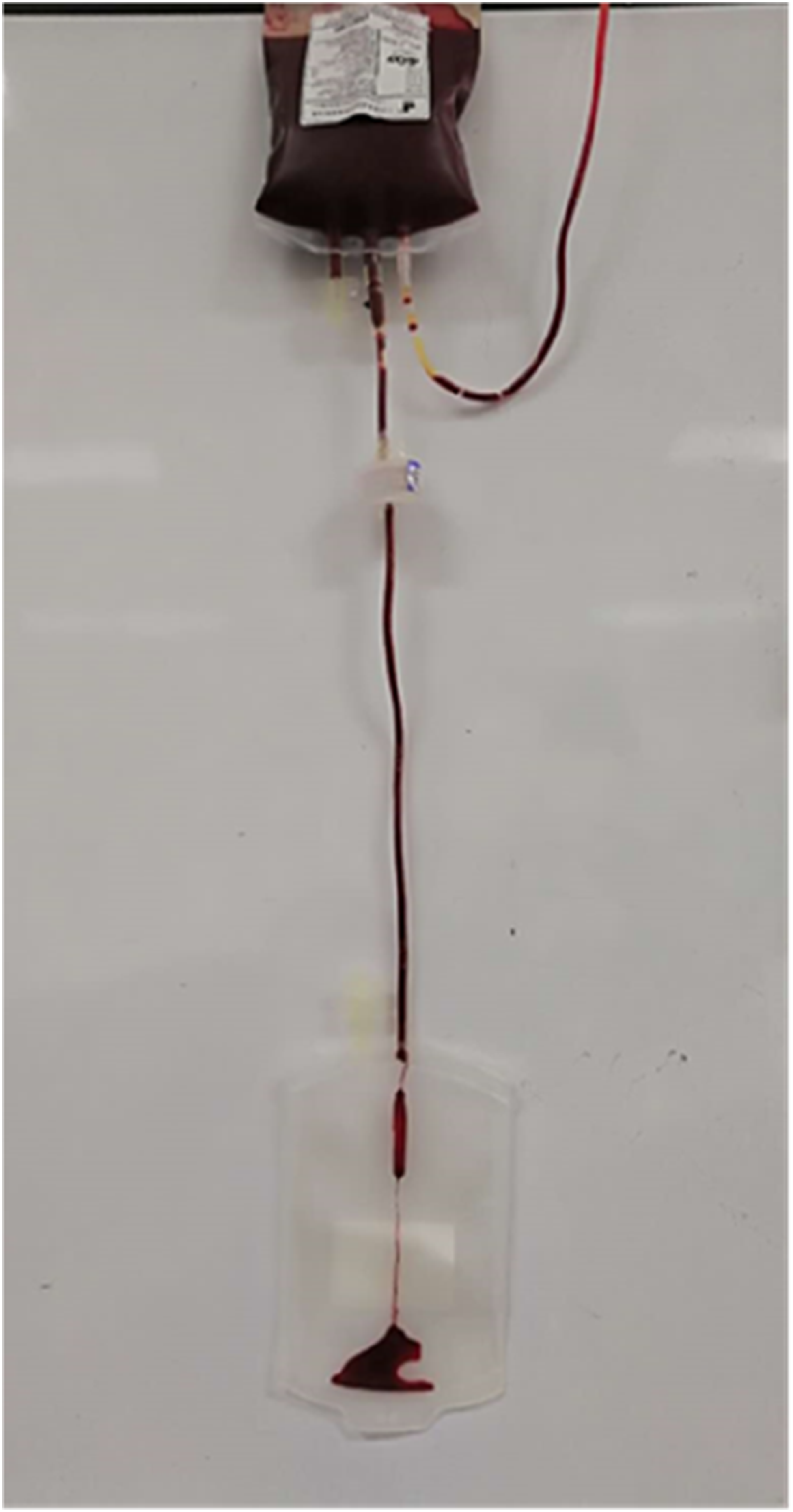
Circular specimens (50 mm diameter) were cut from both PBS and CS/PBS fibrous membranes and assembled into disposable leukocyte filters using 50 mm inner-diameter microporous filter holders (Figure 1). The filter assembly was vertically suspended with its upper end connected to a whole blood bag and lower end attached to a collection bag. After releasing the hemostat clamp below the blood bag, blood was allowed to flow through the filter under gravity. Upon complete filtration into the receiving bag (Figure 2), the clamp was secured. All procedures were performed at ambient temperature with sterilized equipment, where materials underwent steam sterilization in an autoclave (Pressure steam sterilizer). The testing procedure was conducted under China Medicine standard YY0329-2024 Leukocyte reduction filters for single use. Microporous filter. WBC filtration.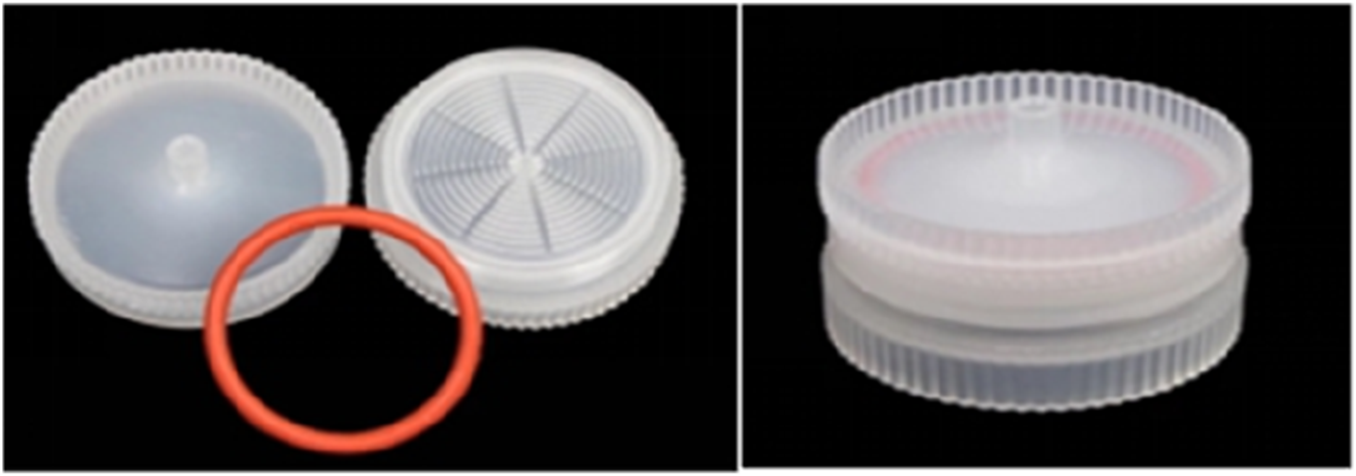
To evaluate the filtration efficiency of the fiber membrane, the number of leukocytes and erythrocytes in the blood before and after filtration was counted by microscope, and the cell concentration was calculated. The method was as follows: the blood before and after filtration was diluted with PBS buffer by gradient dilution method; then drop the diluted blood into counting area on the blood cell counting plate, and use the microscope to count the cells located in the middle and four corners of the counting chamber (80 small squares in total) in turn. Finally, the erythrocyte and leukocyte concentrations before and after filtration were calculated according to equation (1), and the WBC removal rate and the RBC recovery rate were calculated according to equations (2) and (3). The blood filtration experiment was repeated 5 times, and the average of the five structures was the final result.


Results and discussions
Micro-morphology of PBS and CS/PBS fibers
To observe and compare the morphology of PBS and CS/PBS fibers, SEM images of PBS fiber membranes and CS/PBS fiber membranes prepared under the same spinning conditions were selected. The average diameter of PBS fibers was determined to be 1726 nm with a coefficient of variation in diameter of 33.5%. In contrast, the average diameter of CS/PBS fibers was 402 nm with a diameter variation coefficient of 50%. Figure 3 illustrates that the incorporation of CS led to a reduction in fiber diameter while increasing the coefficient of variation in diameter. Additionally, PBS fibers exhibited a flatter and smoother morphology compared to CS/PBS fibers. The SEM images of the PBS and CS/PBS fiber.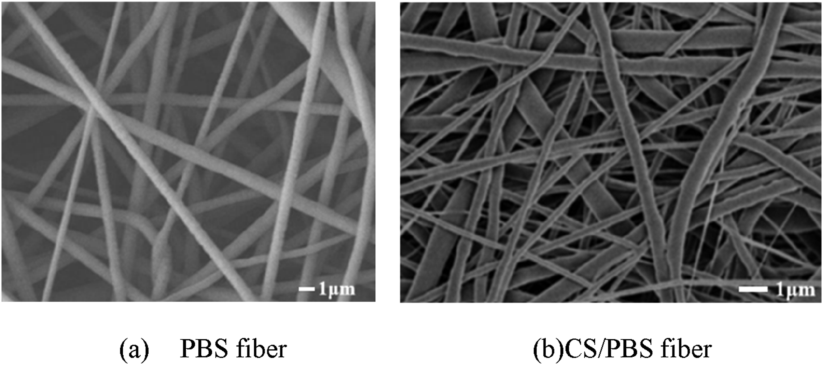
Pore sizes and distribution
The size of pores plays a crucial role in determining the filtration efficiency of electrospun fiber membranes, rendering it a pivotal structural characteristic. In this investigation, a series of single-factor experiments were conducted using PBS as the solute and HFIP as the solvent to methodically examine the impact of solution volume, voltage, and receiving distance on the diameter of membrane pores (refer to Table 1). The pore diameters of the PBS fiber membranes produced in the 24 experimental trials were assessed and are presented in Figure 4. Average pore size variation with electrospinning parameters.
Figure 4(a) illustrates a correlation between the pore size of the fiber membrane and the volume of the spinning solution. Keeping all other conditions constant, an increase in the spinning solution volume results in a reduction in the average pore size of the fiber membrane within a specific range. This phenomenon occurs because thicker membranes are formed when larger quantities of spinning solution are utilized, leading to a more compact distribution of fibers and smaller average pore sizes. In contrast, thinner membranes produced with smaller amounts of spinning solution displayed a more scattered distribution of fibers and larger average pore sizes. As illustrated in Figure Figure 4(b), a decreasing trend in average pore size with increasing voltage was observed at receiving distances of 10 cm and 13 cm. In contrast, an increasing trend in average pore size with increasing voltage was noted at a receiving distance of 12 cm. For receiving distances of 11 cm and 14 cm, no consistent pattern between average pore size and voltage variations was evident. As depicted in Figure 4(c), at a fixed voltage of 12 kV, there is a positive correlation between the average pore size and the receiving distance. However, for voltages of 14 kV, 16 kV, and 18 kV, no consistent pattern in the relationship between average pore size and receiving distance was observed. The results indicate that although the receiving distance and spinning voltage have an impact on the average pore size of the fiber membrane, there is no clear regularity. Thus, adjusting these parameters to control pore size proves less effective. Therefore, maintaining constant collecting distance and spinning voltage, and varying the spinning solution volume provide a more operationally feasible and direct approach for pore size control.
Mechanical properties
Two types of fiber membranes, PBS membrane and CS/PBS, were compared for their mechanical properties under identical spinning conditions (spinning speed of 1 ml/h, voltage of 16 kV, receiving distance of 12 cm, and spinning solution volume of 2 ml). The PBS membrane demonstrated superior breaking strength (322 cN) and tensile elastic modulus (2.6 MPa) compared to the CS/PBS membrane, which exhibited lower strength (223 cN) and modulus (1.7 MPa). This suggests that the PBS membrane possesses higher strength, while the CS/PBS membrane is characterized by greater flexibility. The addition of CS to the CS/PBS membrane led to a reduction in fiber diameter and an increase in the coefficient of variation, consequently lowering its strength. However, finer fibers typically exhibit enhanced flexibility, rendering the CS/PBS membrane relatively softer.
Wetting capability
The permeability of fiber membranes significantly influences blood adsorption and filtration rates, crucial parameters for filter materials. This study assessed wettability by measuring contact angles. The PBS nanofiber membrane exhibited a contact angle of 62.8°, while the CS/PBS composite membrane showed complete wetting (0° contact angle) due to rapid droplet absorption. The enhanced wettability of the fiber membrane upon the addition of CS can be attributed to the hydrophilic nature of CS, known for its exceptional biocompatibility and adsorption capabilities.
Blood filtration performance
Average pore size of PBS and CS/PBS membranes.

GSM (grams per square meter). Tested according to ASTM D3776 standard test methods for mass per unit area (weight) of fabric.
Filtration efficiency of PBS fiber membrane.

Filtration efficiency of CS/PBS fiber membrane.

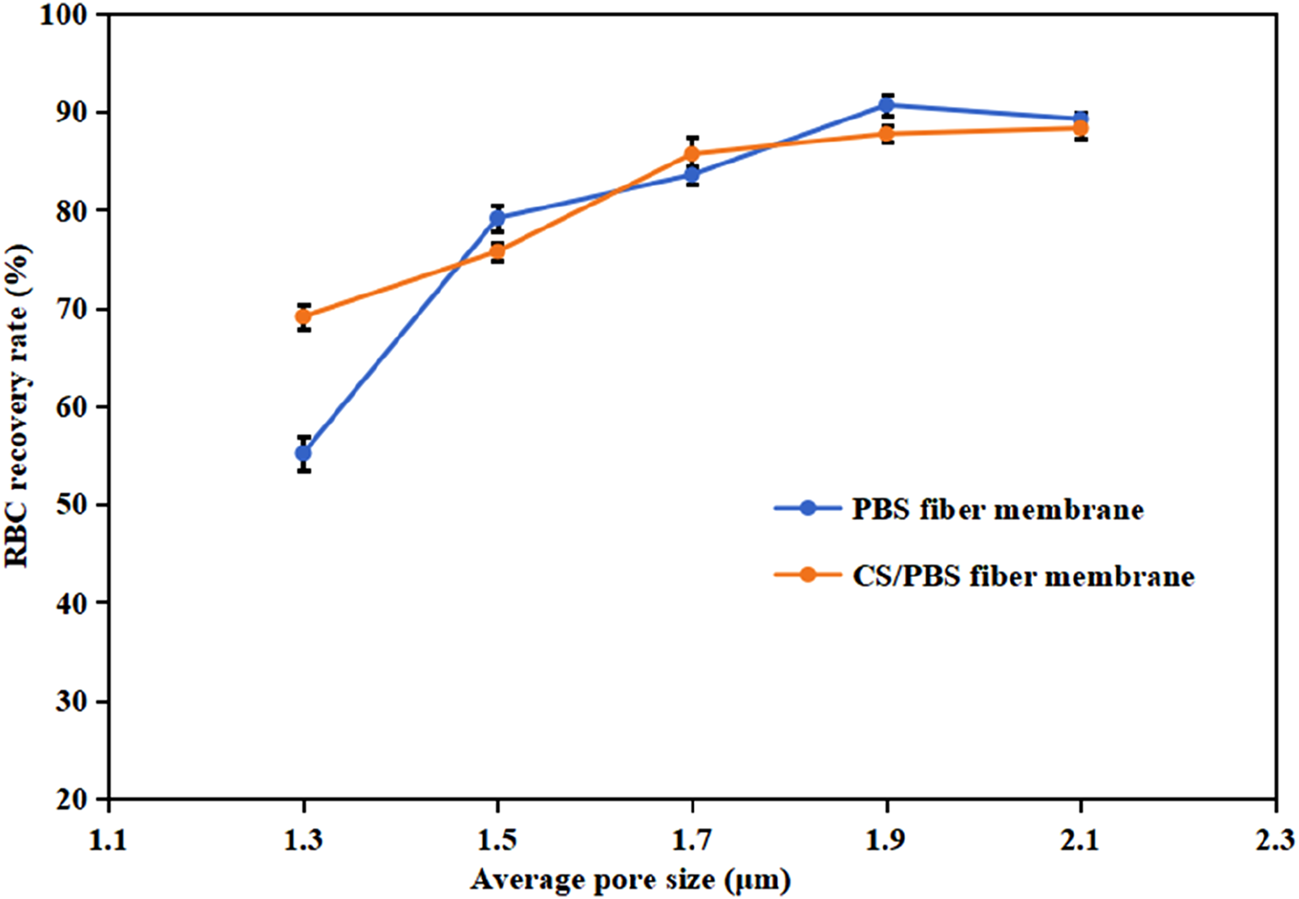
Relationship between RBC recovery rate and average pore size of fibrous membrane.
The results in Figure 5 indicated that RBC recovery rate increased with the increase of average pore size, whether it was PBS fiber membrane or CS/PBS fiber membrane. This was mainly because larger pore size facilitates the passage of red blood cells, thereby improving the recovery rate of red blood cells. But when the average pore size reached 1.9 μm, RBC recovery rate tended to stabilize. This was because when the pore size reaches a certain threshold, the number of red blood cells that can pass through the filter material tends to stabilize. RBC that did not pass were likely to be adsorbed by the fiber membrane or trapped between the nanofibers due to the stacking effect. Additionally, Figure 5 indicated no significant difference in RBC recovery rates between PBS and CS/PBS membranes. Under some conditions, PBS membranes even exhibit marginally higher RBC recovery. This phenomenon may be attributed to the stronger wettability capability of CS/PBS, which enhances RBC adsorption during filtration, thus resulting in slightly lower recovery compared to PBS fiber membranes. Thus, surface adsorption likely plays a role in filtration, in addition to pore size. At the same time, it can be seen that the filtration time of CS/PBS fiber membrane was shorter than that of PBS fiber membrane, which was due to the good wettability of CS/PBS membrane.
In essence, with an average pore size of 1.9–2.1 μm, the nanofiber membrane demonstrated complete removal of white blood cells and a red blood cell recovery rate nearing 90%. The RBC recovery rate of commercial filters is approximately 90%.13,18,19 In comparison to commercial Leukocyte filters, this filtration material achieved full elimination of WBCs and RBC recovery rate comparable to that of commercially available filters. Therefore, the nanofiber membrane devised in this investigation proves advantageous in addressing the particular requirements of individuals receiving blood transfusions.
Conclusion
In this investigation, electrospun membranes composed of polybutylene succinate (PBS) and chitosan (CS)/PBS were successfully fabricated for the purpose of filtering blood leukocytes. The experimental findings indicate that electrospun PBS and CS/PBS nanofiber membranes, featuring precisely controlled pore sizes ranging from 1.3 to 2.1 μm, achieve complete removal of leukocytes with 100% efficiency during blood filtration. This surpasses the performance of conventional melt-blown filters, which typically retain residual white blood cells at levels between 105 and 106 per liter. The optimal pore size range of 1.9–2.1 μm facilitates high recovery rates of red blood cells, ranging from 88% to 90.65%, by capitalizing on the size-based exclusion of rigid leukocytes (6–20 μm) while allowing the passage of deformable red blood cells (6–9.5 μm). Moreover, the incorporation of CS enhances the wettability of the fiber membrane and reduces filtration time. These nanofiber membranes offer an effective solution for leukodepletion in vulnerable patient populations, such as organ transplant recipients and pregnant women. This investigation advocates for electrospun nanofiber membranes to be considered as the next-generation materials for blood filtration. Future research will focus on assessing the safety of PBS and CS/PBS nanofiber membranes as blood filtration materials, including evaluations of cytotoxicity, biocompatibility, antibacterial properties, among others, as well as on advancing the large-scale production and clinical implementation of these fiber membranes.
Footnotes
Funding
The authors disclosed receipt of the following financial support for the research, authorship, and/or publication of this article: This work was supported by Special Fund for The Construction of High-level Teachers in Beijing Institute of Fashion Technology; (Grant No: BIFTXJ202218); Beijing Natural Science Foundation; (Grant No: 2252033).
Declaration of conflicting interests
The authors declared no potential conflicts of interest with respect to the research, authorship, and/or publication of this article.