
Abstract
In this study, antimicrobial and deodorizing nanofibrous composite membranes based on natural substances were developed for use in feminine sanitary napkins. Plant-derived natural essential oils, such as lemongrass oil and May Chang oil, were incorporated into polyvinyl alcohol nanofibers using emulsion electrospinning. The fiber morphology, oil distribution, and pore size distribution of the nanofibrous composite membranes were examined. The antimicrobial and deodorizing effects, tensile properties, and release behavior of the functional ingredients from the composite membranes were investigated to examine their use in sanitary napkins. Core–sheath nanofibers, in which lemongrass oil or May Chang oil was uniformly distributed within the fiber core, were fabricated using various oil contents. The essential oil-loaded nanofibrous composite membranes contained pores ranging from 0.130 to 1.349 µm in size. The release profiles of the essential oils from the composite membranes over an 8 h period demonstrated a continuous release of citral and limonene. Composite membranes containing either lemongrass oil or May Chang oil exhibited outstanding antimicrobial effects against vaginal pathogens, such as Candida albicans and Staphylococcus aureus, despite their low oil content. Deodorizing effects against ammonia gas were observed at oil contents higher than that required to achieve antimicrobial effects. The composite membranes exhibited lower Young’s modulus and greater elongation at break values under wet conditions compared to dry conditions. Our findings demonstrate that antimicrobial and deodorizing nanofibrous composite membranes based on plant-derived essential oils have the potential for use in feminine hygiene products.
Keywords
Introduction
Feminine hygiene products are an essential part of women’s daily lives and are important in maintaining women’s health and hygiene. Sanitary napkins are disposable, absorbent hygiene products that constitute a substantial share of the global female hygiene market. The global sanitary napkin industry was valued at US$25.6 billion in 2022, which is expected to steadily grow in the next 5 years. 1 Sanitary napkins, also known as sanitary pads, have a multilayered structure typically comprising a cover stock, an acquisition and distribution layer, an absorbent core, elastic wings, a back sheet, and a siliconized paper. 2 The primary function of sanitary napkins is to absorb and retain menstrual liquid discharge, but growing consumer awareness and expectations have increased the demand for other functionalities, such as odor prevention, antimicrobial properties, and enhanced comfort.
A survey regarding sanitary napkins 3 demonstrated that the current main problems include leakage and odor, indicating the need for leakage-proof, odorless sanitary napkins. According to the survey respondents, antibacterial activity was the most desirable additional functionality that should be added to sanitary napkins. In addition, the respondents stated that they preferred sanitary napkins composed of natural materials to avoid the health risks associated with using synthetic materials. Using feminine sanitary products may cause irritant contact dermatitis, leading to skin infections. 4 Therefore, feminine sanitary napkins with antimicrobial and deodorizing effects must be developed based on natural substances to satisfy consumer needs and minimize undesired skin effects.
Essential oils are volatile natural oils extracted from the flowers, fruits, leaves, or roots of plants using extraction techniques such as steam distillation. 5 These oils comprise different molecules, such as monoterpenes, sesquiterpenes, and phenols, 6 which possess antimicrobial effects, enhance the immune system, and provide stress relief.7–9 In particular, essential oils such as lemongrass oil and May Chang oil are effective against Candida albicans, a type of vaginitis-causing fungus.10–12 Certain essential oils possess a deodorizing effect on various substances, including ammonia gas.13,14 Therefore, it would be beneficial to investigate if these two types of essential oils can be used to produce sanitary napkins with antimicrobial and deodorizing effects.
Electrospun nanofibrous membranes are extremely thin, lightweight, and flexible. Furthermore, they have an exceptionally high surface-area-to-volume ratio and contain numerous pores in the micrometer range that enable the transport of air and water vapor.15,16 Based on the aforementioned reasons, these membranes are excellent candidates for use in sanitary napkins, ensuring hygiene and comfort. Yadav et al. 17 developed cellulose acetate electrospun nanofibers with high absorbency for feminine hygiene applications, focusing primarily on the moisture management and absorption properties of sanitary napkins. Certain feminine sanitary napkins on the market utilize nanofibrous membranes as breathable but liquid-impermeable backsheets.
Although there are numerous studies regarding incorporating different types of essential oils into nanofiber matrices and focusing on imparting bioactivities such as antimicrobial, anti-inflammatory, and antifungal properties to nanofibers,18–25 studies have primarily focused on pharmaceutical and biomedical applications such as wound dressings, scaffolds for tissue engineering, and targeted/controlled delivery, or active food packaging applications. Essential oil-loaded nanofibers should be further investigated for use in sanitary napkins to address the drawbacks of existing sanitary napkins, which could expand the potential applications of electrospun nanofibrous materials. However, to the best of our knowledge, the development of antimicrobial and deodorizing nanofibrous membranes based on plant-derived essential oils as functional layers for sanitary pads has not been reported thus far. To accomplish this, essential oils that possess the functionalities required for hygiene products need to be identified, and the effectiveness of these functionalities after being encased within the nanofibers must be investigated.
Essential oils can be encapsulated within a polymeric fiber matrix via emulsion electrospinning, which is an innovative technique for producing core–sheath nanofibers using a single-nozzle electrospinning setup by employing an emulsion of two immiscible liquid phases as the working liquid.26,27 Core–sheath nanofibers are produced as a result of the stretching and coalescence of the emulsion that occurs during electrospinning. The solvent in the surface region rapidly evaporates, and the viscosity of the continuous phase increases, moving the emulsion droplets towards the center and promoting coalescence, resulting in the core. 28 Emulsion electrospinning has many advantages, especially for encapsulating functional materials such as bioactive compounds, drugs, and enzymes into polymer fibers without using a coaxial electrospinning apparatus to form core–sheath structures, which enables the sustained and controlled release of functional materials. Emulsion electrospinning is significantly promising for applications in food, biomedicine, and pharmaceuticals, where the toxicity of organic solvents can be critical because it can encapsulate lipophilic compounds within hydrophilic polymers while avoiding the use of organic solvents.29,30 Using essential oil-loaded nanofibrous membranes in hygiene products would enable the gradual release of essential oils encapsulated within the fiber core through the polymer matrix, allowing the sustained release of functional components while using the sanitary napkins.
In this study, we aimed to develop antimicrobial and deodorizing nanofibrous composite membranes by incorporating lemongrass oil and May Chang oil into the fiber core via emulsion electrospinning and to investigate their potential as a functional layer in feminine sanitary napkins. Polyvinyl alcohol (PVA) was used as the polymer matrix because it is nontoxic, biocompatible, and biodegradable. Furthermore, it is used in biomedical applications such as wound dressing, drug delivery, and contact lenses. 31 Lemongrass oil- and May Chang oil-loaded PVA nanofibers were fabricated with various oil contents to examine their antimicrobial and deodorizing effects. The fiber morphology, oil distribution, and pore size distribution of the composite membranes were examined. The release of lemongrass oil and May Chang oil from the composite membranes was assessed over an 8 h period to examine their release profiles during the use of sanitary napkins. The antimicrobial and ammonia-deodorizing properties of lemongrass oil- and May Chang oil-loaded nanofibers were evaluated and compared. The tensile properties of the composite membranes containing the lemongrass and May Chang oils were examined under dry and wet conditions.
Materials and methods
Materials
Lemongrass oil (extracts of Cymbopogon flexuosus) and May Chang oil (extracts of Litsea cubeba) were purchased from Neumond Co. (Germany). PVA (>99% hydrolyzed, Mw = 89,000–98,000) was purchased from Sigma-Aldrich Co. (USA). Tween 80 (Kao Co., Japan) was used as a surfactant to produce a stable oil-in-water (o/w) emulsion. Nile red (Sigma-Aldrich Co., USA) was used as the lipophilic dye for confocal laser scanning microscopy. Citral (Sigma-Aldrich Co., USA) was used as the standard substance for the thermodesorption gas chromatography–mass spectrometry (TDS–GC–MS) analysis.
Fabrication of lemongrass oil- and May Chang oil-loaded nanofibers
Emulsion preparation
First, PVA solutions were prepared by dissolving the polymer in distilled water at 80°C for 6 h. Lemongrass oil or May Chang oil was then added to the PVA solutions, along with a certain amount of Tween 80 (a surfactant). The composition of the emulsion will be specified later. The emulsion was stirred using a vortex mixer at 1000 rpm for 1 h.
Emulsion formulations containing lemongrass oil and May Chang oil for imparting antimicrobial activity.

Emulsion formulations containing lemongrass oil and May Chang oil for imparting deodorizing effects.

Emulsion electrospinning
Electrospinning was conducted in a single-nozzle electrospinning setup (NCC-ESP200R2, NanoNC Co., Korea) comprising a syringe pump, high-voltage power supply, nozzle, and grounded collector. The electrospinning setup was equipped with a computer-aided system that controlled the nozzle and collector to move lengthwise and crosswise, respectively, enabling the production of uniform nanofibrous membranes. Each emulsion, comprising the essential oil, PVA solution, and surfactant, was loaded into a syringe with a metal needle. Electrospinning was conducted under various spinning conditions to optimize the processing conditions for producing core–sheath nanofibers with essential oil as the core and PVA as the sheath. The syringe pump delivered the emulsions at controlled flow rates of 0.2, 0.5, 0.8, 1.2, and 1.8 mL/h; furthermore, 23-gauge (0.33 mm i.d.) and 25-gauge (0.26 mm i.d.) needles were used. The applied voltage and the tip-to-collector distance were maintained at 25 kV and 20 cm, respectively. When a high voltage was supplied to the needle, the electrospun fibers were deposited onto the Teflon sheets placed on the metal collector. The electrospun fibrous membranes obtained were separated from the Teflon sheets for further analysis.
Morphological characterization
The surface morphology of the electrospun nanofibers was examined using field-emission scanning electron microscopy (FE-SEM; JSM-7800F, JEOL Ltd, Japan). Prior to the observation, the nanofibers were sputter-coated with platinum for 90 s. The average fiber diameter was determined using the ImageJ software (National Institutes of Health, USA) by analyzing 100 random fibers from five different SEM micrographs (averaging n = 20 fibers per image).
The morphology of the composite nanofibers was further characterized by transmission electron microscopy (TEM) and confocal laser scanning microscopy (CLSM). A transmission electron microscope (JEM1010, JEOL Ltd, Japan) was used to observe the inner structure of the nanofibers at an accelerating voltage of 80 kV. To visualize the distribution of the essential oils within the nanofibers, Nile red, a lipophilic fluorescent dye, was used to label the essential oils prior to electrospinning. The essential oils were dyed using 0.01 g/mL Nile red prior to emulsification. Emulsions containing the Nile red-labeled oils were electrospun, and the electrospun composite nanofibers were observed using CLSM (CLSM 700, Carl Zeiss, Germany). The excitation and emission wavelengths were 555 and 585–700 nm, respectively.
Stabilization of PVA-based nanofibers
PVA is a water-soluble polymer with good biocompatibility and nontoxicity; therefore, it is widely employed in biomedical applications. However, owing to its intrinsic water solubility, it requires additional treatment to be used in moist environments, such as sanitary products. In this study, heat treatment was performed to stabilize the PVA-based nanofibers in moist environments because we aimed to avoid using toxic crosslinking agents such as glutaraldehyde or formaldehyde.
Essential oil-loaded PVA nanofibers were thermally treated at various temperatures (150, 160, 170, and 180°C) for different time periods (1 min and 5 min) to optimize the heat treatment conditions for the composite fibers. Following heat treatment, the composite fibers were immersed in distilled water at 36°C (thermophysiological temperature) for different durations (1 h and 4 h) to examine the effects of heat treatment. After drying the fibrous membranes, their integrity was observed using FE-SEM.
Pore size distribution measurement
The pore size distributions of the nanofibrous composite membranes were measured using a capillary flow porometer (CFP-1100A, Porous Materials Inc., USA). The largest pore diameter, smallest pore diameter, mean pore diameter, and pore size distribution were determined for the specimens with a web area density of 10 g/m2.
Essential oil release studies
Prior to assessing the release of the lemongrass and May Chang oils from the composite membranes, the chemical composition of the oils was characterized by a thermal extractor (TE, Gerstel, Germany) and TDS–GC–MS. The TDS–GC–MS system comprised a thermal desorption system (TDS2, Gerstel, Germany), gas chromatograph (GC, 6890N, Agilent, UK), and mass selective detector (MSD, 5975, Agilent, UK). A Tenax tube loaded with each essential oil was attached to the TE to collect 1 L of the air sample. Nitrogen gas was used as the carrier gas (flow rate: 39 mL/min) at the thermophysiological temperature (36°C) for 25 min. The collected gas was injected into a gas chromatography (GC) column using helium as the carrier gas (flow rate: 1 mL/min). The initial column temperature (50°C) was maintained for 5 min and then increased by 5°C per minute from 50 to 220°C; this temperature was maintained for 10 min. The temperature was then increased by 10°C per minute to 250°C and maintained for 5 min.
We used TDS–GC–MS with the same experimental setup to investigate the essential-oil release profiles from the lemongrass oil- and May Chang oil-loaded nanofibers. The calibration curve of the essential oil compound was prepared; subsequently, the concentration of volatile organic compounds (VOCs) released from the composite nanofibers was back-calculated from the predetermined calibration curve of the essential oil compound based on the peak area of the GC chromatograms. The release behavior of the fibers containing each essential oil was assessed four times, that is, 2, 4, 6, and 8 h after the heat treatment of the fibers, considering the wearing time of the sanitary napkins. The web area density of the nanofibers was 10 g/m2, and the experiments under each condition were performed in triplicate.
Antimicrobial property assessment
The antimicrobial activities of the lemongrass oil- and May Chang oil-loaded nanofibers were assessed according to ASTM E 2149-13a (the standard test method for determining the antimicrobial activity of antimicrobial agents under dynamic contact conditions). In this test, an inoculated buffer solution was added to the flasks with or without the sterile specimens and agitated in a shaker for a specified contact time. Subsequently, the aliquots were collected from the solution, serially diluted, and plated onto nutrient agar plates. After incubating the agar plates for 24 h, the number of colonies in the agar broth was counted to calculate their reduction rate. Two representative microorganisms were used: Candida albicans (ATCC 10231, Eumycetes) and Staphylococcus aureus (ATCC 6538, a Gram-positive bacterium). The reduction rates of the test organisms after contact with the specimens for 24 h were determined as follows:
Ammonia deodorization property assessment
The ammonia deodorization properties of the lemongrass oil- and May Chang oil-loaded nanofibers were examined using the gas detector tube method. A 10 cm × 10 cm specimen was placed in a 5 L Tedlar bag attached to a gas detector tube (model 3La, Gastech Corp., Japan), and ammonia gas (3 L) was injected. After 2 h, the gas concentration was measured, and the ammonia deodorization efficiency was determined as follows:
Tensile property assessment
The tensile properties (Young’s modulus, tensile stress, and elongation at break) of the nanofibrous composite membranes containing the essential oils were examined using a universal testing machine (Instron 5543, Instron Co., USA). Because the membranes were intended for use in sanitary napkins, their tensile properties were assessed under both dry and wet conditions. The tensile properties were tested according to ASTM D 5035 (the standard test method for breaking force and elongation of textile fabrics (strip method)). Five specimens were prepared with a length of 15 cm, width of 2.5 cm, and thickness of approximately 0.1 mm, as measured by a digital thickness gauge. The web area density was 10 g/m2, and the applied extension rate was set to 3 mm/min.
Results and discussion
Fiber morphology analysis
Nanofibers containing low concentrations of essential oils
The surface morphologies of the composite nanofibers produced from various emulsion formulations (emulsions A–E in Table 1) and the processing conditions are shown in Tables S1 and S2 (Supplementary Information), which demonstrate similar fiber morphologies for the same emulsion formulation prepared with each oil. For emulsion A (PVA: 10 wt%, essential oil: 3.57 wt%, and surfactant: 0.71 wt%), the fiber diameter was relatively thin but inconsistent, and several beads were observed regardless of the oil type. In emulsion B (PVA: 11 wt%, essential oil: 3.93 wt%, and surfactant: 0.79 wt%), the number of beads decreased, and the emulsion produced relatively straight composite nanofibers with smooth surfaces. In the PVA mass fraction range 12–14 wt% (emulsions C, D, and E), the beads disappeared, but thicker fibers were produced with the appearance of significantly fine fibers, demonstrating a significant deviation in the fiber size. As the PVA mass fraction in the emulsion increased, the number of beads decreased, whereas the fiber diameter increased. Based on fiber size and surface morphology, suitable emulsion formulations and spinning conditions for producing uniform composite nanofibers containing each oil were determined. For lemongrass oil, emulsion B (PVA: 11 wt%, essential oil: 3.93 wt%, and surfactant: 0.79 wt%) yielded bead-free composite fibers with an average diameter of 152 ± 55 nm at a feed rate of 1.2 mL/h, using a 25-gauge needle, voltage of 25 kV, and tip-to-collector distance of 20 cm, as shown in Figure 1(a). For May Chang oil, emulsion B (PVA: 11 wt%, essential oil: 3.93 wt%, and surfactant: 0.79 wt%) produced bead-free composite fibers with an average diameter of 124 ± 50 nm at a feed rate of 0.8 mL/h, using a 23-gauge needle, voltage of 25 kV, and tip-to-collector distance of 20 cm, as shown in Figure 1(b). The emulsion formulations and spinning conditions were used for further experiments. (a) SEM image of the electrospun lemongrass oil–PVA nanofibers from emulsion B (PVA: 11 wt%, essential oil: 3.93 wt%, and surfactant: 0.79 wt%), produced with a solution feed rate of 1.2 mL/h, voltage of 25 kV, and tip-to-collector distance of 20 cm through a 25-gauge needle; (b) SEM image of the electrospun May Chang oil–PVA nanofibers from emulsion B, produced with a solution feed rate of 0.8 mL/h, voltage of 25 kV, and tip-to-collector distance of 20 cm through a 23-gauge needle.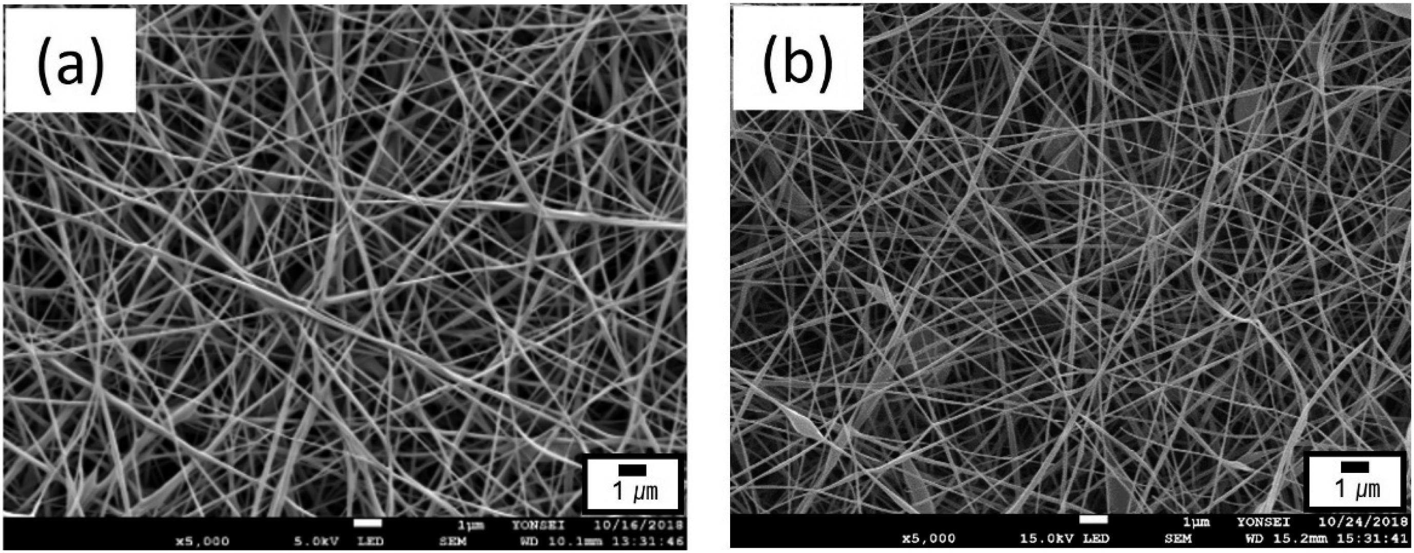
The internal morphologies of the lemongrass oil- and May Chang oil-loaded composite nanofibers were characterized using TEM (Figure 2(a) and (b)), indicating the formation of well-aligned core–sheath structures. The formation of the core–sheath configuration of the emulsion electrospun fiber was associated with the volatility difference between the two liquid phases.28,30 During electrospinning, the solvent of the polymer solution in the near-surface region rapidly evaporated, leading to an increase in the viscosity of the continuous phase compared to that of the dispersed phase. The emulsified oil droplets migrated inward towards the interior of the polymer jet owing to the viscosity gradient and rapid elongation of the jet. Subsequently, the oil droplets coalesced and formed the core, whereas the polymer from the continuous phase formed the sheath, resulting in a core–sheath fibrous structure. The lemongrass oil-loaded fiber had a core–sheath structure with an outer diameter of 101 nm and an inner diameter of 85 nm (Figure 2(a)). The May Chang oil-loaded fiber exhibited a core–sheath structure with an outer diameter of 103 nm and an inner diameter of 89 nm (Figure 2(b)). The lemongrass and May Chang oils were uniformly covered with the PVA polymers comprising the sheath. TEM analyses demonstrated that core–sheath fibers with a sheath thickness of approximately 7–8 nm were successfully fabricated. TEM images of the core–sheath nanofibers containing (a) lemongrass oil and (b) May Chang oil.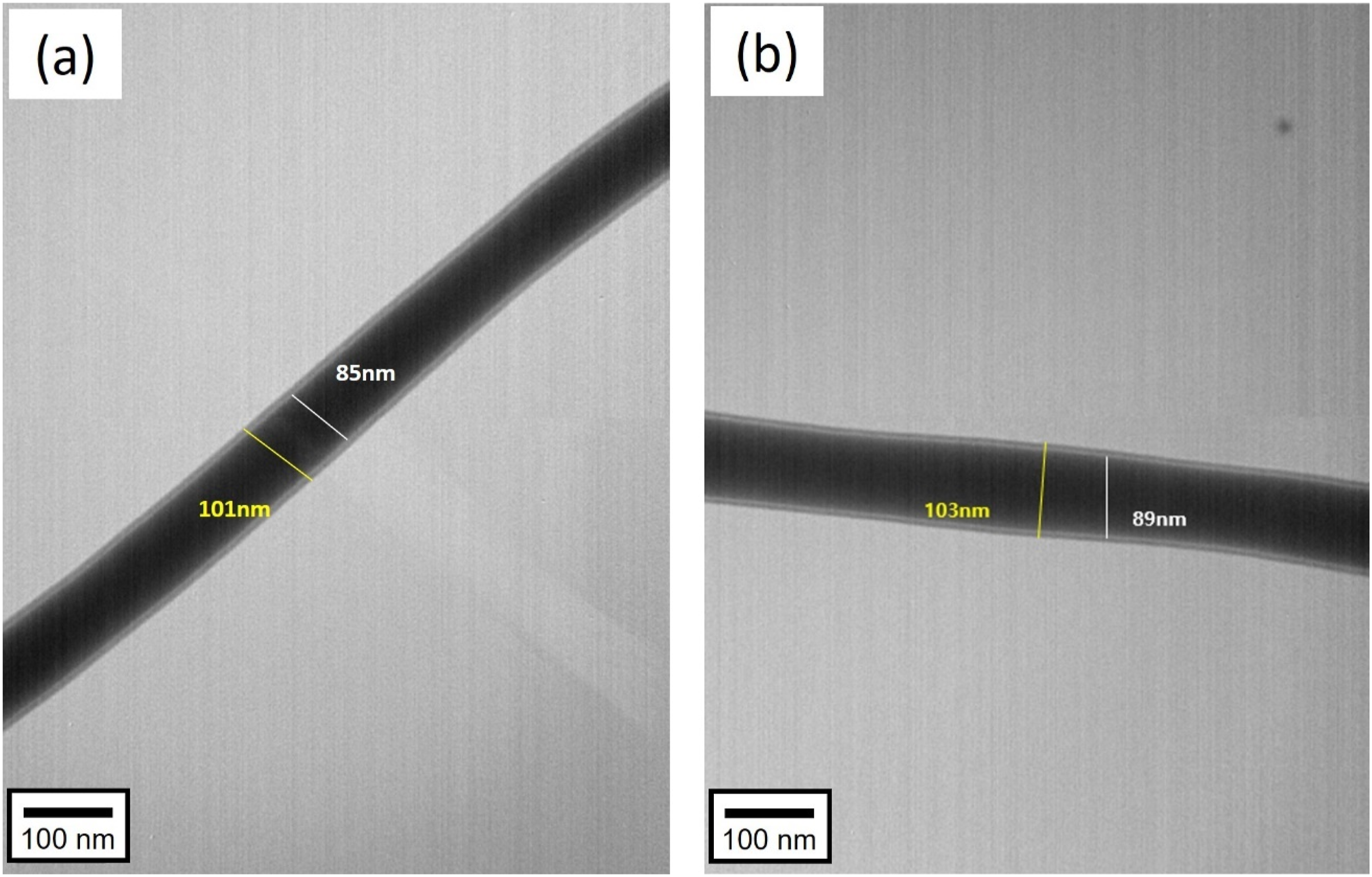
Furthermore, an in-depth analysis of the essential-oil distribution within the nanofibers was performed. Nile red was used to dye the essential oils prior to electrospinning, and the electrospun nanofibers were analyzed by CLSM. Figures 3 and 4 present the nanofibers containing the Nile red-dyed lemongrass oil and May Chang oil, respectively, and their corresponding CLSM images. The essential oils were uniformly distributed in the fiber core, as indicated by red. Thus, the formation of core–sheath nanofiber structures containing lemongrass and May Chang oils was confirmed. CLSM images of the core–sheath lemongrass oil–PVA nanofibers: (a) differential interference contrast (DIC) image, (b) fluorescence image showing the emission fluorescence of Nile red-labeled oil, and (c) overlay of the DIC and fluorescence images. CLSM images of the core–sheath May Chang oil–PVA nanofibers: (a) differential interference contrast (DIC) image, (b) fluorescence image showing the emission fluorescence of Nile red-labeled oil, and (c) overlay of the DIC and fluorescence images.

Nanofibers containing high concentrations of essential oils
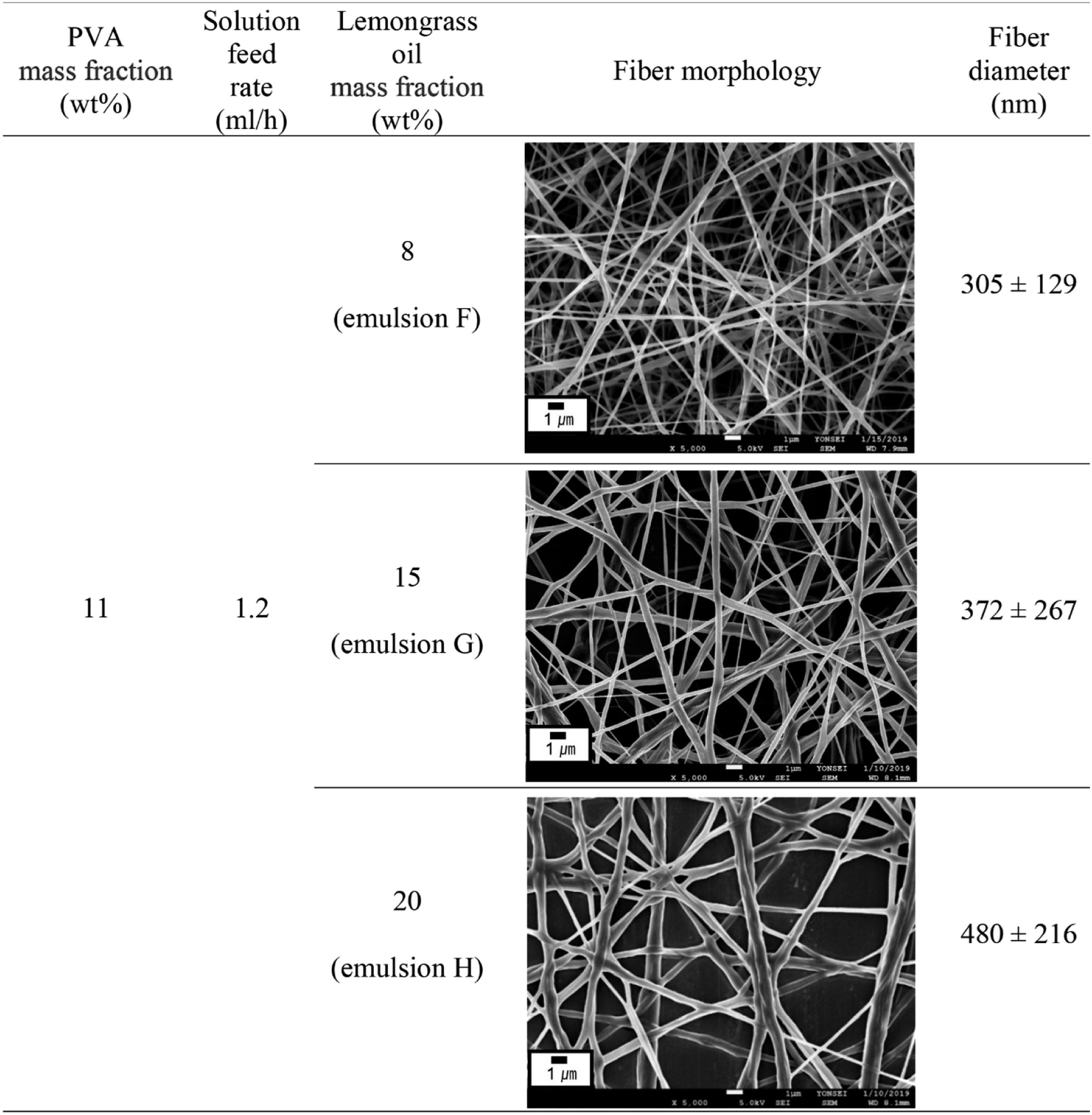
SEM images of lemongrass oil–PVA nanofibers obtained from emulsions containing high oil contents.

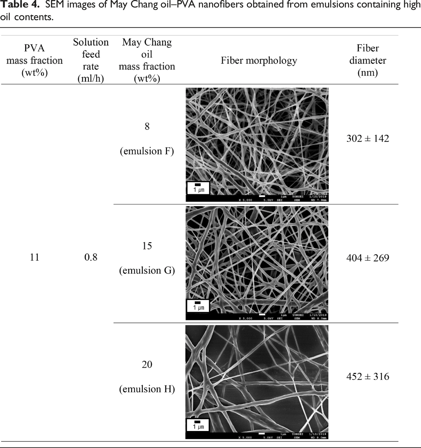
SEM images of May Chang oil–PVA nanofibers obtained from emulsions containing high oil contents.
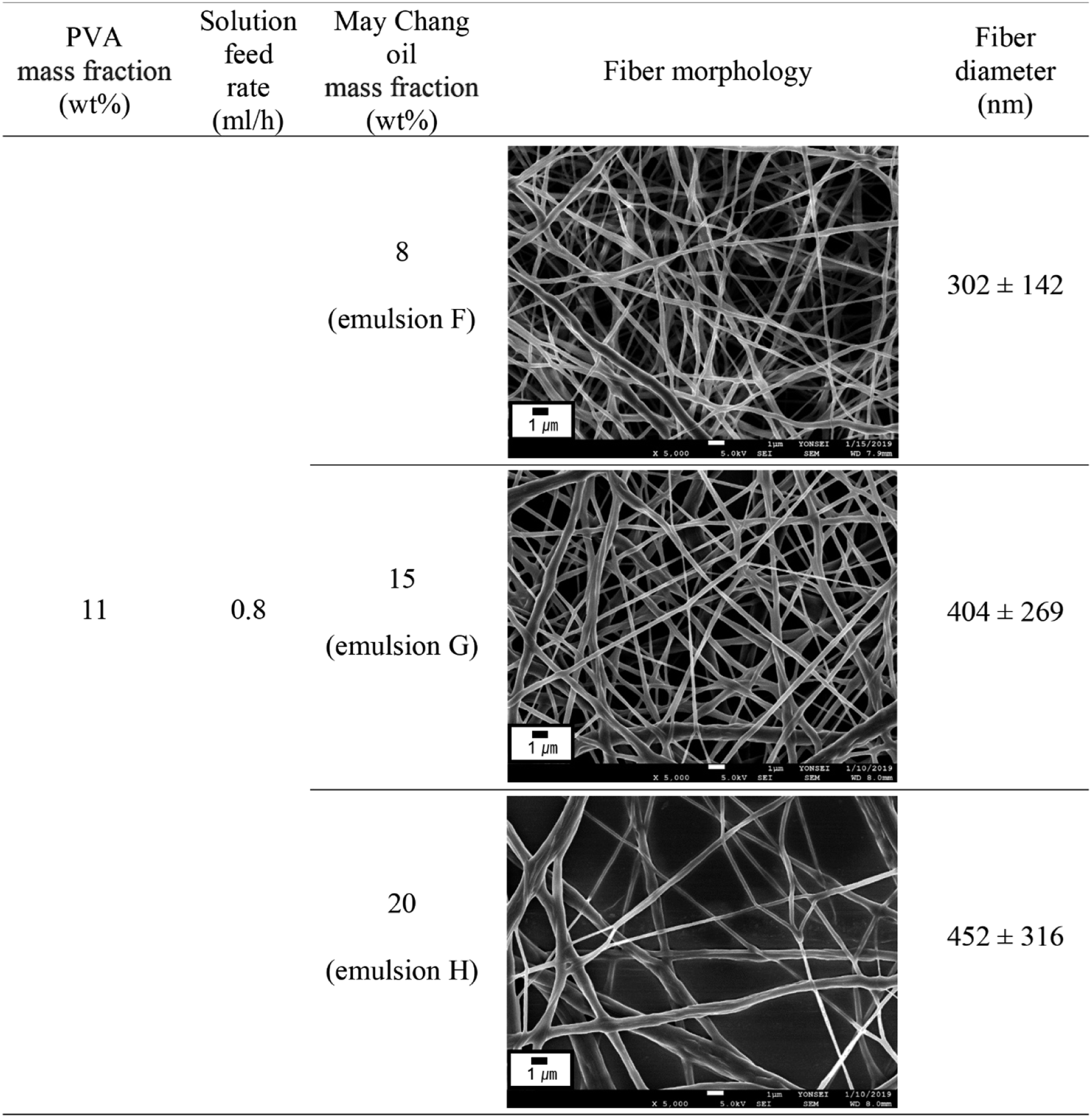
CLSM was used to examine the distribution of essential oils in the composite fibers with a high oil content. Emulsion H, which contained the highest essential oil content, was used. Figure 5(a) and (b) present the CLSM images of the composite fibers containing Nile red-dyed lemongrass oil and May Chang oil, respectively, indicating that the essential oils were evenly dispersed throughout the fibers and that the core–sheath fibers were successfully generated despite the high oil content. CLSM images of the core–sheath nanofibers from emulsion H containing (a) lemongrass oil and (b) May Chang oil.
Effects of heat treatment
Tables S3, S4, S5, and S6 present the membrane structures of each essential oil after being submerged in water under various thermal treatment conditions. For the composite nanofibers containing lemongrass oil (Tables S3 and S4), the samples without heat treatment completely disintegrated when exposed to a moist environment. Conversely, when the samples were heat-treated at 150°C, different fiber morphologies were observed depending on the duration of the heat treatment. When the membranes were heated for 1 min (Table S3), the fibers disintegrated in water and lost their fibrous structures. However, when the duration of the heat treatment was increased to 5 min (Table S4), the fiber morphology was partially retained. When the heat-treatment temperatures were increased to 160, 170, and 180°C, the membranes retained their fiber morphology in water regardless of the heat-treatment and water-immersion durations (Tables S3 and S4). For the composite nanofibers containing May Chang oil (Tables S5 and S6), the untreated samples were completely dissolved in water. When heat-treated at 150°C, the samples partially dissolved when impregnated in water, and certain fibers fused into bundles. However, heat treatment at 160, 170, and 180°C resulted in a stable fiber morphology in water, as observed in the composite nanofibers containing lemongrass oil. The heat treatment of PVA above its glass transition temperature has been demonstrated to stabilize the structure by increasing its crystallinity.32,33 Hydrogen bonding is induced during heat treatment, resulting in the formation of crystallites, and the physical crosslinking network that forms among the crystallites during heat treatment enhances the water resistance of PVA.32,33
Based on these results, the heat treatment conditions for the PVA-based composite membranes containing lemongrass oil or May Chang oil were optimized at 160°C for 1 min, which were consequently applied to all the samples to evaluate the release of the essential oils and the performance of the composite membranes.
Pore size distribution of the nanofibrous composite membranes containing essential oils
To analyze the pore size, the nanofibrous composite membranes containing the essential oils were fabricated using emulsion B with a web area density of 10 g/m2 and subsequently heat-treated at 160°C for 1 min. The pore size was determined by measuring the change in the flow rate as a function of the pressure under a nitrogen gas flow through the dry and wet samples. The through-pore diameters were measured in the most constricted part of the pore.
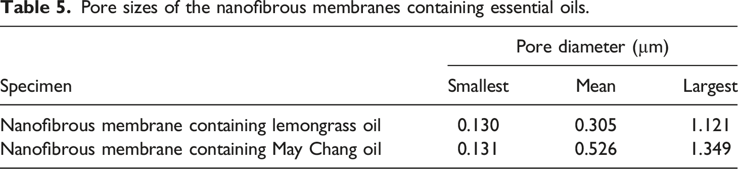
Figure 6 presents the pore size distributions of the nanofibrous membranes containing lemongrass and May Chang oils. As shown in Table 5, the mean pore diameter of the lemongrass oil-loaded nanofibrous membranes was determined to be 0.305 µm. The size of the pores ranged from 0.130 to 1.121 µm. For the May Chang oil-loaded nanofibrous membranes, the pore size ranged from 0.131 to 1.349 µm, and the mean pore diameter was determined to be 0.526 µm. Thus, using the essential oil-loaded nanofibrous membranes as a layer in sanitary napkins may not hinder the comfort of the user owing to their porous structures. Pore size distribution of the nanofibrous membranes containing (a) lemongrass oil and (b) May Chang oil. Pore sizes of the nanofibrous membranes containing essential oils.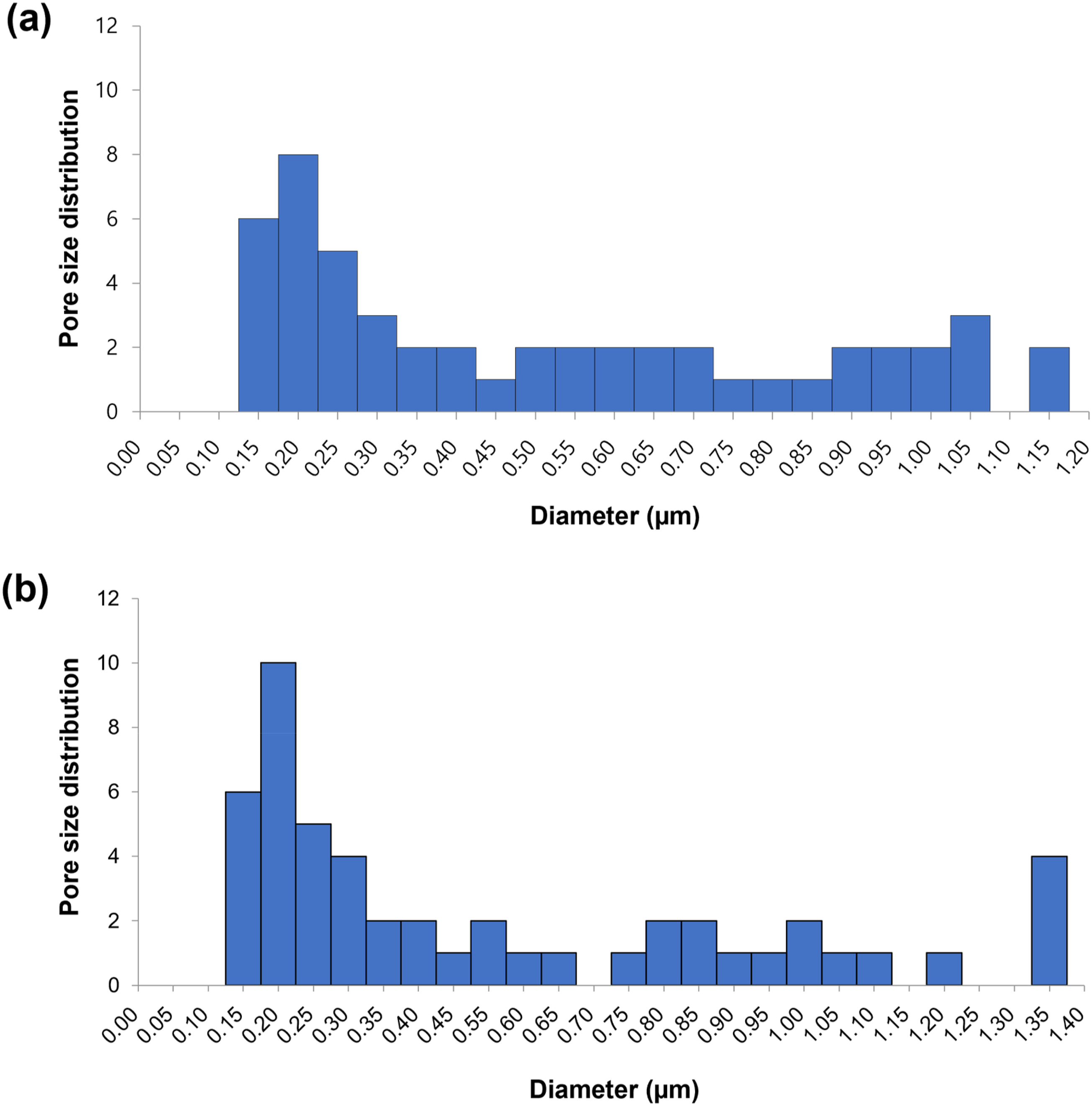
Essential oil release studies
Chemical characterization of essential oils
The VOCs released from the lemongrass and May Chang oils were investigated using TDS–GC–MS at 36°C (human body temperature). The major components of lemongrass oil were neral (33.39%), geranial (19.93%), and ocimene (10.98%). Citral, which is a mixture of two isomeric forms, including geranial (α-citral) and neral (β-citral), constituted 53.32% of the oil. Citral was reported to be the most abundant component of lemongrass oil, accounting for more than 50% of the oil.34,35 Citral has strong antimicrobial activity against pathogenic bacteria and fungi.11,35,36 Silva et al. 11 reported that lemongrass oil and its major component, citral, demonstrated strong antifungal activity against Candida albicans species. The primary components of May Chang oil were neral (26.13%), limonene (21.08%), and geranial (14.69%). Similar findings were reported in a study on the bioactivity of the Litsea cubeba (May Chang) essential oil, where geranial (27.97%), neral (23.57%), and limonene (18.82%) were identified as the main constituents of May Chang oil. 37 Limonene is a naturally occurring monoterpene, and its antifungal activity against Candida albicans and antibacterial properties against various bacterial species have been reported. 38 Therefore, we believe that the incorporation of these two essential oils into feminine hygiene products can impart antimicrobial properties.
Release of essential oils from nanofibrous composite membranes
Release profiles of the major chemical components emitted from lemongrass oil–PVA nanofibrous membranes over time.

Release profiles of the major chemical components emitted from May Chang oil–PVA nanofibrous membranes over time.

For the lemongrass oil–PVA nanofibrous membranes, the amount of TVOCs and NVOCs increased during the initial 4 h of the testing period, after which the released amounts gradually decreased during the remainder of the testing period (Table 6). Citral (geranial and neral) was the most abundantly released component, and the three major chemical components (geranial, neral, and limonene) exhibited release patterns similar to those of the TVOCs over time. This finding indicates that the lemongrass oil-loaded nanofibrous composite membranes release a considerable amount of NVOCs during the initial 4 h of the testing period, followed by a gradual release during the remaining testing period.
The release profiles of May Chang oil were relatively different from those of lemongrass oil. Table 7 presents the release profiles of May Chang oil from the composite nanofibrous membranes over time. The May Chang oil–PVA nanofibrous membranes demonstrated a steady reduction in the amount of TVOCs and NVOCs released throughout the testing period. The three main chemical components (geranial, neral, and limonene) demonstrated release patterns comparable to those of the TVOCs over time.
The release of essential oils from the composite fibers is presumably governed by diffusion. The driving force for diffusion is the concentration gradient of the solute, that is, the concentration gradient of the oil between the reservoir and release environment. The release characteristics are influenced by the ability of the oil molecules to diffuse through the polymer matrix as well as the chemical interactions between the oil molecules and the polymer sheath. This is because different oil molecules have varying chemical interactions with the polymer sheath, resulting in different diffusion rates. Further investigations are needed to determine the cause of the different release profiles of the lemongrass and May Chang oils.
Overall, the release profiles of the three major components (geranial, neral, and limonene) demonstrated that the nanofibrous membranes loaded with lemongrass oil continuously released the bioactive agents for more than 6 h and presented higher and more stable release profiles than the May Chang oil-loaded nanofibers. The release of bioactive agents from lemongrass oil-loaded nanofibers after 8 h was greater than that of May Chang oil-loaded nanofibers after 6 h. Therefore, in terms of the release behavior of bioactive agents, the lemongrass oil-loaded nanofibrous membranes performed better than their May Chang oil-loaded counterparts owing to the higher release of bioactive agents, considering the recommended usage time (2–3 h) and maximum wear time (4 h) of sanitary napkins, as well as the regular sleeping time.
Antimicrobial properties of nanofibrous composite membranes containing essential oils
The antimicrobial activities of the lemongrass oil- and May Chang oil-loaded nanofibrous membranes were evaluated against Candida albicans (ATCC 10231) and Staphylococcus aureus (ATCC 6538). Candida albicans is a major cause of vaginosis in women. 41 Staphylococcus aureus is a common opportunistic pathogen that can cause a variety of human infections. 42 The incidence of Staphylococcus aureus commensalism in the vagina has been reported to be higher during menstruation. 43 Thus, Candida albicans (ATCC 10231) and Staphylococcus aureus (ATCC 6538) were used as representative pathogens to test the antimicrobial activity of the composite membranes for use in sanitary napkins. For the evaluation, the nanofibrous membranes prepared from emulsion B (containing 11 wt% of PVA, 3.93 wt% of lemongrass oil or May Chang oil, and 0.79 wt% of surfactant) with a web area density of 10 g/m2 were used.
Antimicrobial activity of the electrospun nanofibrous membranes containing lemongrass oil and May Chang oil against Candida albicans and Staphylococcus aureus.

Results were collected after a contact time of 24 h.
aNR (no reduction).
Deodorizing properties of nanofibrous composite membranes containing essential oils
The deodorizing activity of the lemongrass oil- and May Chang oil-loaded nanofibrous membranes against ammonia gas was assessed using the gas detector tube method. The mass fractions of the essential oils used to evaluate the deodorizing effect ranged from 3.93 wt% to 20 wt%, as shown in Table 2. Nanofibrous composite membranes with a web area density of 10 g/m2 were used for evaluation.
Figure 7 illustrates the deodorization efficiency of the lemongrass oil- and May Chang oil-loaded nanofibrous membranes based on the oil content. Overall, the deodorizing activity increased as the oil content increased. The nanofibrous membranes fabricated from the emulsion with an oil mass fraction of 3.93 wt% (emulsion B) exhibited no deodorizing effect for either the lemongrass or May Chang oils. At an oil mass fraction of 8 wt% (emulsion F), the lemongrass oil- and May Chang oil-loaded nanofibrous membranes exhibited deodorization efficiencies of 10% and 3%, which increased to 13% and 17% at an oil mass fraction of 15 wt% (emulsion G), respectively. As the oil mass fraction increased to 20 wt% (emulsion H), the deodorization efficiency substantially differed between the lemongrass oil- and May Chang oil-loaded nanofibrous membranes; that is, the lemongrass oil-loaded nanofibrous membranes exhibited a deodorization efficiency of 66%, whereas that of the May Chang oil-loaded nanofibrous membranes was 39%. Lemongrass oil-loaded nanofibrous membranes fabricated from emulsion H (oil mass fraction of 20 wt%) demonstrated the highest deodorizing efficiency (66%) after 2 h; under these conditions, the concentration of the ammonia gas decreased from 100 to 33 ppm. These findings indicate that the composite membranes containing lemongrass oil possess a superior deodorizing efficiency against ammonia gas compared to those containing May Chang oil. Nakasugi et al.
16
demonstrated that the essential oil from black cumin (Nigella sativa L.) seeds provides a substantial deodorizing effect on methyl mercaptan, which is a major cause of oral malodor. Nanashima et al.
17
investigated the deodorizing properties of kuromoji (Lindera umbellata) essential oils using ammonia, hydrogen sulfide, methyl mercaptan, and isovaleric acid. The essential oil eliminated 50% of the ammonia and 97% or more of the isovaleric acid. Although earlier studies16,17 have shown that certain essential oils have a deodorizing effect, this study demonstrates that essential oils encased in the fiber core remain effective for the deodorization of ammonia. These findings may also apply to adult incontinence pads and sanitary products. Deodorization efficiency of the core–sheath nanofibers containing lemongrass oil and May Chang oil at various oil contents.
Tensile properties of nanofibrous composite membranes containing essential oils
Tensile properties of the electrospun nanofibrous composite membranes containing lemongrass oil and May Chang oil.

For the lemongrass oil-loaded nanofibrous membranes, the Young’s modulus, tensile stress, and elongation at break values were 66.67 MPa, 0.75 MPa, and 2.60% under dry conditions and 4.38 MPa, 0.76 MPa, and 21.72% under moist conditions, respectively. For the May Chang oil-loaded nanofibrous membranes, these values were 39.46 MPa, 0.49 MPa, and 2.16% under dry conditions and 5.08 MPa, 0.66 MPa, and 20.02% under moist conditions, respectively.
The Young’s moduli of the nanofibrous composite membranes under moist conditions were considerably lower than those observed under dry conditions. A lower Young’s modulus indicates that a material is more flexible and deforms more easily under stress. Furthermore, both composite membranes demonstrated an increase of approximately tenfold in elongation at break values under wet conditions. The greater flexibility and elongation of the membranes under moist conditions are desirable and can enhance comfort in sanitary napkin applications.
Conclusions
Feminine sanitary products are important disposable hygiene products closely related to feminine health. The feminine hygiene industry has recently shifted towards lighter, thinner, safer, and more comfortable products. In this study, lemongrass oil and May Chang oil were incorporated into PVA nanofibers, and their antimicrobial and deodorizing activities, tensile properties, and release behavior of the essential oils were investigated to examine their use as a functional layer in feminine sanitary napkins.
Emulsions with varying mass fractions of lemongrass or May Chang oils ranging from 3.57 wt% to 20 wt% were electrospun, and core–sheath nanofibers containing various oil contents were fabricated using emulsion electrospinning. A core–sheath structure, in which the essential oils were uniformly distributed within the fiber core, was formed at low and high oil contents, as confirmed by TEM and CLSM. The emulsion formulations and electrospinning conditions were optimized to produce core–sheath nanofibers. The essential oil-loaded PVA membranes were heat-treated under various conditions to increase their water resistance. Heat treatment at 160°C for 1 min effectively stabilized the PVA-based membranes in moist environments. The pore size distribution of the essential oil-loaded nanofibrous membranes with a web area density of 10 g/m2 demonstrated that the membranes contained pores ranging from 0.130 to 1.349 µm in size, which would facilitate the transport of moisture and air. The release profiles of VOCs from the composite membranes containing lemongrass oil or May Chang oil over an 8 h period showed that citral (geranial and neral) and limonene, which are biologically active antimicrobial components of the essential oils, were continuously released from both composite membranes, and that the lemongrass oil-loaded membranes released more bioactive agents than those containing May Chang oil. The nanofibrous membranes fabricated from an emulsion containing 3.93 wt% lemongrass oil or May Chang oil demonstrated strong antimicrobial effects against Candida albicans and Staphylococcus aureus. The ammonia deodorizing efficiency of the composite membranes increased as the concentration of the essential oil increased. The deodorization efficiency of the membranes fabricated from an emulsion containing 20 wt% lemongrass oil was 66%, which was significantly higher than that of membranes containing the same concentration of May Chang oil. The composite membranes containing the essential oils exhibited a substantially lower Young’s modulus and greater elongation at break values under wet conditions than those obtained under dry conditions, implying that the increased flexibility and elongation under damp conditions would be beneficial for sanitary napkin applications. These results indicate that the nanofibrous membranes containing lemongrass oil performed better than those containing May Chang oil, as they released more bioactive agents during the 8 h testing period and possessed superior deodorizing activity against ammonia gas. Our findings demonstrate that ultrathin, lightweight nanofibrous membranes containing lemongrass oil have the potential for use in feminine sanitary napkins with enhanced hygiene and comfort.
Supplemental Material
Supplemental Material - Development of essential oil-containing antimicrobial and deodorizing nanofibrous membranes for sanitary napkin applications
Supplemental Material for Development of essential oil-containing antimicrobial and deodorizing nanofibrous membranes for sanitary napkin applications by Hanul Lee and Seungsin Lee in Journal of Journal of Industrial Textiles
Footnotes
Declaration of conflicting interests
The author(s) declared no potential conflicts of interest with respect to the research, authorship, and/or publication of this article.
Funding
The author(s) disclosed receipt of the following financial support for the research, authorship, and/or publication of this article: This work was supported by the Basic Science Research Program through the National Research Foundation of Korea (NRF) funded by the Ministry of Education, project NRF-2016R1D1A1B03930882.
Supplemental Material
Supplemental material for this article is available online.
References
Supplementary Material
Please find the following supplemental material available below.
For Open Access articles published under a Creative Commons License, all supplemental material carries the same license as the article it is associated with.
For non-Open Access articles published, all supplemental material carries a non-exclusive license, and permission requests for re-use of supplemental material or any part of supplemental material shall be sent directly to the copyright owner as specified in the copyright notice associated with the article.