
Abstract
During current COVID-19 crises, the antimicrobial textiles primarily those utilized in hospital by doctors and paramedical staff have become increasingly important. Thus, there is an unmet requirement to develop antimicrobial textiles for infection control and hygiene practices. Metallic nanoparticles exhibit great effectiveness towards resistant microbial species making them a potential solution to the increasing antibiotic resistance. Due to this, nanoparticles particularly copper and silver have become most prevalent forms of antibacterial finishing agents for the development of antimicrobial textiles. This review is mainly focused on the significance of copper and silver nanoparticles for the development of antimicrobial textiles. The comparative analysis of the antibacterial effectiveness of copper and silver nanoparticles as well as the possible physical and chemical interactions responsible for their antibacterial action are explained. The negative impact of pathogenic microbes on textiles and possible interactions of antimicrobial agents with microbes have also been highlighted. The significance of nanotechnology for the development of antimicrobial textiles and their applications in medical textiles domain have also been discussed. Various green synthesis and chemical methods used for the synthesis of Ag and Cu nanoparticles and their application on textile substrates to impart antimicrobial functionality have also been discussed. The various qualitative and quantitative standard testing protocols utilised for the antimicrobial characterization of textiles have also discussed in this review. The developed Cu and Ag coated textiles could be effectively applied in the field of hospital textiles for the preparation of antibacterial scrub suits, surgical gowns, panel covers, protective clothing, bedding textiles, coveralls, wound dressings, table covers, curtains, and chair covers etc.
Keywords
Introduction
Due to the excellent antimicrobial properties of silver (Ag) and copper (Cu) copper nanoparticles have proven to be in high demand in a variety of applications. 1 The Ag nanoparticles are believed to be a strong antibacterial agent and is used in many commercial items, including those used in the textile sector for the prevention of microbiological contamination. Numerous studies have suggested Ag NPs as efficient antibacterial agents for avoiding or preventing germs on fabric, such as fungi, virus, and bacteria. 2 The Ag-NPs coated textiles have proven excellent materials as wound dressings. The antibacterial properties of silver ions, which have been employed historically, have recently attracted considerable attention. This is due to the fact that some bacillus species have shown an increased antibiotic-resistant behaviour. The strong antibacterial effectiveness of Ag nanoparticles is reported to be effective towards approximately 650 different species of bacteria. 3 The mechanism underlying silver nanoparticles’ antibacterial effect has been the subject of numerous studies. Studies revealed that after being exposed to silver, Escherichia coli microbes are damage, as evidenced by the appearance of “pits” in their cell walls. The antibacterial properties of silver nanoparticles are dependent on chemisorbed Ag+, which is easily generated on the surface of Ag nanoclusters because of their great sensitivity to oxygen. However, detailed research is required to comprehend the method by which silver ions from silver nanoclusters are delivered to the bacterium3,4 Copper has been utilised as an antibacterial agent, conductive materials for wires, electron systems, heat and mass transfer systems, and electromagnetic shielding items. Deposition of Cu-NPs on textiles to develop multifunctional textiles has recently attracted a lot of interest. Electromagnetic shielding, antistatic, UV-protective, and antibacterial textiles can all be fabricated by depositing Cu-NPs on textile materials. 5
The antimicrobial activity of copper and silver nanoparticles has been widely reported. However, the antimicrobial activity of silver and copper coated textiles and their comparative performance has not been reported. In the current study, Various green synthesis and chemical methods used for the synthesis of Ag and Cu nanoparticles and their application on textile substrates to impart antimicrobial functionality have also been discussed. The various qualitative and quantitative standard testing protocols utilised for the antimicrobial characterization of textiles have also discussed in this review. The developed Cu and Ag coated textiles could be effectively applied in the field of hospital textiles for the preparation of antibacterial scrub suits, surgical gowns, panel covers, protective clothing, bedding textiles, coveralls, wound dressings, table covers, curtains, and chair covers etc.
Significance and applications of metallic nanoparticles and NPs coated textiles
Metallic nanoparticles have garnered significant interest owing to their numerous intriguing and distinctive characteristics. Among the all metal nanoparticles that are studied to date, silver nanoparticles are well-recognized for their broad array of applications, including in optical devices, electronic parts, highly stretchable control systems, localised surface plasmon resonance, sensor systems, and pharmaceutics, in addition to their outstanding catalytic and therapeutic properties. 6 The application of metallic nanoparticles for the development of bioactive textiles has become an area of active research owing to their substantially increased demand after COVID-19 crises. The biomedical goods will probably be the largest application of bioactive textiles in near future as they can be used as surgical protective clothing for prevention or wound dressings for therapy. They are suitable for surgical lab coats, or as wound dressings for therapy because they can absorb intoxicants from the skin and deliver bioactive drugs onto the skin. They have also found huge applications for medical textiles such as sutures, bandages, bioactive textiles which include scrub suits, surgical gowns, panel covers, protective clothing, bedding textiles, coveralls, wound dressings, table covers, curtains, and chair covers etc. 7
Comparative performance of copper and silver
Silver and copper are two well-known noble metals that can be found in their natural or “elemental” state at the earth’s crust. Both metals are bright, appealing, and have good corrosion resistance. These metals are soft, ductile and can be twisted into tiny wires. As a result, nanowires and particles are easily found. Furthermore, the draping and bending properties of materials remained unaffected by nanocoating of these metals. Early on, copper was the most common metal, particularly after smelting processes and trade rendered it easily accessible. After aluminium and iron, copper is still the third most significant commercial metal. 1
Copper is a common early metal also known as “red metal” that has a density of 8.94 g/cm3, 63.57 atomic weight and an atomic number of 29. Copper has valences of +1 (cuprous) and +2 (cupric), with +2 (cupric) being the most prevalent.
8
It has two naturally occurring isotopes with mass numbers of 65 (31%) and 63 (69%). In general, auto-oxidation of cuprous salt produces cupric salts easily, as given in equation (1).

Although silver is corrosion resistant, it is rapidly tarnished by sulphur-containing compounds such as mustard, e.g. and rubber. Silver is extremely resistant to oxidation. Tarnished silver, on the other hand, only comprises black Ag2S.
A black-colored layer of silver sulphide (Ag2S) is formed when the silver is tarnished in the air. The following chemical formula describes how hydrogen sulphide (H2S) reacts with Ag to produce silver tarnish:
2Ag(s) + H2S(g) → Ag2S(s) + H2(g)
Ag2S could be easily polished by rubbing it against a slurry of sodium bicarbonate, NaHCO3.9,10
H2S+Na2CO3⟶NaHS+NaHCO3
In general, all metals have the possibility to corrode in a variety of environments. Surface corrosion is minor, but thin coating layers are particularly vulnerable. Generally, thickness loss is determined by the loss in thickness annually. This form of corrosion affects copper-coated materials the most, with an average corrosion rate of 0.02 mm/year. This form of corrosion could be addressed by treating the material with a thick layer of copper. Pitting is a form of corrosion that occurs when the copper coating on the protective layer is degraded by cyclic bends in the textile materials. 11 Surface contaminations containing sulphide contamination or copper alloys on copper-coated fabrics also facilitate pitting. Another factor that accelerated corrosion is the occurrence of cracks on the surface of the plated-copper layer. 12 Copper-silver and copper-nickel alloys are the strongest corrosion-resistant metals. Another type of corrosion that develops on copper alloys and is capable of causing harm is erosion corrosion. 13 This is especially widespread in case of nanoparticles, which are more advantageous in terms of providing antibacterial characteristics.
Copper and silver are both metals that kill a wide range of microorganisms. All bacterial strain categories, including Gram-negative and Gram-positive bacteria, are referred to as broad spectrum. Even in nano form, silver metal is harmless until silver ions are discharged. In wet environments, Ag ions are discharged, causing cell wall damage. In a moist condition, silver antibacterial action is more efficient at a warmer temperature compared to room temperature. 14 Copper, on the other hand, has antibacterial properties at all humidity and temperatures levels. 15 This distinction emphasizes the significance of the copper coatings, as antibacterial action must be achievable at a typical hospital room temperature. The two ionic states of Cu are responsible for its unusual behaviour. Experiments were conducted in response to the inflammatory response induced by the therapeutic impact. 16 The antibacterial effectiveness of Cu and Ag nanoparticles is higher in their transitions state i.e. Ag+ and Cu++ as compared to their oxides (AgO, CuO). This could be explained on the fact that the metallic nanoparticles are most stable in their transition states. Copper and silver nanoparticles Another experiment was carried out to test bis (glycinato) copper, a skin-identical compound (II). The experiment was carried out on the skin of a cat. 17 Electron micrograph images of the skin indicated that the exposed area was stained with copper. Additionally, atomic absorption findings indicated 47 ppm copper. A single study compared the EMI shielding, electrical resistivity, and antibacterial characteristics of silver and copper nanoparticle-coated textiles. Both textiles (Ag and Cu NPs coated) exhibited no significant variation in all characteristics. 18 Ali et al. 19 conducted a comparative study to investigate the washing durability of the silver and copper coated textiles. They established that electroless deposition technique is the most efficient approach to coat silver of copper nanoparticles on textiles for the development of highly durable multifunctional textiles. Their findings revealed that both silver and copper nanoparticles have excellent retention on the cotton fabric and are not removed during the laundry even after multiple washing cycles. 19
Necessity of antimicrobial agents
Even with clean skin, microbes are nearly constantly present on the human body, with a mean number of 100–1000 microbes/cm2. At this level, a variety of microbes, including fungi, bacteria, and yeast, can infect the wearer. These microbes have a number of undesirable side effects, including foul odours and stains caused by material discolouration. As a result, the microbial growth on textiles has a variety of negative consequences, not only for the textile but also for the wearer. Textiles provide an appropriate medium for the adhesion, transmission, and multiplication of infection-causing microbial species due to their properties and accessibility to the human body.21,22 Because of their porosity and hydrophilic character, natural fibres are more prone to microbial attack than synthetic fibres. Natural fibres' structure holds nutrients, water, and oxygen, resulting in an ideal enriched culture for rapid microbial multiplication. Furthermore, direct touch with the human body provides warmth, nutrition, and humidity creating the ideal conditions for microbial growth. Fungi and bacteria are the most harmful pathogens. Algae can grow on textiles if they are kept wet for long periods of time. Fungi cause discoloration, fibre damage and staining in textiles, as well as an unpleasant odour and a sticky, slime feel. Microbes may also be induced by the structure and chemical processes of the textile materials. The difficulty is exacerbated by the warm and humid surroundings. Although synthetic materials do not support microbial proliferation, low molecular weight polluted chemicals used in textile finishing may give sufficient nutrition for mildew microbe growth. This causes an unpleasant odour, and staining in the fabric, as well as a decrease in textile strength. For all of these factors, it is critical to keep microbial growth on textiles to a minimum during usage and preservation23,24
Interaction of microbes with textile fibers
The most prevalent natural polymer on Earth is found in cotton fibres, which are made up of cellulose and hemicellulose. It is connected by a linear, continuous chain of D-glucose monomers that condense through b (14)-glycosidic bonds, ranging in length from 15 or less to 7000–10,000. Cotton is vulnerable to microbial deterioration because cellulose is one of the abundant renewable sources. Different degradation pathways might result for various types of microorganisms involved. Fungi can enter cotton fibres through fissures on their surface or at their ends, which causes deterioration to spread from the fibres’ interior to outer layers. In contrast, the breakdown of cotton by bacteria starts at the fibre surface and progresses all the way to the interior layers of the fibres. The primary factor behind cotton’s general weight and strength loss is the depolymerization of cellulose. 25 Products made of cotton that have biodegraded have issues with colour, odour, aesthetic appeal, and hygiene. 26
The reaction of natural and synthetic fibres to microbial growth differs significantly. Both may operate as appropriate substrates, but the mechanisms are fundamentally different in each scenario. Because natural fibres hold water well and microbial enzymes can easily hydrolyze their polymer bonds, they are prime targets for microbial degradation. 27 Microbes attack the majority of cellulosic and protein fibres in general. Cotton, flax, wool, jute, and silk, are the most prone to microbial attack, according to reports. 28
Microbes grow more slowly on synthetic fibres than on natural fibres as their polymeric framework does not store as much water. These fibres, on the other hand, increase the retention of state sweat in the intercellular spaces, where microorganisms proliferate quickly. Synthetic fibre socks, for example, have been reported to be more prone to foot infection than natural fibre socks. For example, bacterial proliferation in fabric increases with the increase in polyester content in textile materials. 28 If finishing treatments like polysiloxane and polyethylene emulsions are used on synthetic fibres, they become prone to microbial deterioration. These finishing chemicals allow microorganisms to breakdown the polymer into ‘chewable bites,’ thereby triggering the hydrolysis cycle. Even the toughest polyurethanes could be broken down in this manner. Under conductive circumstances, microbial damage has been observed on polyester, polypropylene, and nylon, synthetic fibres. 29
However, the textiles may not only operate as substrates for bacterial growth, but they may also behave as active agents in the multiplication of microorganisms, which is a subject of greater concern. Viruses can survive for up to 16 h on fabrics such as washable wool suit, cotton shirting, polyester/cotton shirting, terry towel, and nylon jersey. Viruses can survive and spread more easily in synthetic fibres than in cotton. The virus could be removed physically from the contaminated fabric during launderings. However, they are not completely inactivated as their presence was detected in the laundry drain. 30 This physical removal is aided by detergents that lower surface tension. As a result, viral transmission can easily occur during regular cold-laundering procedures. Furthermore, some bacteria can live on washed cloth. Textiles can meet all of these bacterial growth criteria, resulting in a variety of unfavourable side effects. The microbial presence and their proliferation can lead to health issues, odour, and eventually fabric damage. 31 Staining and loss of functional qualities such as tensile strength and elasticity can occur as a result of bacteria attacking the additives used in textiles. The production of malodor is one of the most significant negative effects. When bacteria proliferate, they metabolise nutrients available in the environment, such as perspiration and soiling, to form odor-causing chemicals. For example, Gram-positive bacterium Staphylococcus aureus is thought to produce 3-methyl-2, hexanoic acid, which is responsible for the characteristic body odour. 32
Bacteria, among several other reasons, transform human perspiration into odorous compounds including amines, carboxylic acids, and aldehydes resulting in an unpleasant odour. P. Vulgaris is a Gram-negative bacterium that can metabolise urea to generate ammonia, which is the source of smell in infant diapers. 32 Gram-positive and Gram-negative bacteria are two types of bacteria. A lipopolysaccharide-containing outer membrane surrounds the peptidoglycan cell wall that encases Gram-negative bacteria. In contrast to Gram-negative bacteria, which have an outer membrane, Gram-positive bacteria have layers of peptidoglycan that are many times thicker. 33
Interaction of antimicrobial agents with microbes
Antimicrobial drugs interact or function in two ways: they either kill or hinder the microbial growth34,23 The mode of action of antimicrobial agents could involve the following reasons, • damaging the cell-wall of microbes • restricted cell-wall synthesis • Altered cell-wall permeability • Inhibiting the metabolic processes • Interacting with plasma membrane • Inhibiting the essential protein synthesis • Restricting the nucleic acid replication and transcription • Inhibiting the enzymatic activity
Several antimicrobial compounds used in textiles have been studied in the food and cosmetics fields. These compounds are absorbed into textile substrates at lower quantities than other chemicals. It is necessary to ensure that these compounds are not only persistently efficacious but also safe for the body and the external environment. Active finishes are materials that contain effective antibacterial chemicals that act within the genome, on pathogenic cells, or in metabolic processes. 35 However, because of the very unique nature of their action, it is critical to distinguish among antimicrobials and other active compounds with a wide range of applications. Antimicrobial compounds come in a variety of forms, each with its own mechanism of action. Oxidising antibacterial agents which includes peroxy compounds, aldehydes, and halogens are compounds that target the cell membrane, enter the cytoplasm, and disrupt the essential enzymes of microbes. 36 On the other hand, coagulants, typically alcohols, deform protein molecules irreversibly. Due to the availability of free electrons, radical formers such as peroxy compounds, halogens, and isothiazones, are extremely reactive. These chemicals react with almost all organic materials, particularly oxidising thiols in amino acids. These chemicals are extremely harmful to nucleic acids, even at low quantities. 20
Antibacterial action of metallic nanoparticles is thought to be due to their large specific surface area and nano scale particle size. Both metal ions and nanoparticles are thought to have antimicrobial properties. Since nanoparticles possess higher specific surface area through which they interact with microorganisms, their effectiveness rises dramatically as their size decreases. 37 Besides the direct antibacterial action of the metallic nanoparticles, their huge specific surface area allows for a significant rise in the concentration of metallic cations emitted from the surface of the particle, resulting in greater biocidal activity. The main harmful effect of nanoparticles is considered to be the oxidative stress, 38 which causes damage to DNA, lipids, and proteins of pathogens. Higher solubility of metallic oxide nanoparticles has also been shown to improve their antibacterial efficacy, 39 suggesting that the increase in concentration of metallic ions emitted from the nanoparticles surface has a significant impact on their action and toxicity. The photo-catalytic destruction of bacteria by TiO2 gives it antimicrobial characteristics40,41 The toxicity of nanoparticles and metallic is induced by their slow release from the surface, according to the suggested antimicrobial mechanism; this is also true for textile materials treated with noble metals and metallic oxide nanoparticles. When compared to extremely biocidal nanoparticles, micron-sized metals did not cause cell harm. 27 The comparable size of nanoparticles and biomolecules including proteins and polynucleic acids contributes to their antibacterial effects. Chemical interactions, physical interactions, or a combination of both may be responsible for nanoparticles antibacterial properties.
Chemical interactions
Chemical interactions can occur between
(i) Cu++ ions and components in the interior of the cell (ii) Cu++ ions and constituents in the cell interior (transport of Cu++ ions across the membrane), (iii) The effects of Cu++ ions on the physical characteristics of resealed human erythrocyte membranes exhibited the attraction at the lipid–protein interface. Whereas Cu++ ions had no discernible effects on the structure of cells, cell polar heads and hydrocarbon chain configurations were disturbed at low concentrations of Cu2. However, they were restored at high concentrations. As a result, the interaction between this cation and the phosphatidylcholine phosphate groups found in the outer leaflet of the erythrocyte membrane may provide the basis for the ordering effect. This binding may also alter the electric field of the membrane along with reducing membrane mobility. These two impacts ought to have an impact on how membrane proteins, in particular ion channels, function. These findings support the hypothesis that Cu2+ ions prevented Na from crossing epithelial cell membranes. 42 Alteration in oxidative phosphorylation and osmoregulation may occur as a result of this, as well as abnormalities in the structural changes of proteins and nucleic acids. Ultimately, microbes exposed to hazardous levels of some nanoparticles upregulate genetic makeup involved in the removal of reactive oxygen species (ROS), leading to oxidative stress. 43
Physical interactions
Physical interactions can be as follows:
(i) Physical interruption of cellular membranes transport mechanism by copper nanoparticles, (ii) Physical degradation to cellular components by copper NPs due to abrasive action, (iii) Penetration of copper nanoparticles through cellular membranes to interact with the inner membrane, (iv) immediate electrostatic contact between copper nanoparticles and bacterial cell membrane constituents
Combination of the physical and chemical interaction
A combination of the physical and chemical interaction as described above:
The copper toxicity is caused by a variety of mechanisms, and it is impossible to explain with certainty that which pathway is active towards certain bacteria. The redox characteristic is a very well-established and common mechanism for causing deadly oxidative cell damage. Different mechanisms are continuously being investigated. In particular, copper behaves differently on cells depending on the environment and growth conditions. Figure 1 concludes description of all the important mechanisms. Copper is able to enter cells through unknown routes. The cell cytoplasm converts copper to Cu+, which then participates in Fenton type responses and produces extremely reactive hydroxyl radicals. As a result, non-specific reactions occur with nucleic acids, lipids, free amino acids, and proteins. Copper-glutathione compounds, which act as copper-donors for metalloenzymes, are formed under anaerobic environments. Cu+ ions displacing iron from iron-sulfur complex proteins of microbes is the most common toxicity mechanism.
45
The mechanisms involved in the toxicity of copper nanoparticles.
44

Chemical and green synthesis of Cu-NPs coated textiles and their antimicrobial properties
Chemical methods for the synthesis of Cu-NPs
Antibacterial functionalization of cotton or other types of textile materials with copper nanoparticles can be categorized in two methods i.e. in-situ (one-step process) and ex situ (two-step process).46,47 As can be seen in Figure 2, the in-situ approach involves synthesis of nanoparticles in the presence of fabric. In this process, nanoparticles are directly grown inside the textile material. Perelshtein and colleagues
48
used ultrasound to irradiate a copper acetate solution in which a 100% cotton fabric bandage was dipped, and then a reducing agent (ammonium-based) was added for the synthesis of CuO onto the cotton fabric. The researchers confirmed that the nanoparticles in the fabric related to the monoclinic crystalline structure of CuO using X-ray diffraction (XRD). This in-situ process of ultrasound functionalization not only produces textiles with excellent antibacterial effects towards Staphylococcus aureus and Escherichia coli bacteria, but it also ensures that they are durable to launderings. Different steps involved in the in-situ functionalization of textiles with nanoparticles.
49

Dip-coating is another approach for in situ antibacterial functionalization. Using this approach, some researchers performed various fabric pre-treatments prior to their functionalized. For example, Markovic et al. 50 used a solution of non-ionic detergent to wash cotton fabric before dipping them in oxalic acid solutions of various concentrations to alter their surface and generate carboxyl groups that increase Cu+2 ion binding. Nabil et al. 51 alternatively hydroxylate cotton fibres with an aqueous solution including hydrogen peroxide (H2O2) and ammonium hydroxide (NH4OH) in order to generate hydroxyl (-OH) groups which might attach or be substituted with some other chemical products introduced after the CuO functionalization. The group led by Rezaie rinsed a wool fabric samples using a non-ionic detergent solution for the elimination of contaminants before further processing. 52 The researchers then dipped and stirred the fabric samples in solutions of copper precursor salts, including copper sulphate or acetate followed by treating the fabric samples with alkaline solutions of sodium hydroxide or sodium borohydride obtained from industrial sources or solutions derived from natural origin, as performed in Rezaie’s findings, who has used the supernatant obtained after dissolving the burnt leaf or stem ashes of Seidlitzia rosmarinus plant, for the synthesis of CuO nanoparticles in the presence of fabrics. Another significant surface modification of cotton fabric to generate carboxyl functional groups using oxalic acid which increases the uptake of copper ions in the in-situ antibacterial functionalization of fabric with CuO nanoparticles was demonstrated by Markovic et al. 50 According to the study, cotton textiles containing more oxalic acid absorb more Cu ions, which could be seen in SEM images and corresponded to excellent antibacterial activities towards Gram-negative and Gram-positive bacteria. 50
In another investigation, the cotton was functionalized with CuO nanoparticles using an exhaust dyeing approach. For functionalization, sodium hydroxide (NaOH) and copper acetate were also used. The bacteriological results confirmed that the functionalized cotton fabric inhibited the growth of Escherichia coli; in addition, it was suggested that a possible path for the cotton fabric and CuO nanoparticles interaction is through the OH-groups present in the cellulose, implying that a complex-fiber bonding might have become a source for the CuO nanoparticles.
53
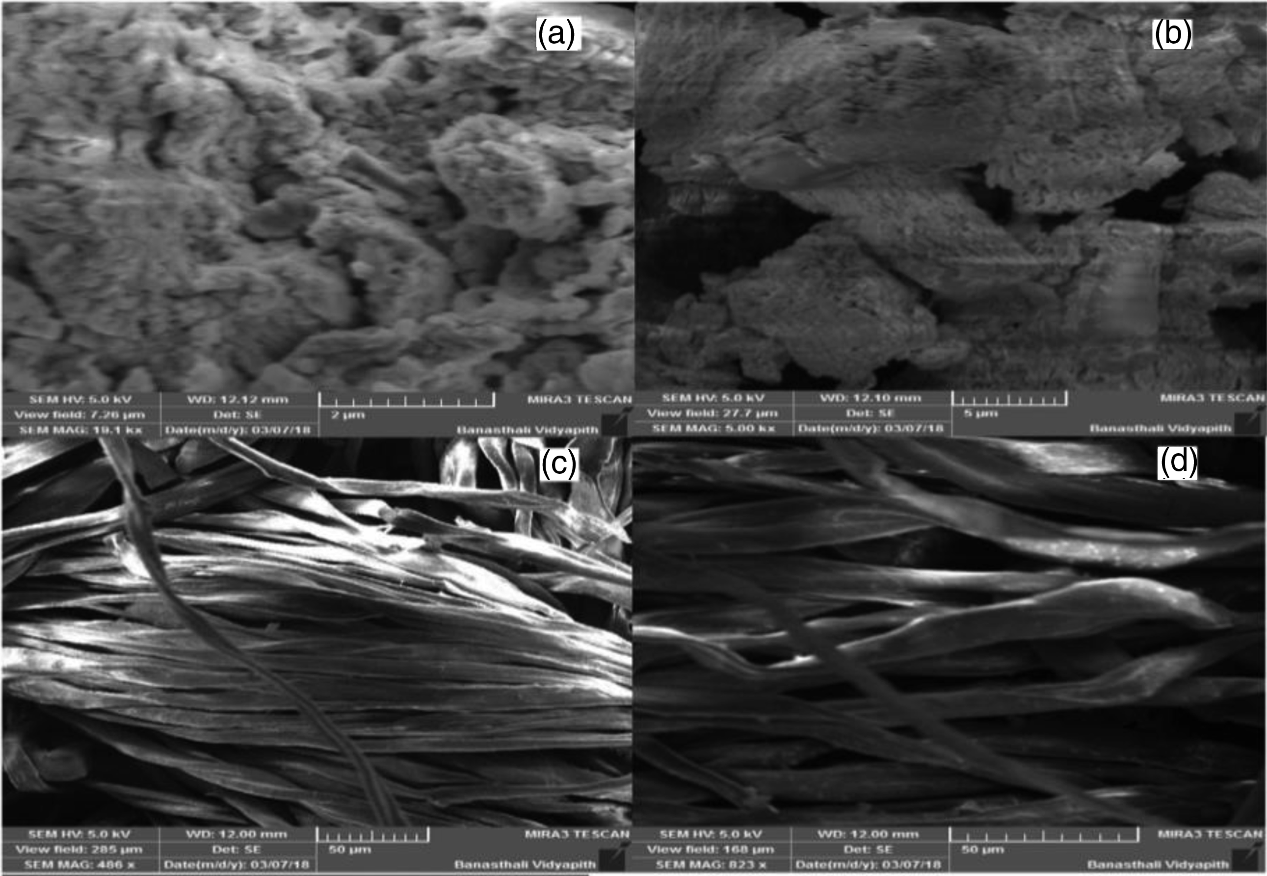
The morphological structure of cotton fibre before as well as after the functionalization treatment could well be assessed in this study using SEM images (Figure 3). In raw fabric, that is, one without any nanoparticles on its surface, can be observed in the non-functionalized cotton fabric shown in Figure 3(a). There are randomly scattered nanoparticles on the cotton fibre in the functionalized cotton fabric (Figure 3(b)). SEM images of untreated cotton fabric 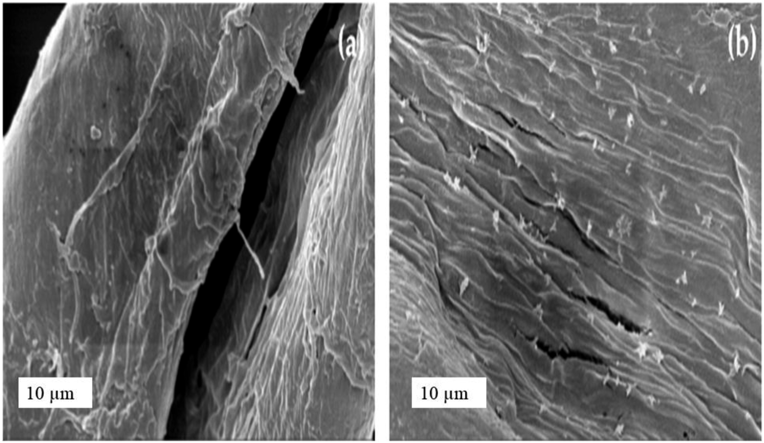
Various chemical methods used for the synthesis of Cu-NPs for antibacterial textiles.

In ex-situ functionalization approach, 2 processes are involved. In first step, nanoparticles are synthesized. Then, the synthesized nanoparticles are applied to textiles in the second step.
47
The ex-situ approach is preferred for the pilot-scale industrial applications. The second stage of the ex-situ functionalization approach is depicted in Figure 4. The schematics showing the second stage of ex-situ functionalization of textiles with nanoparticles.
49

The preparation of CuO nanoparticles through chemical precipitation approach and followed by their impregnation on the surface non-woven and woven textile materials are reported by Thampi et al. 60 CuO nanoparticles were prepared using a mixture of polyethylene glycol and copper nitrate solutions (first stage of the ex-situ process). These nanoparticles were used to produce colloids in which the fabric was dipped, agitated, followed by drying, which is the second step in the ex situ technique. A polymeric matrix constructed of polyaniline was also utilised to immobilise CuO in order to limit the leaching of copper ions from functionalized fabrics into the environment. To achieve that immobilisation, a particular quantity of synthesised CuO was introduced to dissolved aniline, and the resulting solution was applied on the fabric using same impregnation technique described for pure CuO nanoparticles. The functionalized fabric revealed excellent antibacterial action towards S. aureus and E. coli. This study is the best demonstration of two step of ex situ functionalization approach.
Vasantharaj et al. 61 demonstrated similar type of functionalization using the ex situ approach. For the synthesis of CuO nanoparticles, a copper sulphate solution was used as precursor. An aqueous extract from the leaves of Ruellia tuberosa that lower the copper toxicity were used. Following that, these nanoparticles were employed to treat cotton fabric. The researchers confirmed that the fabricated nanoparticles were made of CuO using FTIR (Fourier transform infrared spectroscopy) and UV-visible spectroscopic techniques. Three varying concentrations i.e.25, 50, and 75 g/mL of synthesized CuO nanoparticles were used for the determination of antibacterial activity. The as-synthesized CuO nanoparticles were then applied onto cotton fabric. The treated cotton fabric had remarkable bacterial inhibitory effects against S. aureus and E. coli.
Various ex-situ methods to synthesize CuO NPs for antibacterial textiles.

Green synthesis methods for copper nanoparticles
Due to the growing concerns about sustainability, extensive research on green synthesis of metallic nanoparticles for antibacterial surface functionalization of textiles are currently in progress. The green creation of metallic nanoparticles (particularly copper, gold and silver) utilising plant sources (plant extracts, attenuated plant tissues, and live plants) has gained a great deal of interest as a viable alternative to chemical approaches.
69
Green synthesis, which makes use of natural capping, reducing, and stabilising agents, has been shown to decrease the intensity and frequency of harmful effects in consumers.
70
The green synthesis of copper nanoparticles has been widely reported in literature. Turakhia et al.
71
reported the synthesis CuO nanoparticles form the leaf extract of Carica papaya plant using reduction process and 0.01 M solution of CuSO4 was used as precursor. SEM and TEM images were used to precisely analyse the morphological characteristics of CuO nanoparticles. The size of nanoparticles was discovered to be >100 nm in size, which may have accounted for their specific antimicrobial activities (Figure 5(a)). As seen in the SEM image, the CuO nanoparticles appear to be uniformly distributed over the surface of cotton fabric (Figure 5(b)) SEM images of CuO nanoparticles 
The antibacterial activity of untreated and cotton fabric treated with synthesised CuO nanoparticles was assessed using agar well diffusion method against Gram-positive bacteria like Staphylococcus epidermidis, Bacillus subtilis, Bacillus cereus, and Bacillus megaterium, as well as Gram-negative bacteria including Salmonella typhimurium, Escherichia coli, Klebsiella oxytoca, and Klebsiella pneumonia. The treated fabric had strong antibacterial action towards all test microbes as shown in Figure 6. Antibacterial action of CuO treated cotton towards K. oxytoca, S. typhi, and E. coli.
71

Sharma et al.
72
also reported the green synthesis of copper nanoparticles using the aqueous extract of Tinospora cardifolia leaves as reducing and capping agent and copper chloride solution was used as a precursor. The synthesized nanoparticles were impregnated on cotton fabric using dipping method. The structural and morphological of untreated cotton fabric, copper nanoparticles, and copper NPs treated cotton fabric was examined through SEM analysis. SEM results confirmed that treated fabric contains copper NPs as compared to untreated cotton fabric. It was also observed that the as-synthesized copper NPs were spherical in shape as shown in Figure 7(a)–(d). The NPs treated cotton fabric revealed excellent antibacterial activity i.e. 74% and 99.99% bactericidal action towards Gram negative and Gram-positive bacteria, respectively. The treated fabric was also evaluated qualitatively for antibacterial assessment against S. aureus as well as E. coli and significant zone of inhibitions were observed for coated fabrics as shown in Figure 8. SEM images of Cu nanoparticles ( Zone of inhibitions (ZOI) shown by fabric treated with Cu-NPs against E. coli (a) and S. aureus (b).
72

As a promising non-toxic reducing cum stabilizer, cinnamon bark extract was used by another group of researchers for the one-step fabrication and capping of Cu nanoparticles.
73
As a precursor, copper nitrate solution was employed. Cinnamon’s reducing effect was facilitated by the use of citric acid as a mediator during the synthesis process, which also modified the surface shape of the finished nanoparticles. SEM images, as illustrated in Figure 9, were used to analyse the surface characteristics of synthesized copper nanoparticles. The citric acid-mediated as-synthesized nanoparticles showed a consistent spherical structure. SEM images of citric acid mediated Cu NPs 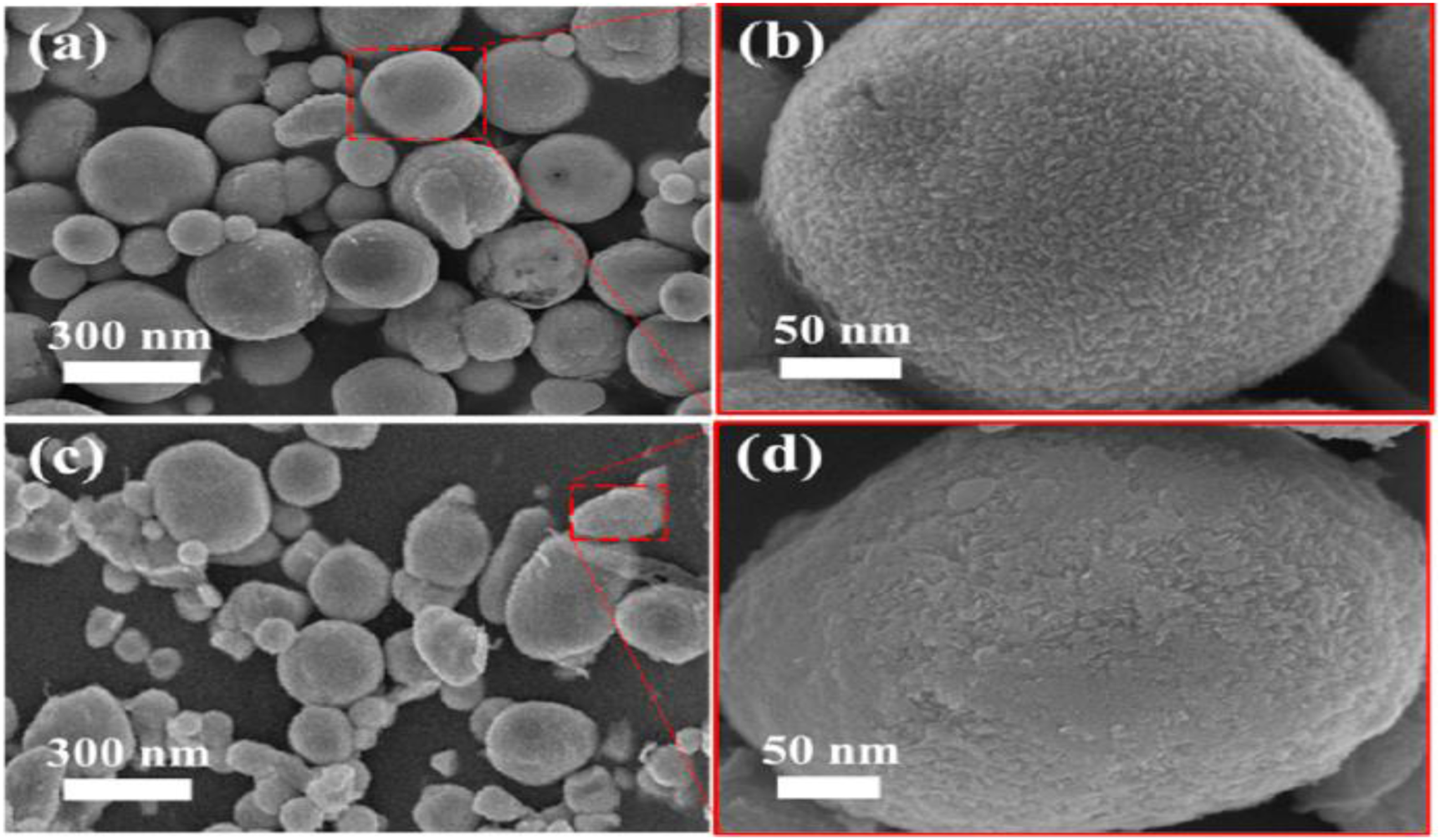
Using the pad-dry-cure procedure, the particulates were applied to cotton fabric, and the antibacterial activity of the treated materials was assessed using the zone of inhibitions test. As shown by Figure 10 the treated fabric samples displayed excellent zone of inhibitions. Antibacterial activity (ZOI) of Cu NPs treated fabric against E. coli 
Vasantharaj et al.
74
have referenced the usage of Ruellia tuberosa aqueous leaf extract for the preparation of Copper nanoparticles. The precursor needed to synthesise copper nanoparticles was copper sulphate (CuSO4) solution. The existence of rod-shaped copper oxide nanoparticles with sizes ranging from 20 to 100 nm was established by TEM investigation. The polydispersed nature of the nanorods supported the existence of the phytochemical substances from Ruellia tuberosa (Figure 11(a) and (b)). Using SEM analysis, morphological assessment of untreated and treated cotton fabrics with copper oxide nanoparticles (CuO) was performed. The distribution of CuO nanoparticles throughout and on top of the thin cotton textiles was clearly and uniformly seen in the SEM data (Figure 12(a)). Untreated cotton fabric has been used as a control to demonstrate the absence of copper oxide nanoparticles (Figure 12(b)). The pathogenic microbes such as Klebsiella pneumoniae, Staphylococcus aureus, and Escherichia coli, were vulnerable to the copper oxide nanoparticles due to their strong antimicrobial actions. The bactericidal action towards all test microbes has also been exhibited by copper oxide nanoparticles treated cotton fabric as clear ZOI can be seen in Figure 13. TEM images of rod-shaped biologically synthesized copper nanoparticles.
74
SEM images of untreated cotton fabric The ZOI of untreated and CuO NPs treated cotton fabrics against E. coli 


Using R. cordifolia bark extract and aqueous copper sulphate (CuSO4) solution as a precursor, Vinothkhanna et al. also reported biological synthesis of copper oxide nanoparticles (CuO-NPs).
75
They examined the antimicrobial potential of the synthesised nanoparticles. The EDX (energy dispersive X-ray) analysis confirmed the presence of the elements in copper oxide nanoparticle. The morphological examination of particles was performed through SEM analysis. The CuO nanoparticles generated by R. cordifolia extracts exhibit rough agglomerates and erratic spherical morphologies (Figure 14). The mean particle size distribution of copper oxide nanoparticles produced from R. cordifolia green was 50.72 nm as can be seen in Figure 15, which is equivalent to the size (50 nm) of copper nanoparticles produced from Sida acuta extract.
66
SEM images of CuO nanoparticles synthesized from R. cordifolia plant extract.
75
Average particle size for the CuO nanoparticles synthesized from R. cordifolia plant extract.
75


Gram-negative bacterial strains such as Pseudomonas aeruginosa and Escherichia coli as well as Gram-positive Bacillus cereus and Staphylococcus aureus were both significantly inhibited by cotton fabric coated with synthesised copper oxide nanoparticles due to its strong antibacterial activity. The SEM images for the live bacteria and death bacterial cell after contact with CuO NPs treated cotton fabric as well as untreated (control) cotton fabric has been shown in Figure 16. SEM images showing live and dead bacterial cells for both untreated and treated cotton fabrics.
75

Chemical and green synthesis of Ag-NPs coated textiles and their antimicrobial properties
Chemical methods for the synthesis of Ag-NPs
Silver nanoparticles are one of the most effective metal-based antimicrobial and therapeutic drugs that has seen extensive use in a variety of industrial fields.
76
According to reports, silver is highly adaptable and can inhibit about 650 different bacterial species .
2
Ag grafted textile production has advanced for use in household, medicinal, and other applications. It is used on bandages, surgical instruments, and surgical thread in medical applications. It is utilised for underwear, sporting, and military uniforms in addition to for home garments.
77
When compared to other metallic nanomaterials which includes ZnO, CuO, and TiO2, its antibacterial efficacy is outstanding.
78
Pre-activation and sorption are typically the two phases involved in the integration of silver nanoparticles into textile materials through in-situ deposition.
79
To make it easier for silver nanoparticles to diffuse into fibres, pre-activation expands the fibre matrix inside the textile. By completely dissociating Carboxylic (COOH) groups in cellulose during the pre-activation stage, the alkali-based substance or reducing agent utilised improves the affinity of silver nanoparticles binding to fabric. This approach has the benefits of being simple, affordable, and suitable for moderate reaction settings.
80
Sodium hydroxide (NaOH),
80
trisodium citrate,
81
compounds based on sulphur,
82
and potassium hydroxide (KOH)
83
are some of the most commonly use reducing agents that are employed during the pre-activation stage. The silver nanoparticles have also been applied on textiles in the form of colloidal solutions of nanoparticles. Montazer et al.
84
documented the development of antibacterial cotton textile using colloidal solution of silver nanoparticles. They applied colloidal silver NPs solution on the fabric by using the butanetetracarboxylic acid (BTCA) as stabilizer through impregnation method in two different concentrations i.e. 100 ppm and 200 ppm. From the SEM images, as shown in the Figure 17, it was demonstrated that treated cotton fabrics had nano silver particles on their surface. Escherichia coli and Staphylococcus aureus, two pathogenic bacteria, were used to assess the antimicrobial properties of the treated cotton fabric samples. The treated fabric was quite effective against both test microorganisms.
84
SEM images of untreated cotton fabric 
In another method, a colloidal suspension of silver (Ag) nanoparticles was produced using polyvinyl pyrrolidone (PVP) as reducing agent with Tollens’ reagent [Ag(NH3)2]+ through UVC irradiation. Ag+ was reduced to Ag2O with the use of ultraviolet radiation. The nylon fabric was treated with the prepared colloidal solution of silver nanoparticles using a conventional pad-dry-cure approach. Further details concerning the morphological structure and size distribution of the produced Ag nanoparticles have been revealed using the transmission electron microscopy. SEM images (Figure 18) demonstrate spherical shape of all prepared Ag nanoparticles solution. The treated unwashed, and washed fabric were tested for their antibacterial activities. After 20 washes, the treated fabric containing 200 ppm of the aforementioned solution had effectively reduced 99.2% of the S. aureus and E. coli microbes. The outcomes demonstrated that the synthesised silver nanoparticles had inhibitory properties similar to the reference solution of silver nitrate and superior to or at least equal to the other documented synthetic silver nanoparticles.
85
SEM images of as-prepared colloidal solution of silver nanoparticles.
85

Another group or researcher also prepared the silver nanoparticles through chemical method by using silver nitrate (AgNO3) solution as precursor and glucose was used as reducing agent. Silver nanoparticles were prepared, dried, and then analysed. By using the Pad-dry-cure approach, the produced nanoparticles were applied on cotton fabric that had previously been treated with sulphated B. cyclodextrin. The successful coating of synthesized silver nanoparticles on cotton fabric was confirmed by the SEM images of coated and uncoated cotton fabric as shown in the Figure 19. According to the reduction in CFU/ml testing method, the treated and untreated cotton fabrics were tested against Staphylococcus aureus and Escherichia coli bacterial strains. Excellent antibacterial action i.e. >90% against both bacterial strains, was revealed by the treated fabric.
86
SEM images of untreated cotton fabric 
Xue et al.
87
proposed an in-situ growth of silver nanoparticles on the cotton fabric. They used [Ag (NH3)2]+ complex as a precursor and glucose as a reducing agent for the in-situ growth of Ag nanoparticles on cotton fibric. Superhydrophobic cotton textiles were produced by further modifying the fibres treated with silver NPs and hexadecyltrimethoxysilane. Scanning electron microscopy (SEM) images revealed that the treated cotton fabrics were uniformly covered in silver nanoparticles, as seen in Figure 20. The fabric treated with silver nanoparticles revealed excellent antibacterial activity when tested against Gram-negative Escherichia coli bacterial strain.
87
SEM images of untreated cotton fabric 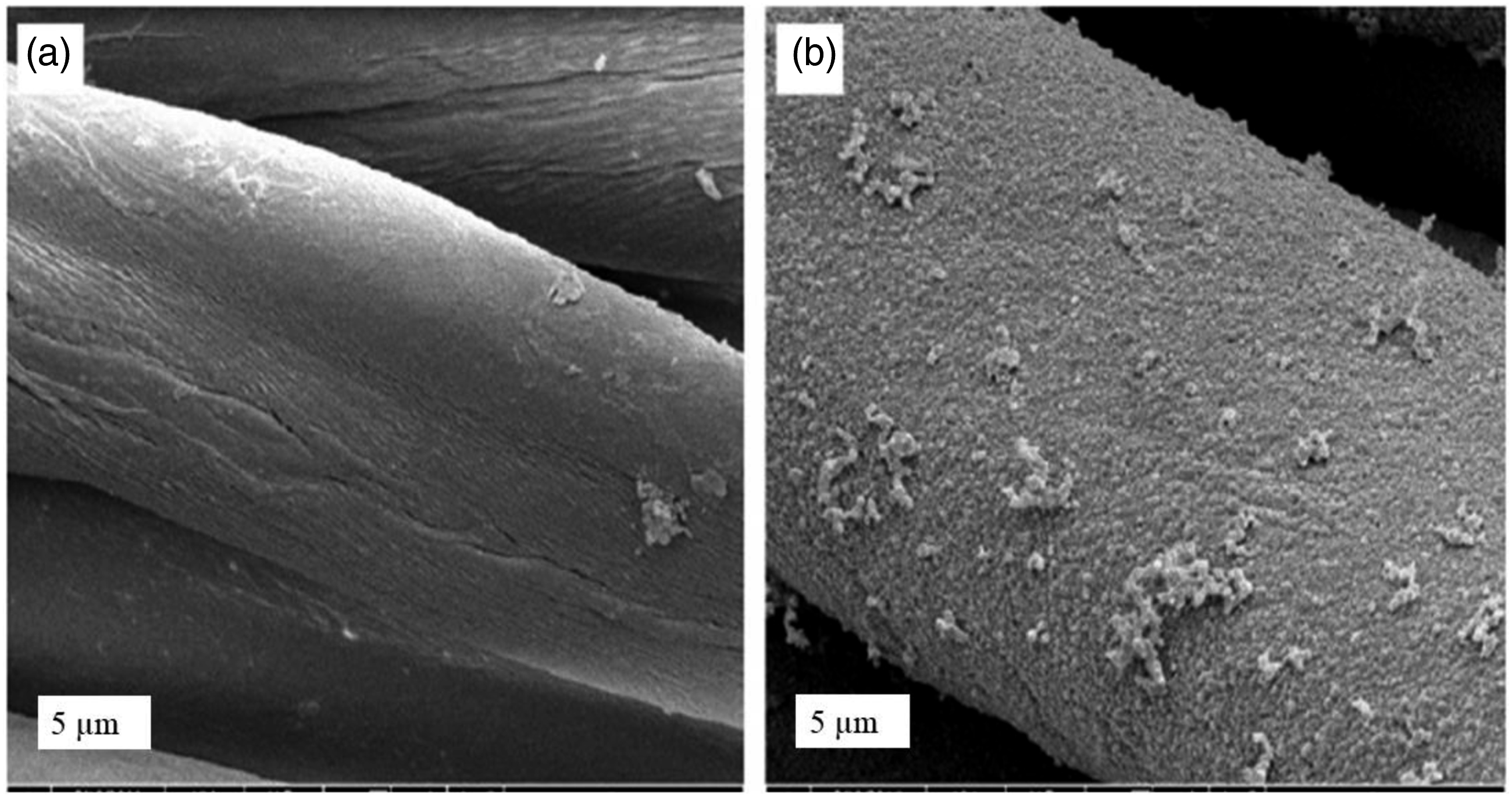
Various methods for the synthesis and application of Ag NPs.

Green synthesis methods for silver nanoparticles coated antibacterial textiles
Due to the use of toxic chemicals and solvents during the chemical synthesis of nanoparticles, the prominence of green synthesis of the nanoparticles have increased manifolds in recent years. The major goal of the current study was to successfully produce nano-silver using xanthan gum as reducing agent due to its high reducing and stabilising properties. Using a basic one-step fast synthesis method, the hydrolysed xanthan gum had been employed to reduce Ag+ to AgO in nanoscale level. The cotton fabrics were subsequently treated with the synthesized Ag nanoparticles by employing the pad-dry-cure process. More details concerning the morphological structure and mean size distribution of the synthesised Ag nanoparticles have been revealed using the transmission electron microscopy. TEM images (Figure 21) demonstrate spherical shape of Ag nanoparticles. The antibacterial activity of both treated unwashed and treated washed samples were evaluated. Even after 20 washes, the treated cotton fabrics exhibit excellent biocidal activity against Staphylococcus aureus. The benefit of this technology is that it could develop highly durable antibacterial cotton fabrics without the need for harsh conditions like the presence of organic solvents.
88
TEM images of the as-synthesized silver nanoparticle using xanthan gum.
88

In another investigation, the medicinal potential of silver nanoparticles produced from Rosmarinus officinalis plant extract on several bacterial pathogens was described by the researchers. The ability of Rosmarinus officinalis plant extract to produce silver nanoparticles has been established. These nanoparticles were highly stable in the solution. However, further studies are needed for their application on textiles to develop antibacterial textiles.
92
Syafiuddin et al.
93
proposed a novel environmentally friendly technique to deposit Ag nanoparticles permanently on the surfaces of nylon, cotton, and polyester fabric by employing the purified extract of Muntingia calabura leaves. Silver nanoparticles have been deposited on the fibres of the fabric, as evidenced by the appearance of a darker colour of the fabric. The SEM micrographs, as depicted in the Figure 22, provided additional evidence of the successful application of synthesised nanoparticles. The inhibitory effect of nylon, cotton, and polyester fabrics is superior to that of Chromobacterium haemolyticum and then superior to that of Bacillus cereus as compared to the Escherichia coli.
93
SEM micrographs of untreated cotton fabric and fabric treated with Ag nanoparticles.
93

It has been shown that cotton fabric treated with Ag nanoparticles that were produced using a biological method and extracts of Acalypha indica plant is effective against microorganisms. It was observed that Ag nanoparticles have a size of 100 nm. Electron microscopy was used to confirm the morphological structure of the silver nanoparticles that had grown on the cotton fibres. As shown in Figure 23, the antimicrobial property of cotton fibres coated with silver nanoparticles was assessed against four bacterial species, including Klebsiella pneumonia, Candida albicans, Bacillus cereus, and Pseudomonas aeruginosa, and (a-d). The findings indicate that adding % leaf extract to cotton fibres will have excellent antimicrobial effect.
94
Antibacterial activity of Ag NPs coated fabric against Klebsiella pneumonia 
Biological synthesis of silver nanoparticles from plant sources.

Evaluation methods of antibacterial fabrics
A number of testing techniques have been developed to evaluate the effectiveness of antibacterial textile materials. The two primary categories of these testing standards are quantitative analysis methods and qualitative analysis methods. Below is a brief discussion of the procedures used to conduct these tests.
Qualitative testing protocol
JIS L1902-2002 (Japanese Industrial standards), AATCC 147–2004 (American Association of Textile Chemists and Colorists), and SN 195,920–1992 (Swiss Norm) are some of the types of qualitative antibacterial tests commonly known as agar diffusion or disc diffusion tests. 103 Even though these evaluations are only qualitative, they are simple to carry out and are most effective when screening a large number of samples for antibacterial activities. To carry out this experiment, microbial cells are injected onto nutrient media plates, which are then injected on the textile samples for direct contact. The as-prepared plates are examined for bacterial growth from around test specimens and beneath the textiles after being incubated at 37°C for 18–24 h (zone of inhibition). The lack of bacterial activity right beneath the textile swatch indicates antimicrobial efficacy. The absence of a zone of inhibition occurs due to the inability of the antimicrobial agent to permeate into the agar media if it is highly covalently linked to the textiles. A zone of inhibition (ZOI), which diameter provides a rough indication of the potential for antibacterial properties, is formed if the antibacterial agent diffuses in the agar media. Equations are used to determine the diameter of the zone of inhibition. All of the aforementioned procedures follow the same methodology, although each method uses a different calculation method. The AATCC-147 approach is recommended because it provides data that are relatively reliable in terms of the diameter of the inhibitory zone. 104
Quantitative testing protocols
JIS L1902-2002, AATCC 100–2004, SN 195,924–1992, and ISO-20,743 transfer technique are the main quantitative assessment procedures employed for the antibacterial assessments. 105 These methods take much longer than qualitative antibacterial testing, but they provide exact quantitative assessment for the antibacterial fabrics. Approximately 1 mL of the bacterial suspension is entirely absorbed onto test textile swatches of various sizes as suggested by the respective norms. This step guarantees direct contact between the tested samples and the microorganisms. The textile sample that has been loaded with bacterial suspension is incubated for the prescribed period of time according to standard procedures. By using the serial dilution approach and subsequent plating of prepared dilutions on nutrient agar plates, the exact number of injected and recovered bacterial colonies is counted. Antibacterial activities are quantified in terms of log reduction or percentage reduction by comparing the number of injected and recovered bacterial colonies at 0 h and after specific incubation duration. Appropriate control samples, such as textile samples that have experienced the same processing phases excluding the antibacterial finishing, must be examined in each experiment to confirm that the acquired reduction in microbial population is actually attributable to the antibacterial finishing. The choice of an appropriate calculation equation may be essential. All methods are used for quantitative evaluation, but the ISO-20,743 approach is thought to be the most effective for assessing the textiles resistance to microbes. 106 This is because it is impossible to simulate hospital textiles using any other approach because a certain amount of inoculum (often 1 mL) is entirely absorbed onto the fabric which is practically not possible for hospital textiles. Due to this reason, the ISO-20,743 transfer method is the most appropriate for evaluating antibacterial activities of textiles because in this procedure the textile swatches are placed on the infected agar plates for specific time (60 s) and is subsequently detached from the agar surface for further examination.
Minimum inhibitory concentration assays
Broth tube dilution tests 107 and Liofilchem strip test procedures are used to determine the minimum inhibitory concentration (MIC) of antimicrobials. 108 The MIC value represents the lowest concentration at which the test microorganisms cannot grow. In order to assess MIC in fresh growth media, microorganism sub-cultures are subjected to various doses of the antimicrobial agent. The MIC value of that particular active agent is defined as the concentration of antimicrobial at which no growth is seen. The Liofilchem strip test are not economically sound due to the high cost of the strips required for this test. 108 As a result, broth dilution tests are preferable because they do not require any additional materials like strip test methods do.
Conclusion
Metallic nanoparticles have found extensive applications in engineering as well as biomedical domains. Their demand has grown significantly in recent years, and it is not predicted to recede. In this review, the antibacterial potential of copper and silver nanoparticles, different synthesis methods, development of antibacterial textiles for synthesized Cu and Ag NPs and a comparative analysis of their antibacterial characteristics has been discussed. From the extensive aforementioned review, it was concluded that both Cu and Ag NPs are highly efficacious towards a wide spectrum of Gram-positive and Gram-negative bacterial species. However, the antimicrobial potential of Ag nanoparticles was higher when compared to results for Cu-NPs treated fabrics. The SEM results revealed that Cu-NPs are mostly spherical or cubical while Ag-NPs are spherical in shape. It was also observed that the antimicrobial action of cuprous oxide (Cu2O) treated textiles was higher than the cupric oxide (CuO) treated textiles as Cu is more stable in its +2 oxidation state. The developed Cu and Ag NPs treated textiles could be efficiently used as hospital textiles for the production of bed sheets, wound dressings, chair and table covers, coveralls, curtains, protective suit, and surgical gowns. Further, from the comparison of different chemicals methods for the synthesis of Ag and Cu NPs with green synthesis of these NPs it was established that green synthesis methods are efficient in terms of sustainability as these methods do not necessitate any hazardous materials and organic solvents. Therefore, there is a potential for researchers to explore novel plant sources that could be effectively used for nanoparticle synthesis. Also, multiple studies on the green synthesis of nanoparticles have been reported in this review but their application on textiles has been ignored by the researcher. Hence, there is a room for researchers to optimize the application parameters of these synthesized nanoparticles on different textile substrates to develop antimicrobial textiles.
Footnotes
Declaration of conflicting interests
The author(s) declared no potential conflicts of interest with respect to the research, authorship, and/or publication of this article.
Funding
The author(s) disclosed receipt of the following financial support for the research, authorship, and/or publication of this article: This work was supported by the Modular platform for autonomous chassis of specializedelectric vehicles for freight and equipment transportation, Reg. No. CZ.02.1.01/0.0/0.0/16_025/0007293. The research was also supported by the project, Textile structures combining virus protection and comfort reg. c.:cz.01.1.02/0.0/0.0//20_321/0024467.