
Abstract
In this work, the glass fiber–epoxy self-healable composites containing the capsulated healing agent as smart composite structures were fabricated. Then, the effects of various percentage of capsulated epoxy (7, 14, and 21 wt.%) and different damage forces (400, 500, and 600 N) on the healing ability of composites under the flexural loading were investigated. The obtained results showed that by increasing the percentage of microcapsules, the flexural strength of composites was slightly reduced, whereas the healing ability of those was improved. The full recovery of initial flexural strength was obtained in the composite containing 21 wt.% microcapsules under the damage force of 400 N. By increasing the damage force, the healing ability of composites was reduced, because the damage mechanisms were changed from matrix cracking to fiber breakage. The microscopical investigations on the microcapsules/micro-cracks behaviors illustrated the deflecting, debonding, crushing, and buckling phenomena under the flexural loading. Considering with the healing efficiency and initial flexural strength, the self-healable composite containing 14 wt.% microcapsules and 2 wt.% NiCl2(imidazole)4 catalyst had the optimum results.
Introduction
Although polymeric composite structures have attracted many attention due to their superior properties such as very high strength and stiffness to weight ratios, they have a brittle structure, the cracks can easily propagate into them, and consequently, they have a catastrophic failure [1,2]. The mentioned problem leads to introduce the self-healing composites as a new kind of smart materials [3]. The self-healing behaviors can be activated by chemical or physical provocative factors such as the thermal, pH, solvent, and mechanical forces. In the composites with the self-healing ability, the crack healing can be done by different mechanical and materials’ point of view. To study the methods’ ability for recovering the initial mechanical properties, the healing efficiency parameter was introduced [4].
Closing the crack into the composite structures is a mechanical way for recovering the initial properties which can be done by shape memory strips like Ni–Ti alloys [5]. In contrast, filling the crack by polymeric agent is a material way for that, which can be accomplished by the intrinsic and extrinsic methods [6]. The polymeric materials, which are caused to heal the cracks, are known as healing agent. These healing agents in the intrinsic system are divided in three main approaches, which are the reversibility of covalent bonds, supramolecular interactions, and polymer blends systems [7]. In contrast, the healing agents in the extrinsic system are stored into a container such as hollow fibers, microvascular networks, and microcapsules [8].
It should be noted that using these healing methods in the composite structures (composites reinforced with high performance fibers such as glass, Kevlar, or carbon fibers) is too limited, and the most of the researchers have focused to heal the polymeric materials instead of polymeric composite structures. Ghanbari et al. [9] investigated the flexural properties of self-healing composites containing shape memory strips. They used the Ni–Ti strips and 25 wt.% hollow glass fibers containing epoxy 828 in the glass fiber–epoxy composites. First, an initial damage was exerted on the composite. Then, the damaged samples subjected to flexural test after the certain period time of healing. The obtained results showed that the healing efficiency of sample without Ni–Ti strip was 37.9% after seven days, whereas the healing efficiency of composite with three Ni–Ti strips and 4% pre-strain was reached to 78.19%. Wang et al. [10] used the intrinsic system to heal the carbon fiber–epoxy composites by the poly(ethylene-co-methyl acrylate) (EMA) and poly(ethylene-co-methacrylic acid) (EMAA) under the interlaminar fracture toughness and short-beam shear tests. They reported that by adding EMA and EMAA, the healing efficiencies of composites under the interlaminar fracture test were achieved to 46 and 86%, respectively. Also, those composites under the short beam shear test could recover 39 and 36% of initial properties.
In the study of Zhu et al. [11], the polypropylene tubes as hollow fibers containing epoxy and mercaptan healing agent into the glass fiber–epoxy composites were used. The interesting idea in this work was using a foaming agent to increase the fluid of healing agents for diffusing in the damage area during the healing process. The maximum healing efficiency in flexural loading was approximately 95% for the sample including polypropylene tube with the thickness of 750 μm and three parts of foaming agent after seven days in the ambient temperature. Chowdhury et al. [12] capsulated the epoxy as first healing agent by Brown’s method. In the following, they synthesized the hollow urea formaldehyde microcapsules and filled them by polyamine hardener through a vacuum method. After that, they dispersed the microcapsulated epoxy and polyamine hardener into the glass fiber–epoxy composites. The damaging system in this research was low velocity impact test. The obtained results showed that this composite could recover the 40% of initial properties. Neisiany et al. [13] synthesized the core-shell nanofibers by electrospinning method. They encapsulated the epoxy and amine hardener into the styrene acrylonitrile shell and then loaded into the carbon fiber–epoxy composites. The remarkable results in this method were the 90–97% encapsulation yield. Also, the healing efficiency of flexural strength was 94, 97, and 89% for first, second, and third recovery cycles. Patel et al. [14] investigated the healing behavior in the low-velocity impact damage of glass fiber–epoxy composite by microcapsules containing dicyclopentadiene healing agent and microspheres containing Grubbs’ catalyst. The reported healing efficiency for recovering compressive strength by this research was 95%.
In spite of the many published literatures about self-healing behavior of polymers by microcapsulation methods, the dedicated researches for recovering the mechanical properties of composite structures are too limited. Hence, the first novelty of this work is to use the capsulated epoxy and NiCl2.(C3N2H4)4 as healing agents into the glass fiber–epoxy composite structure. It is worth noting that the epoxy was encapsulated by promoted one-stage method, which has been previously published by us in other journal [15]. The second novelty is using the quasi-static penetration method for creating the different damages into the composite structures. Finally, the effects of different percentage of microcapsules and various damage forces on the flexural properties recovery of glass fiber–epoxy composite were surveyed.
Experimental
Raw materials
In order to fabricate the glass fiber–epoxy self-healable composites, the urea (Pardis Petrochemical Company, Iran), methanol, ammonium chloride, formaldehyde solution, ethyl acetate and hydrochloric acid (Dr. Mojallali Industrial Chemical Complex Company, Iran), 1-octanol, resorcinol, sodium carboxymethyl cellulose (NaCMC), nickel chloride and imidazole (C3N2H4) (Merck, Germany), linear alkyl benzene sulfonic acid (LABSA) (Behdash Chemical Company, Iran), epoxy 828 resin, and triethylenetetramine (TETA) hardener (Kumho P&B Chemical, Korea) as well as plain weave glass fibers fabric with the surface density of 400 g cm−2 (Lintex, China) were used.
Preparation of microcapsules and catalyst
The microencapsulation of epoxy was done by promoted Tang’s method as one of the core-shell methods for encapsulation [16]. In this method, the epoxy and urea–formaldehyde were core and shell, respectively. It should be noted that the encapsulation of epoxy was performed based on the one stage in situ polymerization method [15]. Figure 1 represents the schematic flowchart of encapsulation process in this research. In the 1000 ml three-necked flat flask, 300 ml deionized water, 0.3 g NaCMC, 0.05 g LABSA, and 7.5 g urea were stirred by three-blade mechanical stirrer with the rate of 600 r/min. After the dissolution of urea, 1 g resorcinol and 0.75 g ammonium chloride were added. After 10 min, the pH of solution was set to be 3.5 by HCl. Then, four droplet 1-octanole was added. In the following, the mixture of 45 g diluted epoxy by 35 g ethyl acetate was added. In the next step, 18.9 g formaldehyde was slightly added to the solution. With the heating rate of 1°C min−1, the temperature of solution has been reached to 55°C and stabled in this temperature for a period time of 240 min. After cooling the solution, the pH of that was neutralized and then washed by deionized water. After that, the synthesized microcapsules were filtered and air dried.

The schematic flowchart of encapsulation process.
The latent curing agent was synthesized by Dowbenko’s method [17]. A solution of 0.1 mol NiCl2 in the 50 ml methanol was slightly added to the solution of 0.4 mol imidazole in the 50 ml methanol. By heating the solution, the extra solvent was evaporated, and the crystallization of NiCl2(imidazole)4 was obtained.
Fabricate the self-healable composites
Based on other literature [18], although adding the higher percentage of microcapsules increases the healing ability of composites, the viscosity of epoxy matrix is strongly enhanced, so that the epoxy cannot completely wet the glass fibers. By this point of view, the various weight percentages of microcapsules (7, 14, and 21 wt.%) were selected to disperse them into the epoxy resin by three-blade mechanical stirrer. After that, the 2 wt.% NiCl2.(imidazole)4 catalyst was added, and the dispersions of catalyst and microcapsules were continued. In the following, the 10 wt.% TETA hardener was added according to the producer’s recommendation. Based on previous work, the glass fiber–epoxy self-healable composites with ratio of 50:50 wt.% (glass fibers to epoxy) were manufactured with four plies glass fiber fabric by hand lay-up method [19,20]. The schematic of fabricated self-healable composite has been shown in Figure 2. The curing and post curing times for fabricated composites at the ambient temperatures were one and seven days, respectively. After that, the flexural samples with the dimensions of 100 × 15 mm2 were cut and polished as per ASTM D790 standard.

The schematic of fabricated self-healable composite.
The exertion of initial damage and healing process
Based on other literature [4], double-cantilever beam, tapered double-cantilever beam, compact tension, and single-edge notched beam as quasi-static methods are used for studying the self-healing behaviors in polymers, due to predictable crack propagation, small damage volumes, and quantifiable fractures parameters. But these methods are not proper for composite structures. Therefore, the quasi-static penetration test (ASTM D6264) was selected, and set-up of that for flexural samples was designed, which could be seen in Figure 3. Figure 4 shows the force–displacement curve of flexural sample under the quasi-static penetration test. Based on literatures and ASTM D6264 standard [21,22], by applying the load, the sample first had elastic behavior. After that, the first kink was observed, which corresponded to the matrix cracking (approximately 400 N). By continuing the load, the matrix cracking was propagated, and the debonding and delamination phenomena were occurred. At the maximum load (about 600 N), the fiber breakage was happened. In light of the above mentioned, the 400, 500, and 600 N damage forces were selected to create the matrix cracking, delamination, and fiber breakage failure mechanisms, respectively. After that, the healing behaviors of these failure mechanisms in the samples containing 7, 14, and 21 wt.% under the flexural loading were investigated. It should be noted that the damages were created by Hounsfield H25KS machine with the cross head of 1 mm min−1 at the ambient temperature.
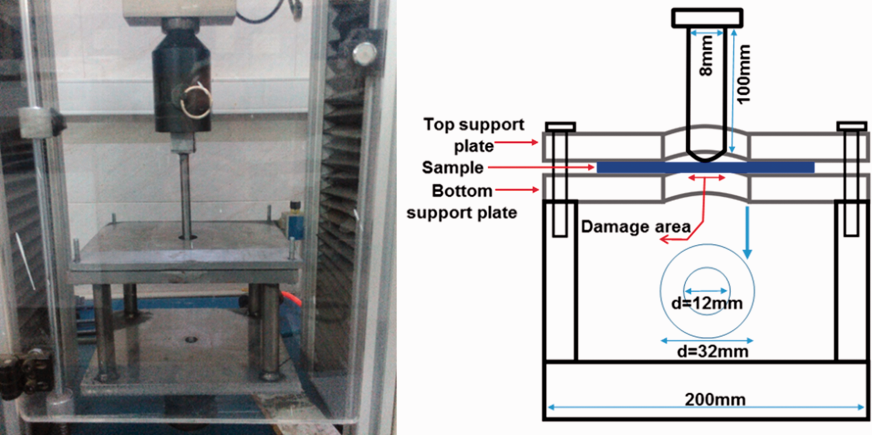
The designed quasi-static penetration test for exertion of initial damages on the self-healable composites as per the ASTM D D6264 standard.
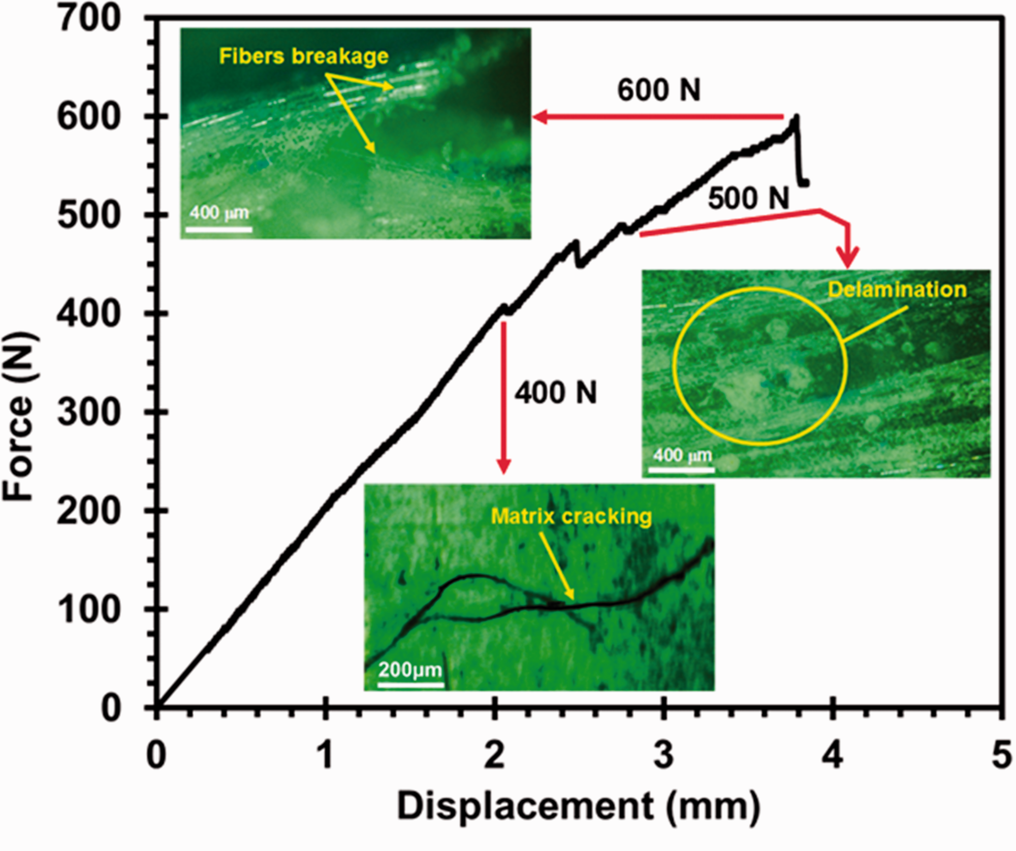
The force–displacement curve of composite under quasi-static penetration test and selecting damage forces.
After creating the initial damages, the samples were set into the oven in the temperature of 130°C for period time of 1 h. In this temperature, the catalyst was decomposed to the imidazole and NiCl2. In the following, the imidazole reacted with epoxy healing agent and polymerized that [23]. For investigating the healing behavior in flexural test, the three samples, virgin, damaged, and healed, were considered. The three-point bending tests were done with the cross head of 3.5 mm min−1 as per ASTM D790 standard by Hounsfield H25KS machine. It is worth noting that for obtaining the accurate results, at least five samples were tested, and the average results were reported. The healing efficiency of composite structures (η) was calculated from equation (1) [4]

Characterization
The microstructure of healed composites and interaction between micro-cracks/microcapsules were characterized by using the field emission scanning electron microscopy (FESEM), Mira3 Tescan-Czech, and optical microscopy (OM), Meiji Techno-Japan.
Results and discussion
Synthesized microcapsules
Figure 5(a) represents the FESEM image of fabricated microcapsules by promoted Tang’s method [16], which has been published in our previous work [15]. The encapsulation efficiency was about 90–95%. The FESEM investigation of microcapsules showed that the fabricated microcapsules had spherical shape with slightly rough surface. The rough surfaces were created by agglomeration of UF nano-spheres on the surface of capsules. This slightly rough surface was the desired shape for interlocking between capsules and epoxy matrix [6]. The size distribution of microcapsules was illustrated in Figure 5(b). The average size and thickness of microcapsules were about 225 μm and 320 nm. Also, the calculated core content was about 71.2%, which was determined by extraction method [15].

The synthesized microcapsules for self-healable composites: (a) FESEM observation and (b) the size distribution of microcapsules.
Flexural properties
Figure 6(a) to (c) shows the flexural curves of virgin, damaged, and healed composites containing different microcapsules contents. The obtained results from flexural tests have been illustrated in Figure 7(a) to (d). Based on Figure 7, the undamaged or virgin sample in the composite containing 7 wt.% microcapsules had 309 MPa flexural strength. By loading the 400 N damage force, the remained flexural strength was 199 MPa. This means that the sample under the 400 N damage force lost approximately 33.6% of its flexural strength. After the healing process, the flexural strength was reached to 286 MPa. By calculating the healing efficiency via equation (1), the 7 wt.% microcapsules-filled composite recovered 79.1% of its initial flexural strength.

The stress–strain diagram of composites containing different percentage of microcapsules under various damage forces: (a) 7 wt.% microcapsules, (b) 14 wt.% microcapsules, (c) 21 wt.% microcapsules (M: microcapsule; V: virgin or undamaged; H: healed; D: damaged; 7, 14, and 21: the percentage of microcapsules; 400, 500, and 600: damage forces).

The extracted data from stress–strain curves: (a) the flexural strength of virgin and damaged self-healable composites, (b) the remained flexural strength after created damages, (c) the recovered flexural strength after healing process, and (d) the calculated healing efficiency.
By applying the 500 N damage force on the composite, the flexural strength was reduced to 171 MPa. Therefore, the 7 wt.% microcapsule-loaded composite under the damage force of 500 N lost 44.5% of its initial flexural strength and only 55.5% of that remained. After setting the composite into the oven for healing process, the flexural strength of that was reached to 252 MPa. The obtained healing efficiency for that under the 500 N damage force was 58.7%. Under the damage force of 600 N, the flexural strength of 7 wt.% microcapsule–glass fibers/epoxy composite was 165 MPa (53.6% of initial flexural strength). The achieved flexural strength for that after healing process was 204 MPa. The healing efficiency of composite containing 7 wt.% microcapsule under the 600 N damage force was approximately 27.1%.
According to Figure 7 and by loading the 400 N damage force on the 14 wt.% microcapsule-loaded composite, the flexural strength had 26.2% reduction. After the healing process, the flexural strength was reached to 293 MPa, and this self-healable composite was able to recover 87.5% of its initial flexural strength. The damage force of 500 N reduced the flexural strength down to 157 MPa (48.0% reduction as compared with undamaged sample). By activating the healing agents at the temperature of 130°C, the 14 wt.% microcapsule-filled composite could recover the 67.1% of flexural strength. The remained flexural strength of damaged composite under the 600 N damage force was 141 MPa. After the healing process, only 42.6% of its initial flexural strength was obtained.
Considering Figure 7, the glass fiber–epoxy composite including 21 wt.% microcapsules had 280 MPa flexural strength. The damage forces of 400, 500, and 600 N decreased the flexural strength down to 191, 131, and 141 MPa, respectively. The remaining flexural strengths of those were 68.2, 46.8, and 50.4%, respectively. After the healing process, the recovered flexural strengths of those were 280, 255, and 209 MPa, respectively. The calculated healing efficiency for damage forces of 400, 500, and 600 N was 100, 83.2, and 48.9%, respectively.
As previously mentioned, the damage force of 400 N was selected to create the matrix cracking. The obtained healing efficiency showed that the glass fiber–epoxy self-healable composites can recover the initial flexural properties, so that, by adding 21 wt.% microcapsule, the healing efficiency reaches to approximately 100%. This means that this healing system can completely heal the matrix cracking. Similar results have been observed by Yin et al. [18]. By loading the 500 N damage force in this composite structure, the delamination and debonding between fibers and matrix were created in the quasi-static penetration [24]. The maximum-obtained healing efficiency for this damage force was 83.2%. By comparing the damage force of 500 N with 400 N, 13% reduction in maximum healing efficiency was observed. It is worth noting that in the 500 N damage force, the damage area was expanded, as compared with 400 N damage force. Therefore, the required healing agent was increased. Also, more time was necessary for filling the damage area, and the catalyst must be capable to fully polymerize the added healing agent [25]. To understand these phenomena, the microscopically investigation is so essential. In the damage force of 600 N, the healing efficiency was finally reached to 48.9% in the sample containing 21 wt.% microcapsules. The low healing efficiency in this damage force was due to fiber breakage phenomenon. Actually, the healing agent only increased the adhesion between fibers and matrix and was not capable to heal the fibers breakage. Similar results were reported by Yin et al. [26].
The pristine glass fiber–epoxy composite had 280 MPa flexural strength. By adding the 7, 14, and 21 wt.% microcapsules in the composites, the flexural strength of undamaged composites was 309, 303, and 280 MPa, respectively. This means that the microcapsules first increased the flexural strength of those which can be attributed to the interaction between microcapsules and micro-cracks [27]. By increasing the percentage of microcapsules, the flexural strength had reducing trend. This trend can be attributed to the agglomeration of microcapsules in the epoxy matrix and trapping the created bubbles during the fabrication process (Figure 8), due to the viscosity increment of epoxy by microcapsules [28]. Considering with healing efficiency and initial flexural strength, it can be said that the composite containing 14 wt.% microcapsules had the optimum results, and this percentage is recommended for other investigations.

The agglomerated microcapsules and trapped air bubbles phenomena.
In order to comprehensively understand the healing ability of fabricated composite in this research work, this composite was compared with the previous related literatures. This comparison was listed in Table 1. As previously mentioned, the healing efficiency is the proper parameter for comparing the healing ability. Based on Table 1, it can be found that the maximum healing efficiency of the composite in this research was 100%. It should be noted that this healing efficiency belonged to damage of matrix cracking. By comparing the imidazole and Grubbs’ catalysts in Table 1, it can be said that the imidazole-based catalyst has higher healing efficiency.
Comparison healing ability between different self-healing systems in the epoxy-based composites.

Microstructure investigation
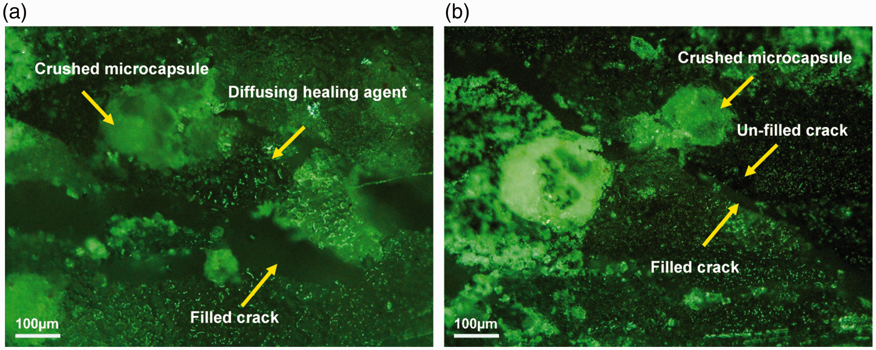
Figure 9 represents the interaction between cracks and microcapsules. The OM micrograph shows crossing the micro-crack in the interface of microcapsule and matrix, crushing, and buckling the microcapsules under the flexural loading. Crossing the crack in the interface is the sign of weak interface between microcapsules and matrix. So that, it can reduce the mechanical properties. The other phenomenon is the buckling of microcapsules, which may increase the toughness of composite structure and prevent the crack growing. In the FESEM micrograph, the diffusing healing agent, deflecting the micro-crack by microcapsules, and debonding the microcapsules can be seen. The deflection of crack by microcapsules is one of the important mechanisms to control the crack behavior. Therefore, the higher flexural strength of composites containing microcapsules than the pristine composite can be attributed to the deflection of micro-cracks. Similar behavior was observed by Tripathi et al. [29] in the pristine epoxy-containing microcapsules.

The interaction between micro-cracks/microcapsules under flexural loading: (a) OM and (b) FESEM micrograph.
After diffusing the healing agent and filling the damage area, the polymerization reaction was started in the temperature of 130°C. In this temperature, the ligand bonds between NiCl2 and imidazole were broken [23]. Then, the imidazole reacted with the epoxy healing agent to heal the created micro-cracks. This reaction could be produced by the polymeric shell with nanometer thickness between fibers and matrix, which could be seen in Figure 10.

The formed polymeric healing agent shell on the surface of plain weave glass fabric into the glass fiber-epoxy composite.
Figure 11(a) and (b) shows the OM micrograph of created micro-cracks under the damage forces of 400 and 500 N, respectively. In Figure 11(a), it could be observed that the crack ruptured the microcapsules. After that, the healing agent diffused the crack area and filled that. Whereas, by seeing Figure 11(b), it can be understood that the healing agent could not be capable to fill the damage area. Therefore, the lower healing efficiency of sample under the 500 N than that of under the 400 N could be attributed to the incomplete filling of the microcracks. Increasing the percentage of microcapsules into the epoxy matrix can be one of the solutions for this issue. For this reason, under the damage force of 500 N, by increasing the microcapsule percentage from 7 to 21 wt.%, the healing efficiency was improved approximately to 70%. However, the healing efficiency was 83.2%, and composite could not completely recover its initial flexural strength under delamination damages. This means that the other influence factor had an important role in the healing process. Figure 12 shows the fracture surface of composite containing 21 wt.% healing agent. As can be seen, in some areas, the healing agent only wets the surface, and polymerization has not occurred. This phenomenon can be attributed to the catalyst percentage. In other words, the catalyst content for reaching to 100% healing efficiency under the delamination area is insufficient [18].
The OM micrograph of created micro-cracks under the damage forces of: (a) 400 N and (b) 500 N into the composite containing 7 wt.% microcapsules.

The fracture surface of composite containing 21 wt.% healing agent under flexural loading after healing process.
Conclusion
In this research, the self-ability of glass fiber–epoxy composites under the flexural loading was investigated. By creating the matrix cracking in the composites containing 7, 14, and 21 wt.% microcapsules, these composites could recover 79.1, 87.5, and 100% of their initial flexural strength, respectively. Also, the fiber breakage phenomenon strongly reduced the self-healing ability of composite, so that the maximum healing efficiency in this damage area was about 48.9%. Microscopically, investigations on the interaction between microcapsules/micro-cracks under the flexural loading illustrated the deflecting, debonding, crushing, and buckling of microcapsules. As the most important finding of the work, it was realized that the self-healable composite containing 14 wt.% microcapsules had the optimum results. Therefore, this percentage is recommended for other investigations.
Footnotes
Declaration of conflicting interests
The author(s) declared no potential conflicts of interest with respect to the research, authorship, and/or publication of this article.
Funding
The author(s) received no financial support for the research, authorship, and/or publication of this article.