
Abstract
Diabetes contributes directly to the development of cardiovascular aortic valve disease. There is currently no drug therapy available for a dysfunctional valve and this urges the need for additional research to identify distinctive mechanisms of cardiovascular aortic valve disease evolution. The aim of this study was to evaluate changes of valvular aortic lesions induced in a hyperlipemic ApoE−/− mouse model by early type 1 diabetes onset (at 4 and 7 days after streptozotocin induction). The haemodynamic valve parameters were evaluated by echography and blood samples and aortic valves were collected. Plasma parameters were measured, and inflammatory, remodelling and osteogenic markers were evaluated in the aortic valves. Next, correlations between all parameters were determined. The results showed early aortic valve dysfunction detected by echography after 1 week of diabetes; lesions were found in the aortic root. Moreover, increased expression of cell adhesion molecules, extracellular matrix remodelling and osteogenic markers were detected in hyperlipemic ApoE−/− diabetic mice. Significant correlations were found between tissue valve biomarkers and plasmatic and haemodynamic parameters. Our study may help to understand the mechanisms of aortic valve disease in the diabetic milieu in order to discover and validate new biomarkers of cardiovascular aortic valve disease in diabetes and reveal new possible targets for nanobiotherapies.
Keywords
Introduction
Aortic valve disease and especially cardiovascular aortic valve disease (CAVD) is a global health burden in all ageing societies. The pathogenic processes in the CAVD evolve from the early phase characterized by valvular endothelial inflammation and thickening of the basal lamina 1 to the progressive phase, when the proinflammatory processes may induce valvular interstitial cells (VICs) to undergo osteogenic differentiation, finally resulting in severe calcification with impaired leaflet motion or aortic valve stenosis, a hallmark of a structural and functional compromised valve. 2
It is known that the presence of diabetes accelerates CAVD, and is predictive of poor prognosis in valve disease and of faster degeneration of implanted bio-prosthetic aortic valves. 3 However, to our knowledge, there are no specific targets or targeted therapies for the treatment of aortic valve disease in diabetes, urging for the need of new cellular and molecular insights.
There is evidence of possible mechanisms involved in diabetes-accelerated atherosclerosis. In a mouse model of combined atherosclerosis and increased susceptibility to type 2 diabetes, 4 the development of aortic stenosis, calcification in the aortic valves, inflammatory infiltrates, upregulation of hypertrophic genes (anp, bnp and b-mhc) in myocardial tissues and osteogenic genes (spp1, bglap and runx2) were detected in aortic tissues. 4 In a recent study, it was reported that bone morphogenetic protein 4 (BMP4), which is a bone-inducing morphogen belonging to the members of transforming growth factor-beta (TGFβ) superfamily, was expressed and enhanced oxidized low-density lipoprotein (oxLDL) uptake at 4 weeks after diabetes onset in aortic atherosclerotic plaques in a mouse model of diabetes-accelerated atherosclerosis. It was suggested that the induction of BMP4 may promote aortic atherosclerotic plaque formation in diabetes. 5
Aortic valve is a lesion prone area for atherosclerotic plaque development, and one of the first affected in hyperlipemic and hyperglycaemic hamsters (HD). 1 In the aortic valve of HD hamsters, structural modifications occurred at a much faster rate than in hyperlipemic or hyperglycaemic animals, and were detected even after 2 weeks of diabetes. 1 The mechanisms initiating CAVD in diabetes are not well understood. Since we aim to investigate CAVD-initiating events and the mechanisms of its progression in diabetes, we hypothesized that aortic valve may be affected structurally and functionally even at shorter time periods of 2 weeks after diabetes onset and we performed studies to verify our hypothesis.
Therefore, this study aims to determine what are the molecular and functional changes relevant for early stage of aortic valve disease induced by diabetes and to establish a correlation between these changes and plasmatic and haemodynamic parameters relevant for diabetes state. The inflammatory process was followed by investigating the activation of valvular cells via enhanced expression of the cell adhesion molecules (CAMs): P-selectin, intercellular adhesion molecule-1 (ICAM-1), vascular cellular adhesion molecule-1 (VCAM-1) and platelet endothelial cellular adhesion molecule-1 (PECAM-1) and proinflammatory cytokines: TGFβ, BMP2 and BPM4 in aortic valve leaflets. Modifications at the level of extracellular matrix (ECM) components and remodelling process was tracked by investigating the expression of fibronectin (FN), laminin and other ECM proteins and the proteolytic enzymes involved in the remodelling of ECM: matrix metalloproteinase-2 (MMP2) and matrix metalloproteinase-9 (MMP9). Osteogenesis was evaluated by assessment of the alkaline phosphatase (ALPL) and other osteogenic molecules: osteocalcin (OCL) and osteonectin. The functional changes of aortic valve in diabetes were evaluated by measuring the haemodynamic parameters using echocardiography at different time points. After collecting the data, correlations between proinflammatory, remodelling and osteogenic parameters, plasma parameters and functional haemodynamic parameters were determined. Our results may help to get more insight into the early pathology of CAVD and may help to identify new possible biomarkers for early CAVD and indicate new targets for therapy.
Materials and methods
Animal models
Male apolipoprotein E knockout (ApoE−/−) mice obtained from the breeding colony of Taconic Biosciences, Inc. (Hudson, NY, USA), 12 weeks of age, were used in all experiments. After an adaptation period of 1 week, mice were injected intraperitoneally (i.p.) for five consecutive days with either a low dose of streptozotocin (STZ) (55 mg/kg body weight) dissolved in 50 mM citrate buffer, pH 4.5, at a final concentration of 20.7 mM to induce type 1 diabetes mellitus (D group – hyperlipemic ApoE−/− diabetic mice), or an equivalent volume of citrate buffer alone (C group – hyperlipemic ApoE−/− mice). After the last STZ or citrate injection, the diet was switched for both groups from standard chow diet to hyperlipemic diet (standard chow supplemented with 1% cholesterol and 15% butter), 6 and animals were followed-up for 4 and 7 days. Consequently, four experimental groups of mice were established: D4, D7 and C4, C7, representing diabetic animals (D) or control animals (C) sacrificed at 4 and 7 days, respectively, after the last STZ or citrate injection. Glucose levels were measured from 3 h-fasted mice on days – 4, 0, 3 and 6 of the experiment, directly from the tail tip using a strip-glucometer, and for the rest of the time, food and water were provided ad libitum. Also, the normal cycle consisted of 12 h light–12 h dark was maintained throughout the experiments. At 4 or 7 days after the last STZ/citrate injection, the echocardiographic measurements were performed and then profound anaesthesia was induced with a ketamine (100 mg/kg body weight)/xylazine (10 mg/kg body weight) cocktail via i.p. injection for subsequent surgical procedures. Blood was collected on 5 mM ethylenediamine tetraacetic acid (EDTA) through ventricular puncture; subsequently mice were perfused with phosphate buffer solution (PBS) and the aortic valves were collected from each animal in the above experimental groups (eight mice per group).
Mice were bred and housed in specific pathogen-free conditions at Institute of Cellular Biology and Pathology (IBPC) ‘Nicolae Simionescu’. All the protocols were approved by the Ethics Committees from IBPC ‘Nicolae Simionescu’ (accredited by the Order No. 789 from 21 February 2008, according to the national Law No. 206 from 27 May 2004) and by the national authority in charge, ANSVSA. Also, all scientific activities performed in this study were conducted in accordance with national, European and international legislation on the use of experimental animals in biomedical research.
Measurements of plasma parameters
The levels of glucose, triglycerides, total cholesterol, low-density lipoprotein (LDL)-cholesterol and high-density lipoprotein (HDL)-cholesterol were determined from plasma (obtained after a 2500g centrifugation of EDTA-collected blood, for 10 min at 4°C) using colorimetric kits from Dialab GmbH, Austria, according to the manufacturers’ instructions. Fetuin A was measured from plasma using an enzyme-linked immunosorbent assay (ELISA) kit (R&D Systems, Minneapolis, MN, USA). Glycated haemoglobin (Cusabio Biotech, Houston, TX, USA) and haemoglobin (BioVision, San Carlos, CA, USA) were determined from erythrocyte lysate following manufacturers’ instructions.
For these experiments, eight ApoE−/− mice were used per experimental group (C4, C7, D4 and D7), and the measurements were made in duplicate.
Echocardiographic evaluation
The aortic valve function of ApoE−/− mice from the four experimental groups (eight mice per C4, C7, D4 and D7 group) were evaluated using a high-resolution ultrasonic imaging system for small animals (Vevo2100). The chests of the mice were shaved off the hair using an electric clipper designed for use with fine hair. During the entire imaging procedure, the mice were under light anaesthesia with 2% isoflurane and were maintained on a heated platform for a constant body temperature. Heart rate and core temperature were continuously monitored.
The echocardiographic data sets were performed using the parasternal long-axis views. Flow velocity across the aortic valve also called transvalvular velocity, and left ventricular outflow tract velocity time integral (LVOT VTI) were recorded using pulsed wave-Doppler (PW-Doppler) mode. VTI of the blood flow wave is defined as a measure of cardiac systolic function and cardiac output, and VEL represents the cardiac output that passes through the aortic valve; cusp separation shows opening the aortic valve in systole. In addition, aortic cusp thickness and separation were performed in B and M modes, respectively. The images were stored in the ultrasound system hard drive and transferred to an external memory hard for off-line analysis. Subsequently, the measurements were made on the images recorded digitally, using VevoLab300 software. Moreover, the images were analysed in a blinded fashion by three investigators. For the cusp separation measurements, the average of the three values was taken into account.
In order to establish the time points for estimation of early changes of aortic valve function induced by diabetes in Apo-E mice on hyperlipemic diet, we performed a preliminary time course study at 1, 2, 4 and 8 weeks from the onset of diabetes (after the last STZ injection). The aortic valve dysfunction induced by diabetes in ApoE−/− mice was evaluated by measuring the following parameters: VTI, transvalvular velocity (VEL), aortic cusp separation and cusp thickness. Our analysis revealed that, even after 1 week of diabetes, the aortic valve function was significantly affected, namely, VTI, the transvalvular velocity, aortic cusps separation and thickness were significantly modified, indicating important structural modifications even after 1 week of diabetes onset. These results determined us to investigate structural and functional correlations in aortic valve at shorter time intervals after diabetes onset. As a result, we have chosen 4 days and 1 week to assess early events in diabetes evolution in the aortic valve of diabetic hyperlipemic ApoE−/− mice (D group) compared to hyperlipemic ApoE−/− mice assigned as control (C group).
Tissue structure and immunofluorescence analysis
After sacrifice, blood collection and perfusion were done by ventricular puncture with ice-cold PBS, the heart was removed and cryoprotected in solutions containing increasing concentrations of glycerol (5% for 15 min, 10% for 1 h, 20% for overnight and 50% until sectioning). After washing in 3% sucrose, approximately 70% of the heart was cut away (from the apex to the base); the samples were snap-frozen in liquid nitrogen and mounted in OCT compound (Neg-50; Thermo Fisher Scientific, Waltham, MA, USA), with the plane surface facing the cryostat holder, so that the aortic root is perpendicular to the knife blade. Serial cryostat sections, 5 μm thick (Leica CM1850), containing the three aortic valvular leaflets were collected on poly-
For immunostaining of murine heart tissue, the aortic valve sections were first incubated with 0.1% Sudan black B in 70% ethanol (30 s) to reduce tissue autofluorescence and blocked with 3% bovine serum albumin (BSA) (1 h). The samples were incubated overnight at 4°C with specific antibodies, in PBS + 1% BSA. The following antibodies were used: P-selectin (1:50; Thermo Fisher Scientific), ICAM-1 (1:100; Thermo Fisher Scientific), VCAM-1 (1:25; Thermo Fisher Scientific), PECAM-1 (1:100; Santa Cruz, CA, USA), TGFβ1 (1:20; Thermo Fisher Scientific), BMP2 (1:200; Thermo Fisher Scientific), BMP4 (1:100; Thermo Fisher Scientific), alpha-smooth muscle actin (αSMA; 1:200; Santa Cruz), S100A4 (1:100; Thermo Fisher Scientific), FN (1:500; Thermo Fisher Scientific), MMP2 (1:100; Abcam, Cambridge, UK), MMP9 (1:500; Thermo Fisher Scientific), ALPL (1:50; R&D Systems), OCL (1:25; Thermo Fisher Scientific) and osteopontin (1:100; Novus Bio, Centennial, CO, USA). After incubation with primary antibodies, the sections were washed and incubated with fluorescein isothiocyanate (FITC)-conjugated or AlexaFluor594-conjugated secondary antibodies for 1 h, at room temperature. Nuclei were counterstained with 4′,6-diamidino-2-phenylindole (DAPI). Stained sections were visualized under a fluorescence microscope (Olympus IX81 equipped with an XC50 camera). For each sample (n = 8), a number of 4–6 images were taken at 400× magnification. Secondary antibodies (anti-mouse, anti-rabbit or anti-goat fluorochrome-coupled antibodies) were used as negative controls to evaluate specific staining of each primary antibody.
Semi-quantification of immunolabelled images
The semiquantitative assessment of immunostained images was analysed using Image J software. Mean fluorescence intensity (MFI) of all stained images was estimated for each fluorophore and normalized to the number of cell nuclei stained with DAPI, to measure a comparable expression for each tested molecule. 7 A total number of 4–6 images were measured for each aortic valve cryosection marked with a given antibody, and a total number of eight samples for each experimental group were analysed.
Statistical and correlation analysis
Mean values of measured parameters (echocardiography parameters, plasma measurements and values of MFI/number of nuclei measured on immunostaining images) and standard deviations were calculated; statistical significance of the differences between groups were measured using one-way analysis of variance (ANOVA) (for echocardiographic measurements), two-way ANOVA (for plasma parameters) or Student’s t test (for immunostaining measurements) in GraphPad Prism 7 software.
For correlation analysis, the Pearson coefficient of correlation (r) was calculated in R version 3.5.1 8 to quantify the association between two measured parameters, for all experimental groups, namely, control and diabetic mouse, after 4 or 7 days on hyperlipidaemic diet. A value of p < 0.05 was considered statistically significant.
Results
Characterization of hyperlipemic diabetic ApoE−/− mice model
At the beginning of the experimental period and after randomization, the blood glucose levels were not different between the C and D groups (136.6 ± 4.1 mg/dL vs 144.3 ± 5.2 mg/dL, respectively, p < 0.05). Importantly, in the C group, plasma glucose levels did not change over the entire experimental period, which included the citrate injection and hyperlipemic diet (Figure 1(a)). Compared to hyperlipemic ApoE−/− mice from the C group, the hyperlipemic ApoE−/− diabetic mice (D group) showed a significant increase in glycaemia at 3 days (219.1 ± 18.2 mg/dL vs 144.3 ± 5.2 mg/dL, p < 0.001) and 6 days post-injection (271.8 ± 25.8 mg/dL vs 144.3 ± 5.2 mg/dL, p < 0.001), which means that diabetes and not the atherogenic diet induced an increase in glycaemia (Figure 1(a)). These data indicate that after only 3 days from the last STZ injection, the glycaemia increased significantly.
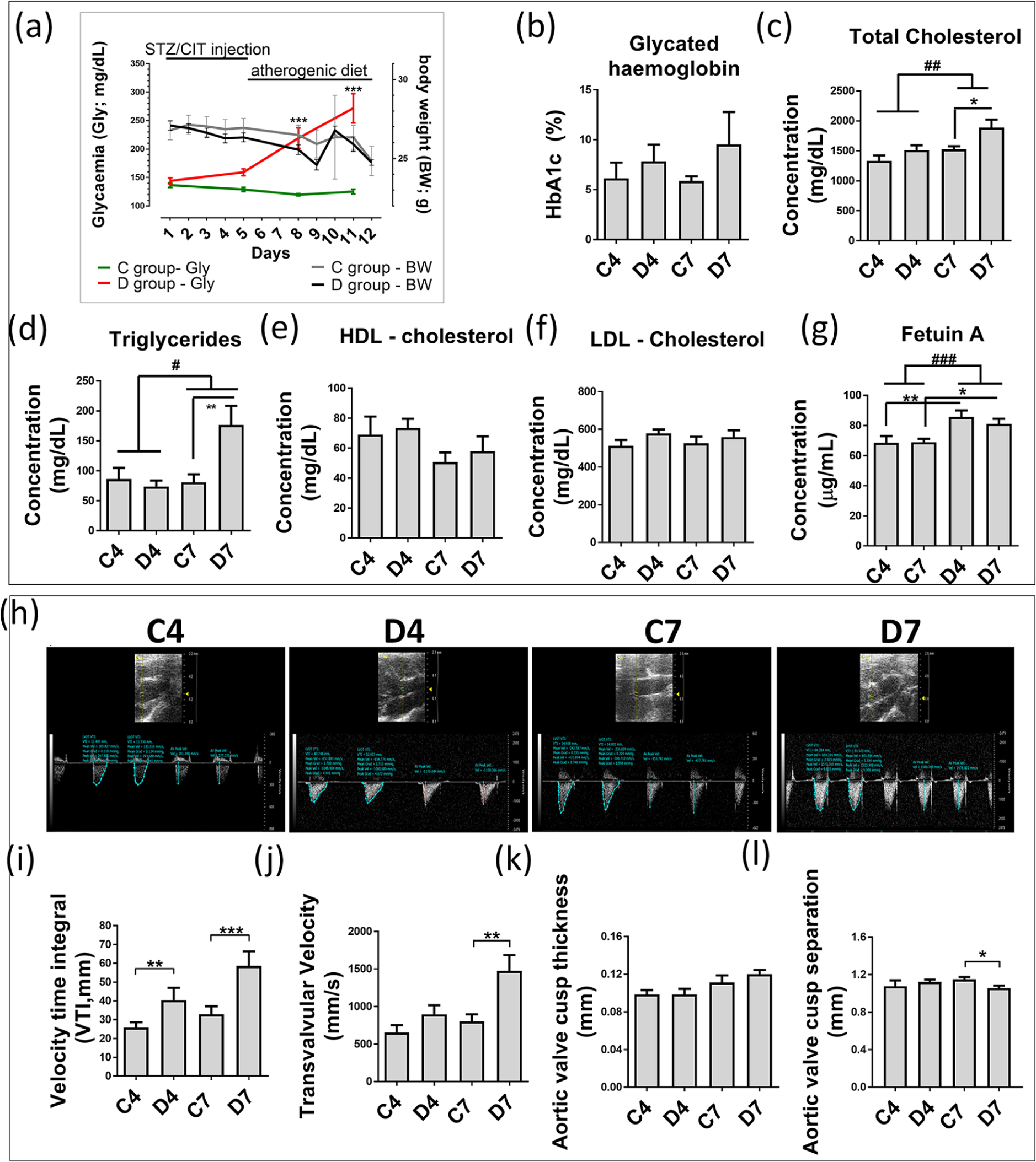
Plasma and echocardiographic parameters measured between hyperlipemic ApoE−/− and hyperlipemic ApoE−/− diabetic mice groups. (a) A schematic representation of the protocol is used to generate the animal models. Evolution of glycaemia and body weight is shown during and after streptozotocin (STZ) – for the diabetic animals (D group), or citrate (CIT) – for the control animals (C group), treatment and atherogenic diet. Approximately, 70% of the mice were diabetic at 3 days after the last STZ administration, defined as fasting glucose >200 mg/dL. The statistical significance was represented as ***p < 0.001 (n = 8). Levels of plasma glycated haemoglobin expressed as percent of total haemoglobin (b), total cholesterol (c), triglycerides (d), HDL cholesterol (e), LDL-cholesterol (f) and fetuin A (g) were measured in the STZ-treated diabetic (D) and citrate-treated non-diabetic (C) groups subjected to atherogenic diet for 4 and 7 days. The statistical significance was represented as #p < 0.05, ##p < 0.01, ###p < 0.001 two-way ANOVA; *p < 0.05, **p < 0.01 one-way ANOVA, Bonferroni post-test (n = 8). (h) Representative echocardiographic PW-Doppler mode images for velocity time integrals and transvalvular velocity. Quantification of velocity time integrals (i), transvalvular velocity (j), cusp thickness (k) and cusp separation (l). The statistical significance was represented as *p < 0.05, **p < 0.01, ***p < 0.001 (n = 8).
No significant change in body weight was observed for the citrate-injected animals from C group, while animals from the D group had significantly reduced body weights at 3, 4 and 7 days after the last STZ injection, Figure 1(a).
Glycated haemoglobin levels did not exhibit statistically significant differences between groups C and D at either 4 or 7 days after the last STZ injection (Figure 1(b)). This may be due to the fact that the diabetes duration in this experiment was very short.
In addition, we found that both cholesterolaemia and triglyceridaemia were significantly increased in diabetic hyperlipemic ApoE−/− mice at 7 days after the last STZ injection (D7 group) as compared to hyperlipemic ApoE−/− mice at 7 days after the last citrate injection (C7 group) (Figure 1(c) and (d)). Also, the duration of the atherogenic diet significantly interacted with diabetes to increase plasma cholesterol and triglycerides concentration (Figure 1(c) and (d)). These changes closely mimic those observed in diabetic dyslipidaemia known to yield a high risk of cardiovascular complications. 9
The plasma HDL cholesterol levels were not significantly altered between treatment groups (Figure 1(e)). Also, LDL-cholesterol was elevated in all experimental groups, but no significant differences were seen between groups (Figure 1(f)).
Because the plasma fetuin A levels were positively associated with diabetes risk, 10 these levels were measured by ELISA in the plasma collected from all animals. Our results showed that, the plasma fetuin A concentrations were significantly increased by STZ injection in both D4 and D7 groups compared to the C4 and C7 groups (Figure 1(g)), suggesting that diabetes has a greater contribution than accelerated atherosclerosis to increasing the circulating levels of fetuin A.
Early functional alterations/modifications induced by diabetes in the aortic valves of hyperlipemic ApoE−/− mice
To see functional modifications in the aortic valve induced by diabetes compared with accelerated atherosclerosis caused by hyperlipemic diet in ApoE−/− mice, the haemodynamic parameters were evaluated by echocardiography in D4 and D7 groups compared with C4 and C7 groups. In this sense, on anaesthetized mice, we measured the following functional parameters: VTI, transvalvular velocity (VEL), aortic cusp separation and cusp thickness. Representative echocardiographic images for VTIs and transvalvular velocity are shown in Figure 1(h).
The results revealed that the mean value of VTI was significantly increased both for D4 group compared to C4 group (by ~1.56 times), and for D7 group compared to C7 group (by ~1.77 times) (Figure 1(i)). Also, the mean value of VEL was significantly higher for D7 group versus C7 group (by ~1.80 times) (Figure 1(j)). Although, we noticed a slight increase in the median value for peak VEL across aortic valve in mice from D4 group compared to the C4 group, this change was not statistically significant. To further evaluate the function of the aortic valve, we investigated aortic cusp thickness and separation by echocardiographic measurements performed in B and M modes, respectively (Figure 1(k) and (l)).
The values obtained for cusp thickness of aortic valves from D7 mice were slightly increased (by ~1.08 times) compared to those measured at C7 mice, but they were not statistically significant (Figure 1(k)).
Results also showed a small but significant decrease in cusp separation values in mice from D7 group compared to that from mice in C7 group. The echocardiographic records of the aortic cusp separation at mice in C4 and D4 groups had similar values (Figure 1(l)).
The decrease of maximal separation of the aortic valve cusps during systole in the progressive stage of diabetes correlates well with the severity of aortic stenosis measured by the increase in VTI and VEL.
Hyperlipemic diabetic ApoE−/− mice develop early stage lesions in the aortic valve
In the diabetic ApoE−/− mice fed with a hyperlipemic diet, we could observe lesions displaying the morphological features of early stage lesions described in previous studies. 11 The initial lesions appeared to be mostly localized in the aortic sinuses and in the base of the leaflets as Oil Red O-stained lipid pools were found accumulating within 4 days of diabetes onset (Supplementary Figure 1, upper panel).
Histological analysis of leaflets showed characteristic valvular ECM composition including collagen (Supplementary Figure 1, middle panel) and glycosaminoglycans (GAGs) (Supplementary Figure 1, lower panel). Calcification was not detected according to Alizarin red staining (data not shown).
Inflammatory markers expression is significantly increased in the diabetic aortic valve in the first week of diabetes
To evaluate modifications associated with inflammation at aortic valve level, cryosections of murine heart tissues were stained for adhesion molecules known to be involved in vascular cells in diabetes: P-selectin, ICAM-1, VCAM-1, PECAM-1. Positive staining was detected for P-selectin, VCAM-1 and ICAM-1 after 4 days of diabetes induction.
The semiquantitative analysis revealed that staining levels in diabetic mice aortic valves were significantly increased over controls for P-selectin (by 1.5-fold) in D4. Moreover, C7 control was significant increased over C4 control (Figure 2(a) and (h)). P-selectin staining was generally associated with the endothelial layer. Significantly increased ICAM-1 staining levels were detected in aortic valves of diabetic mice, both in D4 (by 1.3-fold) and D7 (by 1.7-fold) over C4 and C7, respectively; moreover, a more intense staining was noticed in D7 compared to D4 (by 1.3-fold). Interestingly, in D7, ICAM-1 staining was observed throughout the valve leaflet interstitium and not only associated with the endothelial cells (Figure 2(b)). VCAM-1 staining was observed both in D4 and D7 in diabetic aortic valves mainly associated with endothelial cells; the semiquantitative analysis revealed that the increase was significant only in D4 compared to C4 (by 1.3-fold). In addition, the C7 control was significantly increased over C4 control (Figure 2(c) and (j)). PECAM-1 staining was detected in D4 and D7, on or under the leaflet surface, and in D7 the staining was detected also in the aortic valve leaflet. The semiquantitative analysis revealed a statistically significant increased expression, by 1.3-fold in D4 and 1.4-fold in D7 as compared to the corresponding controls (Figure 2(d) and (k)).
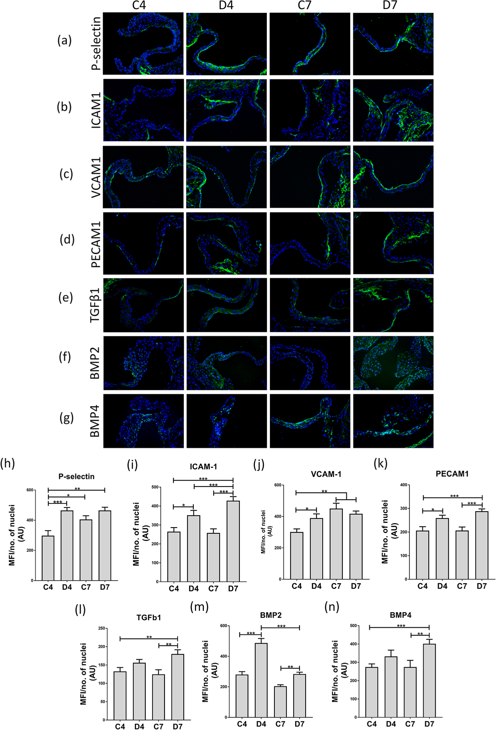
Effect of diabetes on adhesion molecules and TGFβ family members’ expression induced in the murine aortic valve. Representative images of P-selectin (a), ICAM-1 (b), VCAM-1 (c), PECAM-1 (d), TGFβ1 (e), BMP2 (f) and BMP4 (g) expression in aortic leaflets, in control groups and diabetic groups at 4 and 7 days. Aortic valve sections were marked with specific primary antibodies and FITC-coupled secondary antibodies (green staining). Nuclei were stained with DAPI (blue staining). Statistical analysis of P-selectin (h), ICAM-1 (i), VCAM-1 (j), PECAM-1 (k), TGFβ1 (l), BMP2 (m) and BMP4 (n) expression in control and diabetic groups. The statistical significance was represented as *p < 0.05, **p < 0.01, ***p < 0.001 (n = 8).
TGFβ family members expression are increased in aortic valves of hyperlipemic ApoE−/− diabetic mice
Since members from the TGFβ superfamily TGFβ1, BMP2 and BMP4 were detected in stenotic aortic valve leaflets and found to be relevant for CAVD progression, 12 , 13 they were evaluated by immunohistochemistry. Our results showed that TGFβ1 was increased by 1.2-fold in D7 over C7 (Figure 2(e) and (l)), and BMP4 was significantly increased by 1.5-fold in D7 over C7 (Figure 2(g) and (n)). BMP2 expression was significantly increased in both diabetic conditions compared to controls, its level being lower at 7 days (1.4-fold over control) compared with 4 days (1.9-fold over control) after diabetes instalment (Figure 2(f) and (m)). Negative controls displayed no staining (data not shown).
Pro-osteogenic markers expression is increased in the aortic valves of hyperlipemic ApoE−/− diabetic mice
Calcification evolving from inflammatory conditions predominantly results from active osteogenesis within aortic valve leaflets. 14
Aortic valve calcification is an actively regulated process, comparable at many levels with bone mineralization. The expression of molecules associated with physiologic mineralization, such as ALPL, osteopontin 15 and OCL 16 has been described in calcific valve disease. Thus, in this study, the role of diabetic conditions on the expression of osteogenesis-associated molecules, such as ALPL, OCL and osteopontin, was assessed through immunofluorescence staining. The results showed the presence of all molecules in both control and diabetic groups, probably due to the mouse strain (ApoE−/−) and the hyperlipemic diet. We could observe a significantly increased expression of ALPL, in D4 (by 1.7-fold over C4) and D7 by 1.2-fold over C7 (Figure 3(a) and (i)). OCL staining levels were significantly increased over controls in D4 (by 1.9-fold) and D7 by 2.1-fold (Figure 3(b) and (j)). Osteopontin staining was found to be significantly increased over controls, both in D4 (by 1.5-fold) and D7 (by twofold) (Figure 3(c) and (k)).
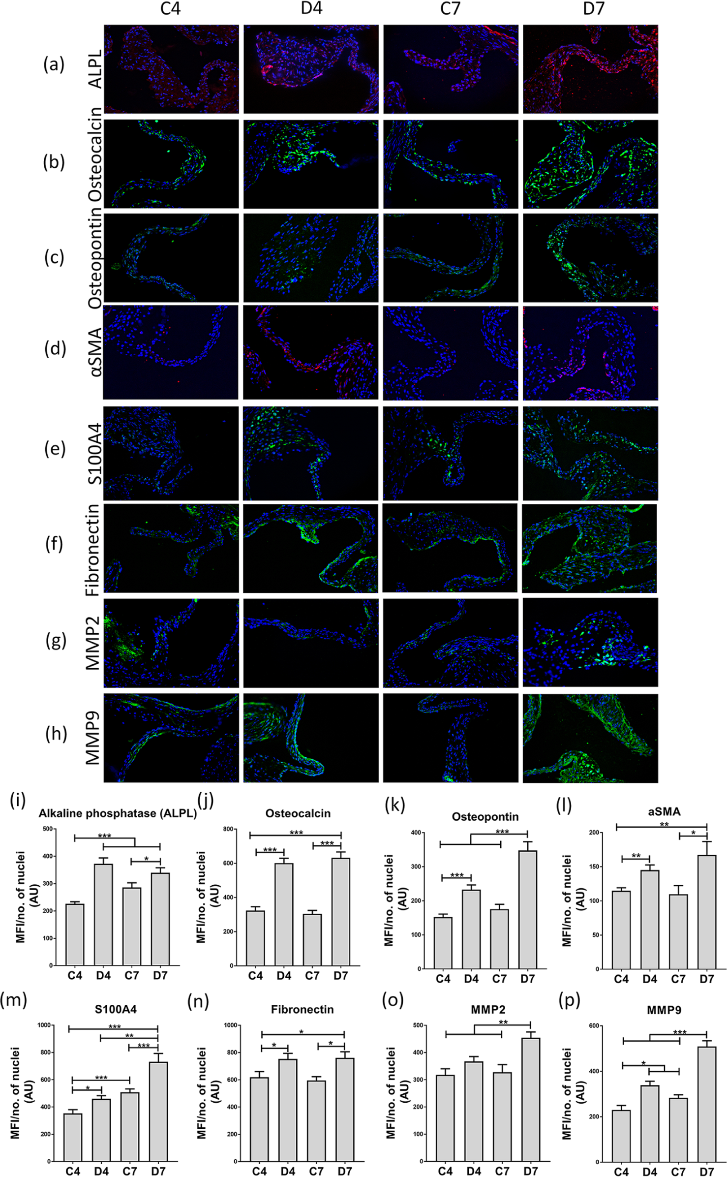
Identification of osteogenesis-associated proteins, cell activation markers and ECM-specific proteins induced by diabetes in the murine aortic valve. Representative images of alkaline phosphatase (ALPL) (a), osteocalcin (b), osteopontin (c), αSMA (d), S100A4 (e), fibronectin (f), MMP2 (g) and MMP9 (h) staining in control and diabetic groups (4 and 7 days). Aortic valve sections were marked with specific primary antibodies and AlexaFluor594 (red) or FITC-coupled (green) secondary antibodies. Nuclei were stained with DAPI (blue staining). Statistical analysis of ALPL (i), osteocalcin (j), osteopontin (k), αSMA (l), S100A4 (m), fibronectin (n), MMP2 (o) and MMP9 (p) expression in control and diabetic groups. The statistical significance was represented as *p < 0.05, **p < 0.01, ***p < 0.001 (n = 8).
Diabetic conditions induce increased expression of valvular cells activation markers
It is known that αSMA is expressed by activated valvular cells; hence, we investigated whether this molecule is expressed in the aortic valve in diabetic conditions. Immunofluorescence staining showed that αSMA is significantly increased in diabetic conditions compared with controls and its expression increases over time, by 1.3-fold in D4 over C4 and by 1.5-fold in D7 over C7 (Figure 3(d) and (l)). S100A4 is an intracellular calcium-binding protein expressed by osteoblastic cells, 17 and its presence in the diabetic aortic valve was assessed by immunostaining. Our data showed that S100A4 is significantly increased in the diabetic group compared with control group, by 1.3-fold in D4 and by 1.4-fold in D7 (Figure 3(e) and (m)).
FN and remodelling molecules are increased in the aortic valve of hyperlipemic ApoE−/− diabetic mice
It was found previously that the anatomical structure of mouse aortic valves differs from that of humans. Mice do not have the trilayer morphology characteristic for human valve, and their leaflets are usually only 5–10 cells thick and do not exhibit segregated layers. 18 Although, the ECM of murine aortic valve comprises the same components, with collagen type 1 and 3 being the main components of ECM, with interweaved fibres of elastin, FN and proteoglycans. Because aortic valve disease is characterized by thickening of valve leaflets and ECM protein modifications, we investigated the effect of diabetic conditions on ECM components visualized by immunofluorescence staining. Our results showed a constant expression of collagen 1, collagen 3, laminin and elastin in the aortic leaflets, regardless of diabetes instalment (data not shown). From the ECM components analysed, the only ECM component affected by diabetes was found to be FN, its expression being significantly increased over time by STZ-induced diabetes, by 1.2-fold in D4 over C4 and by 1.3-fold in D7 over C7 (Figure 3(f) and (n)).
Aortic valve disease is characterized by an upregulated expression of MMPs associated with collagen degradation, such as MMP1, MMP2, MMP3, MMP9 and MMP13. This leads to extensive disorganization of collagen fibres, compromising the structural integrity of the valve. 19 Immunostaining of murine aortic valve sections with specific antibodies for MMP2 and MMP9, showed that MMP2 was significantly induced by 1.4-fold over control only in D7 (Figure 3(g) and (o)) and MMP9 is significantly expressed as early as 4 days, in D4 by 1.5-fold over C4, but increases significantly after 7 days in diabetic conditions, by 1.8-fold (Figure 3(h) and (p)).
Correlations between aortic valve tissue markers and haemodynamic and plasma parameters in hyperlipemic ApoE−/− diabetic mice
To gain insight into the possible associations between haemodynamic parameters, tissue markers and plasma parameters determined in this experimental setting, we performed a correlation analysis (Figure 4 and Supplementary Table 1).
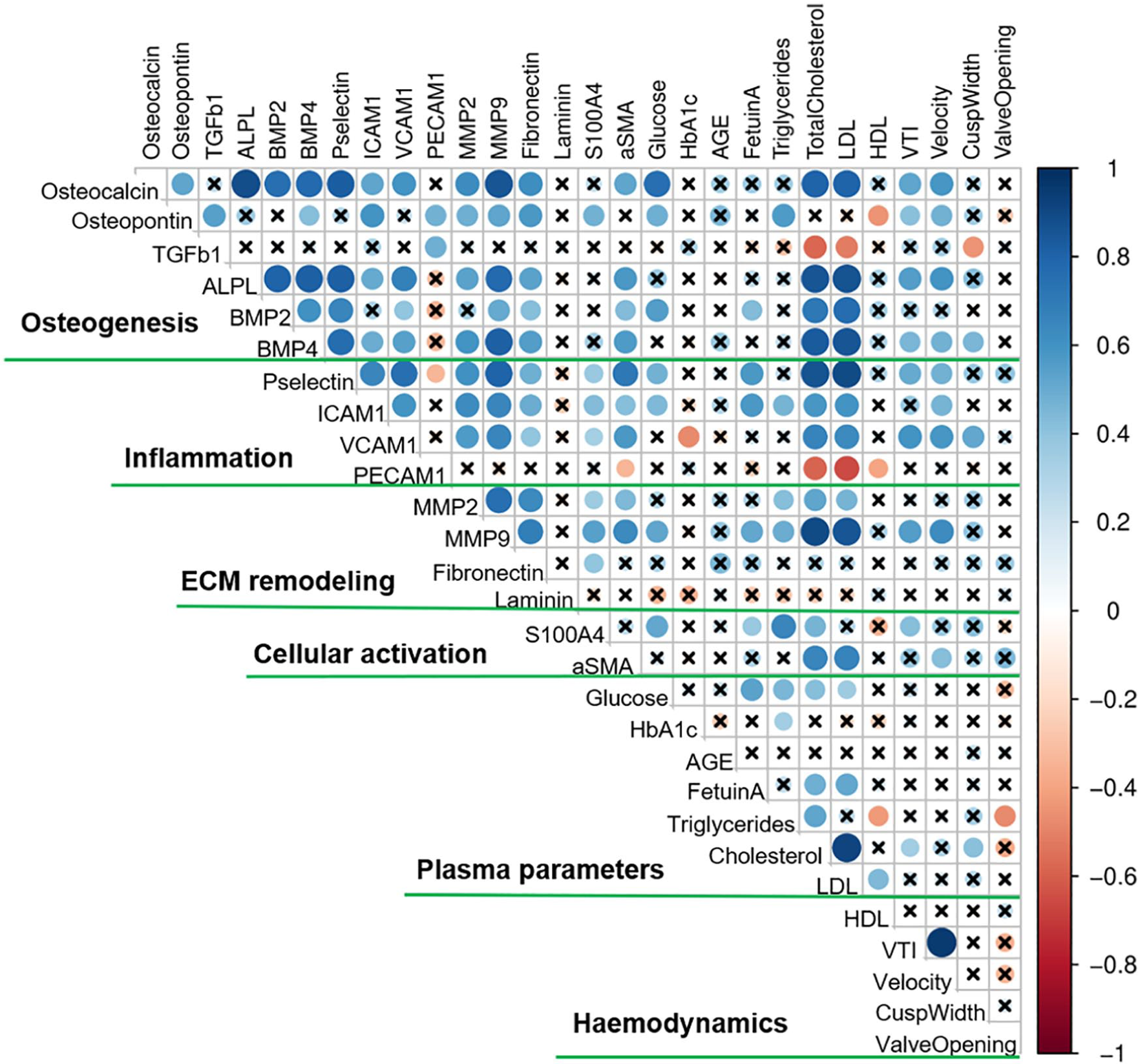
Correlation matrix between pro-osteogenic, inflammatory, ECM remodelling, cellular activation, plasmatic and haemodynamic parameters. Mean values of measured parameters for each experimental group were used for a Pearson correlation matrix conducted using the R package ‘corrplot’. Blue colour represents positive correlation; red colour represents negative correlation; darker colours and larger shapes represent higher association. X represents no statistical difference.
Our analysis revealed that peak aortic jet velocity was highly correlated with inflammatory biomarker VCAM-1, pro-osteogenic markers OCL and ALPL, remodelling enzyme MMP9 and myofibroblastic marker αSMA; peak aortic jet velocity was moderately associated with P-selectin, ICAM-1, BMP4 and osteopontin, suggesting that these molecules may contribute to the early aortic valve dysfunction detected in hyperlipemic ApoE−/− diabetic mice. No correlation was found between velocity and plasma markers.
Glycaemia was significantly correlated with OCL, osteopontin, BMP2, P-selectin, ICAM-1, MMP9, S100A4, fetuin A, triglycerides, total cholesterol and LDL-cholesterol.
Very high correlations (r > 8) were found between – P-selectin and plasma parameters – such as LDL and total cholesterol, osteoblastic markers OCL and ALPL and remodelling enzyme MMP9; BMP2 and ALPL, BMP4 and total cholesterol, LDL, ALPL and MMP9; MMP9 with total cholesterol, LDL, P-selectin; ALPL with total cholesterol, LDL, P-selectin, BMP4, OCL; OCL was positively and significantly correlated with total cholesterol, LDL, P-selectin and ALPL.
High correlations (0.5 < r < 0.8) were found between: P-selectin with fetuin A, VCAM-1, ICAM-1, BMP2, BMP4, MMP2, αSMA, VTI; VCAM-1 with total cholesterol, LDL, P-selectin, ICAM-1, BMP4, OCL, ALPL, MMP9, MMP2, αSMA and the haemodynamic parameters VTI and velocity. ICAM-1 displayed a positive high correlation with total cholesterol, LDL, fetuin A, P-selectin, VCAM-1, osteopontin, OCL, with ALPL, MMP9, MMP2 and with FN. PECAM-1 displayed a negative high correlation with the seric parameters total cholesterol and LDL.
Notably, our analysis detected significant correlations between activation of inflammatory, remodelling and pro-osteogenic pathways in valve tissues with expression of plasma markers and alterations in valve haemodynamics, pointing to molecular associations unique for CAVD initiation in atherosclerosis accelerated by diabetes. Pearson correlation coefficient values and p-values are indicated in Supplementary Table 1.
Discussion
Diabetes leads to accelerated atherosclerosis in both natural aortic valves and in valvular scaffolds/prosthesis, by mechanisms that still need to be uncovered in order to identify new relevant biomarkers for CAVD and to reveal new targets for therapies. Since there is evidence that the aortic valve is one of the first territories affected by diabetes 1 from the second week of diabetes onset, we hypothesized that important changes for CAVD progression may appear even earlier after diabetes onset.
Our preliminary time course studies (data not shown) on aortic valve haemodynamic parameters revealed significant changes from the first week of diabetes; these results prompted us to monitor changes in the aortic valve after 4 days and 1 week after diabetes onset (induced by STZ) in an ApoE−/− mice. To accelerate more the process, we fed the mice also with hyperlipemic diet after diabetes onset and evaluated valvular tissue inflammatory, osteogenic and remodelling markers and plasma and haemodynamic parameters changes, comparing diabetic mice with hyperlipemic mice (designated as control groups). Subsequently, we established correlations between these parameters.
Our data showed the following main findings:
Peak aortic velocity, an aortic valve haemodynamic parameter, was significantly increased by 1.8-fold and aortic cusp separation significant decreased by ~0.918-fold, 7 days after diabetes onset, while VTI was significantly increased in D4, by 1.56-fold, compared with C4 and by 1.77-fold in D7 compared with C7.
Inflammatory markers: P-selectin, VCAM-1, ICAM-1 and PECAM-1 expression was significantly increased in aortic valve tissue during the first week of diabetes; namely, all molecules displayed increased expression compared to controls from 4 days after diabetes onset, while only ICAM-1 and PECAM-1 were further increased after 7 days.
TGFβ family members’ analysis revealed that BMP2 was increased after 4 days of diabetes while BMP4 and TGFβ1 only after 7 days of diabetes.
Osteogenic markers OCL, osteopontin and ALPL were increased after 4 and 7 days of diabetes.
Valvular cells activation/phenotypic type markers: αSMA and S100A4 were increased from 4 days and also after 7 days.
FN and remodelling molecule MMP9 were increased after 4 days, while MMP2 after 7 days; collagen type 1 and 3, laminin and elastin were unchanged in D4 and D7 compared to controls.
Significant important correlations were found between inflammatory, osteogenic and remodelling parameters and haemodynamic and plasma parameters, suggesting specific molecular associations that may contribute to the early aortic valve dysfunction detected in hyperlipemic ApoE−/− diabetic mice.
Our data indicate that aortic valve dysfunction may install early in the time course of diabetes, 1 week after diabetes onset in hyperlipemic ApoE−/− diabetic mice, comparing with hyperlipemic ApoE−/− mice (without STZ injection), indicating that diabetes may accelerate aortic valve dysfunction, subsequently diabetes onset.
The quantitative measurements of valve function by echocardiographic analysis indicated that the transvalvular velocity was significantly higher in diabetic mice versus controls, suggesting the development of aortic valve stenosis. Since this parameter estimates aortic valve stenosis severity, and is the principal parameter to be followed in the evolution of valvular degeneracy, our results may suggest that aortic valve stenosis may develop early in diabetic conditions.
There is evidence that aortic valve dysfunction occurs in diabetes, but after longer time periods, subsequently diabetes onset. 20 However, aortic stenosis development in a shorter period of time (1 week in our study), may be explained by use of a different diet, a hypercholesteraemic diet enriched with fat and a different mouse model, the STZ-induced diabetic ApoE−/− mice.
Important significant high correlations between peak aortic jet velocity and aortic valve tissue biomarkers VCAM-1, OCL, ALPL and MMP9 were found, suggesting that early changes in molecules expression involved in inflammation, osteogenesis and remodelling pathways may be associated to the increased aortic valve dysfunction detected in our model, by mechanisms that need to be uncovered.
However, our analysis revealed some interesting correlations between seric parameters and aortic tissue markers. Our results revealed significant correlations between fetuin A and glucose in the first week of diabetes onset. In humans, serum fetuin A levels are positively associated with diabetes. 21
Fetuin A is presumed to play a role in development of cardiovascular disease. 22 Our data showed a high positive correlation of fetuin A with VCAM-1 and P-selectin with glycaemia, suggesting that fetuin A may be considered also as a biomarker or a possible mediator of aortic valve events occurring in early diabetes onset events associated with endothelial cells dysfunction, by mechanisms that still need to be uncovered.
There are few studies on aortic valve changes in structure and function at early stages after diabetes onset. In order to further uncover molecules and mechanisms which may be involved in accelerated aortic valve stenosis existing in our model, we examined the presence in aortic valve of molecules associated with inflammation, remodelling and calcification, processes shown to be associated with aortic CAVD.
Our data showed that inflammatory and osteogenic markers increased very early, 4 days after diabetes onset, as well as activation markers for valvular cells; moreover, the ECM molecule FN and remodelling enzyme MMP9 were found to be increased also at this very early time-point. However, after 7 days of diabetes onset, P-selectin and VCAM-1 were no more increased compared to controls, while TGFβ1, BMP4 and MMP2 were increased only after 7 days. Our results may indicate that important changes occur very early after diabetes onset in aortic valve tissue, and these molecular changes may vary shortly after subsequent diabetes onset and may indicate successive early stages with specific molecular signatures in CAVD evolution and may point to possible mechanisms involved and indicate possible strategies for therapy.
Concerning inflammatory mediators, we analysed cell adhesion molecules (CAMs) and TGFβ family cytokines expression in aortic valve. Our data indicate a very early upregulation of P-selectin and VCAM-1 by diabetic conditions, a further increase induced by diet at 7 days and no additional increase over control after 7 days of diabetes. By contrast, ICAM-1 and PECAM-1 were upregulated after 4 and 7 days post diabetes induction. P-selectin and VCAM-1 expression was demonstrated in early atherosclerotic lesions of ApoE−/− mice after only 4 weeks of Western diet. 23 In addition, there is evidence that reductions in the expression of P-selectin or ICAM-1 provide direct protection from atherosclerotic lesion formation in a model of ApoE−/− mice. 24 However, there are no data, to our knowledge, on P-selectin, VCAM-1 or ICAM-1 detection in early diabetic lesions in aortic valve in ApoE−/− mice. Our results indicate that these inflammatory mediators appear very early at the level of aortic valve after diabetes onset and may influence by specific mechanisms on lesion evolution.
Our correlation analysis revealed very high positive correlations of P-selectin with total cholesterol and LDL, high positive correlation with fetuin A and moderate positive correlation with glucose, indicating that hypercholesterolaemia is strongly involved in P-selectin expression over the effects of high glucose. Moreover, P-selectin was highly correlated with fetuin A and VTI and moderately correlated with velocity and with functional haemodynamic parameters of the aortic valve, suggesting important contribution of this molecule in the pathophysiology of aortic stenosis in diabetes.
The PECAM-1 plays an important role in many inflammatory processes, including the development of atherosclerosis. 25 Our data showed an increased PECAM-1 expression in diabetic mice aortic valve. However, PECAM-1 was highly negative correlated with seric lipids and positively with TGFβ1; considering PECAM-1 associations with endothelial-to-mesenchymal transformation (EMT) and that we detected PECAM-1 under the endothelial surface in diabetic mice, we may presume that in early diabetes onset, EMT may occur and PECAM-1 expression may be modulated in the process. Further experiments need to be done to clarify the matter. Moreover, our correlation analysis, revealed that P-selectin, ICAM-1 and VCAM-1 were highly correlated, while P-selectin was significant negatively correlated with PECAM-1; ICAM-1 and VCAM-1 were not correlated with PECAM-1, possible indicating independent mechanisms of their regulation in early diabetes in the aortic valve.
TGFβ1, BMP2 and BMP4 are important osteogenic mediators and they were found increased in aortic valve in diabetic mice. Their role in early diabetes in ApoE−/− diabetic mice is not very well known, yet. There is evidence of upregulation of TGFβ as a consequence of degenerative conditions and in direct dependency with glucose concentrations; 26 it is known that BMP signalling is required for the development of aortic valve calcification in vitro and in vivo. 27 Our studies revealed that BMP2 increases very early after diabetes onset (4 days), while TGFβ1 and BMP4 increase after 7 days. Moreover, BMP2 was highly correlated with hyperglycaemia and total cholesterol, indicating that both may contribute to BMP2 upregulation in very early diabetes; BMP2 is also correlated with P-selectin. The possible mechanisms need further to be understood. Moreover, BMP2 and BMP4 were highly correlated, suggesting that BMP2 may contribute to BMP4 upregulation. Our studies indicate that, in the aortic valve of diabetic mice, important inflammatory and osteogenic processes occur with important consequences in the evolution of CAVD.
Since our data showed an increased expression of BMP2 in aortic valve of diabetic mouse, and since it is known that BMP2 is an osteoinductive factor that promotes osteogenic differentiation in a variety of cells and that OCL is a typical calcification marker, we analysed the modulation of osteogenic markers in our experimental setting.
Analysis of osteogenesis-associated molecules revealed that ALPL, OCL and osteopontin were significantly increased both after 4 and 7 days after diabetes onset. Since these are generally accepted as markers for valvular cells differentiation towards osteoblastic phenotype 28 and they were found to be localized in the valve leaflets, we may assume that very early diabetes onset in hyperlipemic ApoE−/− mice may induce an osteogenic process involving OCL, osteopontin and ALPL. These markers were also found to be associated with early CAVD in diabetes induced by high-fat (HF) diet in ApoE−/− mice. 18 In addition, these molecules were detected in early CAVD inflammation and osteogenesis by imaging aortic valves of ApoE−/− mice fed with atherosclerotic diet. 29 Our results extend these data and detect these osteogenic molecules increase expression also in early CAVD in diabetic ApoE−/− mice fed with hyperlipemic diet. In our study, we also found inflammation and osteogenesis to be associated but in early diabetes in hyperlipemic ApoE−/− mice and revealed specific markers in highly association. The mechanisms involved in their generation need further to be uncovered. In addition, we found high positive correlations between these molecules and inflammatory molecules, indicating the association of specific inflammation and osteogenic molecules in the early events/processes occurring in the aortic valve of hyperlipemic ApoE−/− diabetic mice.
OCL and ALPL were both highly correlated with BMP2 and BMP4. There is evidence that BMP2 and BMP4 can induce a significant increase in the activity and expression of alkaline phosphatase and OCL in isolated human valve interstitial cells, 30 therefore we may presume, since OCL and ALPL staining was found all over the valve leaflet, that this mechanism may also function and be accelerated in our model, in diabetic conditions. Further experiments will clarify this matter.
Moreover, P-selectin, BMP4, MMP9, ALPL and OCL were found to be very highly correlated with total cholesterol and LDL, indicating important role for hypercholesterolaemia in their upregulation in valve tissue in diabetic hyperlipemic ApoE−/− mice.
Concerning the activation markers expression of valvular cells, our study revealed that αSMA and S100A4 increased both in the aortic valve after 4 and 7 days of diabetes. Since it is considered that the αSMA-positive cells represent an ‘activated’ population of myofibroblasts that differentiate into osteoblast-like cells 31 and it was shown that S100A4 is expressed by pre-osteoblasts in vitro, 17 we may presume that, in early diabetes increased presence of myofibroblasts and pre-osteoblast phenotypes of VIC may be detected. Moreover, αSMA displayed a positive significant correlation with VCAM-1, BMP4, MMP9, OCL, ALPL, P-selectin, total cholesterol and LDL, indicating that hypercholesterolaemia, inflammatory mediators and osteoblastic molecules may contribute to αSMA upregulation in our model. S100A4 protein displayed a positive significant correlation with triglycerides, glucose and MMP9, indicating that diabetes may enhance S100A4 induction in aortic valve leaflet.
Analysing early modifications of ECM and remodelling molecules in diabetic aortic valve – namely, collagen type 1 and 3, elastin, FN and laminin – our study revealed that only FN was significantly upregulated both after 4 and 7 days of diabetes. FN is one of the abundant ECM proteins and is upregulated in diabetes as well as in endothelial cells treated with high concentrations of glucose. 32 Our results showed that FN was highly correlated with the inflammatory tisular marker ICAM-1, with the pro-osteogenic markers OCL, osteopontin and ALPL, with BMP4 and with remodelling enzymes MMP2 and MMP9, suggesting that early diabetes may induce modifications in ECM proteins that may be associated with particular inflammatory, osteogenic and remodelling molecules and accelerate by mechanisms that need to be uncovered atherosclerosis progression.
Hyperglycaemia directly or indirectly (via oxidative stress or advanced glycation products) increases MMP expression and activity. These changes are associated with histologic alterations in large vessels. MMPs are upregulated in CAVD and may play a role in its pathogenesis. 33 The remodelling parameters analysis in our study revealed that MMP9 increased after 4 and 7 days of diabetes, and MMP2 only after 7 days. MMP9 upregulation was highly positively correlated with most markers investigated: total cholesterol, glucose, fetuin A, VTI, velocity, αSMA, S100A4, P-selectin, VCAM-1, ICAM-1, osteopontin, BMP2, BMP4, FN and MMP2. These strong associations may indicate MMP9 as a molecule with key role in early CAVD in diabetic atherosclerosis in ApoE−/− mice and further studies will clarify this matter.
Limitations
A limitation of this study is the animal model of diabetes. STZ-induced type 1 diabetes model has been used for more than 50 years 34 and it models the natural evolution of the disease as the level of glycaemia control seen in real case human patients is nearly impossible to obtain in mice by insulin injections. However, even with the best available treatments, human patients eventually develop cardiovascular complications due to the inevitable glycaemia and insulin concentration variations from normal. Thus, we believe the observed pathological modifications in our experimental conditions are an accelerated model of cardiovascular complications in human patients.
Another limitation is that this study is mainly descriptive, and as a resource paper, might help to define some new targets and pathways in CAVD. We plan to perform additional studies investigating whether inhibition/activation of these factors could influence prognosis or phenotypes of CAVD in mice to demonstrate that the shown markers might be potential therapeutic targets.
Conclusion
Our results show that the aortic valve function and structure of diabetic ApoE−/− mice was affected early after diabetes onset, from the first week and suggest important molecular contributors to valvular dysfunction. Our data indicate enhanced ongoing active processes of osteogenesis in increased inflamed valves and establish high and significant correlations between the markers identified to be modified. Our study may help to advance the understanding of the molecular mechanisms of early aortic valve dysfunction, before significant fibrosis and calcification occurrence, in diabetes-accelerated atherosclerosis, in order to discover and validate new biomarkers of CAVD in diabetes and reveal new possible targets for nanobiotherapies for inhibition of CAVD progression of valvular lesions.
Supplemental Material
Answers_to_Reviewers_Rev2 – Supplemental material for Diabetes-induced early molecular and functional changes in aortic heart valves in a murine model of atherosclerosis
Supplemental material, Answers_to_Reviewers_Rev2 for Diabetes-induced early molecular and functional changes in aortic heart valves in a murine model of atherosclerosis by Monica Madalina Tucureanu, Alexandru Filippi, Nicoleta Alexandru, Cristina Ana Constantinescu, Letitia Ciortan, Razvan Macarie, Mihaela Vadana, Geanina Voicu, Sabina Frunza, Dan Nistor, Agneta Simionescu, Dan Teodor Simionescu, Adriana Georgescu and Ileana Manduteanu in Diabetes & Vascular Disease Research
Supplemental Material
Supplementary_figure_1 – Supplemental material for Diabetes-induced early molecular and functional changes in aortic heart valves in a murine model of atherosclerosis
Supplemental material, Supplementary_figure_1 for Diabetes-induced early molecular and functional changes in aortic heart valves in a murine model of atherosclerosis by Monica Madalina Tucureanu, Alexandru Filippi, Nicoleta Alexandru, Cristina Ana Constantinescu, Letitia Ciortan, Razvan Macarie, Mihaela Vadana, Geanina Voicu, Sabina Frunza, Dan Nistor, Agneta Simionescu, Dan Teodor Simionescu, Adriana Georgescu and Ileana Manduteanu in Diabetes & Vascular Disease Research
Supplemental Material
Supplementary_Table_1 – Supplemental material for Diabetes-induced early molecular and functional changes in aortic heart valves in a murine model of atherosclerosis
Supplemental material, Supplementary_Table_1 for Diabetes-induced early molecular and functional changes in aortic heart valves in a murine model of atherosclerosis by Monica Madalina Tucureanu, Alexandru Filippi, Nicoleta Alexandru, Cristina Ana Constantinescu, Letitia Ciortan, Razvan Macarie, Mihaela Vadana, Geanina Voicu, Sabina Frunza, Dan Nistor, Agneta Simionescu, Dan Teodor Simionescu, Adriana Georgescu and Ileana Manduteanu in Diabetes & Vascular Disease Research
Footnotes
Acknowledgements
The authors thank Gabriela Mesca for her skilful assistance.
Author contributions
M.M.T. contributed to immunostaining, data analysis, statistical analysis and drafting manuscript; N.A. contributed to echocardiography analysis and drafting manuscript; A.F. contributed to plasma measurements, data and statistical analysis; C.A.C. and G.V. contributed to animal procedures and sample collection; L.C., R.M. and M.V. contributed to acquisition of fluorescence images; S.F. contributed to echocardiography measurements; D.N. contributed to echocardiography analysis; A.S. and D.T.S. contributed to critical revision of the manuscript; A.G. contributed to echocardiography analysis, interpretation of data and drafting manuscript; I.M. contributed to study conception and design, critical revision of intellectual content and writing the manuscript.
Declaration of conflicting interests
The author(s) declared no potential conflicts of interest with respect to the research, authorship and/or publication of this article.
Funding
The author(s) disclosed receipt of the following financial support for the research, authorship and/or publication of this article: This work was supported by the Competitiveness Operational Programme 2014–2020, Priority Axis1/Action 1.1.4/, Financing Contract no. 115/13.09.2016/ MySMIS:104362 and by Romanian Academy.
Supplemental material
Supplemental material for this article is available online.
References
Supplementary Material
Please find the following supplemental material available below.
For Open Access articles published under a Creative Commons License, all supplemental material carries the same license as the article it is associated with.
For non-Open Access articles published, all supplemental material carries a non-exclusive license, and permission requests for re-use of supplemental material or any part of supplemental material shall be sent directly to the copyright owner as specified in the copyright notice associated with the article.