
Abstract
Introduction:
The angiotensin converting enzyme inhibitor ramipril is a standard antihypertensive therapy for many patients. Because angiotensin II may promote inflammation, we were interested in whether basal pretreatment with ramipril may modify renal function and inflammation as well as systemic outcome in experimentally induced sepsis in mice.
Material and methods:
Ramipril (10 mg/kg/day) pretreatment or placebo (NaCl) was given intraperitoneally for 5 days to C57BL6/J mice, followed by either sham operation or cecal ligation and puncture sepsis induction. Real-time polymerase chain reaction and immunological stains were used to evaluate renal gene and protein expression, respectively. Plasma creatinine, neutrophil-gelatinase associated lipocalin, and blood urea nitrogen were used as markers for renal function. A clinical severity score was determined.
Results:
Administration of ramipril before cecal ligation and puncture surgery was associated with reduced renal inflammation but did not improved renal function and structure and even worsened the clinical status of septic mice.
Conclusions:
The data suggest that the effects of ramipril pretreatment are complex. Additional studies including monitoring of hemodynamic parameters are necessary to elucidate the exact mechanism(s) of this observation. In addition, the timing of the ramipril administration could be of importance.
Introduction
Angiotensin converting enzyme (ACE) inhibitors such as ramipril are widely used by many patients. Retrospective clinical observations suggest that preadmission of drugs interfering with the renin-angiotensin-aldosterone system (RAAS) may decrease mortality in patients with sepsis.1,2 However, there are only a few studies in animals investigating the effect of ACE inhibitor treatment prior to experimentally induced sepsis, mainly using LiPS-induced sepsis.3,4 There are intrinsic problems with lipopolysaccharides (LPS)-based models and they may not reflect the situation in humans. 5 Therefore, we were interested in how pretreating mice with ramipril for 5 days may influence renal structure and function in a widely used model of sepsis in mice (cecal ligation and puncture, CLP). 5
Material and methods
Sham operation and CLP surgery
In this study we used wild-type C57BL/6 J male mice (Jackson Laboratories, Main, USA obtained from Charles River Laboratories, Sulzfeld, Germany), 12–16 weeks old, weighing 20–25 g, receiving a standard rodent food and free access to tap water. The animals were housed in regular 12/12 h light/dark cycles at a 23 ± 1 °C temperature. The experimental design was approved by the Animal Committee of the State of Thuringia and were carried out in accordance to the National Institute of Health Guidelines for the Care and Use of Laboratory Animals. Mice received 10 mg/ kg BW ramipril or placebo (saline) by i.p. injection over a total of 5 consecutive days before a sham operation (SOP) or CLP surgery, but nothing on the day of surgery. Age-matched male animals were used in the experimental procedure and were distributed in randomized fashion to four experimental groups as follows: (a) SOP group; (b) CLP-treated mice (CLP group); (c) ramipril + SOP group, (d) ramipril + CLP group. SOP and CLP performed as previously described.6,7 In brief, animals were anesthetized and midline laparotomy was performed, followed by ligation of the cecum and a double puncture. To induce the blood contamination with bacteria, two droplets were pressed out of the wound in the abdominal cavity to trigger the system’s inflammatory response. 6 SOP mice were anesthetized and midline laparotomy was performed but without ligation and puncture of the cecum. Fluid resuscitation was given every 6 h until the end of the experimental procedure by intra-peritoneal application of 25 µl/g body-weight 0.9 % NaCl.
The clinical status of the animals was evaluated every 6 h by applying a Clinical Severity Score (CSS) as previously described. 7 The score uses the parameters of spontaneous activity and reaction to exogenous stimuli and posture, with ranges from one to five for each where five means death of the animal. 7
Then 24 h after SOP or CLP surgery the mice were deeply anesthetized with isoflurane and sacrificed. The kidneys were extracted and paraffin embedded as described elsewhere for further analyses. For analyses of the blood plasma parameters, blood samples were collected, and the isolated plasma samples were kept frozen at -80oC until examined.
Analyses of the renal structure and function
To estimate renal injury a Periodic acid-Schiff (PAS) stain was performed on a 4 µm paraffin kidney sections using a PAS staining kit (Carl Roth GmbH & Co.KG, Karlsruhe, Germany). Tubular injury was evaluated by a scoring system where a score of zero represented no damage and five corresponded to more than 90% tubular damage. Plasma creatinine and blood urea nitrogen (BUN) levels were analyzed on a clinical chemical analyzer (Fuji DRI-CHEM 3500i, Fujifilm, Düsseldorf, Germany) using colorimetric chip assays and are presented in mg/dl. Determination of the plasma concentration of neutrophil-gelatinase associated lipocalin (NGAL) was performed with an NGAL-specific enzyme-linked immunosorbent assay (BioPorto Diagnostics, Gentofte, Denmark). NGAL concentrations are presented in µg/ml.
RNA isolation, reverse transcription and real-time polymerase chain reaction analyses
Renal tissues were homogenized by the aid of SpeedMill P12 homogenizer (Analytic Jena Bio Solutions, Jena, Germany) and total RNA was extracted via RNAeasy kit (Qiagen, Hilden, German. Routinely 1 µg of total RNA was reverse transcribed using the M-MLV reverse transcription system (ThermoFisher Scientific, Invitrogen, Life Technologies, Darmstadt, Germany). The gene specific primers used in real-time polymerase chain reaction (PCR) analyses are as follows: hprt (hypoxanthine phosphoribosyl transferase) forward: 5’-ATCAGTCAACGGGGGACATA-3′, reverse: 5’-AGAGGTCCTTTTCACCAGCA-3′, ngal (lipocalin-2) forward: 5’-CACCACGGACTACAAGTTCGC-3′, 3′ ngal (lipocalin-2) reverse: 5’-TCAGTTGTCAATGCATTGGTCGGTG-3′, tnf-α - forward: 5’-GGCAGGTCTACTTTGGAGTCATTGC-3′, reverse: 5’ ACATTCGAGGCTCCAGTGAATTCGG 3′, renin primers were exactly as described in reference in [29], renin forward: 5’- CCTCTACCTTGCTTGTGGGATT -3′, renin reverse: 5’- CTGGCTGAGGAAACCTTTGACT -3′, IL-1β forward: 5' AAGGAGAACCAAGCAACGACAAAA 3' IL-1β reverse: 5' TGGGGAACTCTGCAGACTCAAACT 3'. The gene expression was estimated using a Q-Tower thermocycler (Analytik Jena Bio Solutions, Jena, Germany), and qPCRsoft 3.4 software (Analytic Jena, Jena, Germany) in a multiplex assay. The relative gene expression was normalized to hprt expression and the relative expression ratio was quantified by ΔΔCT method, where Ratio = 2-ΔΔCT. 8 The mRNA levels in SOP mice was set as 1.
Immunohistology
For immunohistological analyses, we used 2–4 μm paraffin kidney sections with heat-mediated antigen retrieval, as previously described,9,10 followed by inactivation of the endogenous peroxidase activity with 3% H2O2 for 10 min. Next, sections were blocked with 5% BSA for 1 h at room temperature and the primary antibodies were added for overnight at 4°C. The following primary antibodies were used: an anti-CD3 antibody (1:100 dilution) purchased from Dianova (Hamburg, Germany), the detection of the activated caspase-3 was performed by the use of anti-cleaved caspase-3 antibody (Abcam, Cambridge, UK) (1:100 dilution). The anti-HIF2α antibody was from R&D Systems (Wiesbaden, Germany) and used in a 1:200 dilution. The corresponding secondary antibodies, horseradish peroxidase-conjugated (1:500 dilution) or alkaline phosphatase-conjugated (1:500), were purchased from KPL (Gaithersburg, MD, USA). When applicable, the nuclei were counter-stained with hematoxylin (Vector Laboratories Inc.) for 2 min. The microscopic analyses were performed on an Axioplan microscope with AxioVision Rel. 4.6. Software (Zeiss, Jena, Germany). A minimum of six animals per experimental group were investigated. Routinely, five non-overlapping images (magnification 200 x) were taken and the staining was analyzed by a person unaware of the experimental protocol via a semi-quantitative scoring method as previously described. 10 For cleaved caspase-3 staining, the number of positive tubuli per field (200 x magnification) in the cortex region was counted and graphically presented.
Statistical analyses
Results were evaluated by the Kruskal-Wallis one-way analysis of variance on ranks test, followed by the Mann-Whitney rank sum-test to analyze the differences between two groups; the t-test two-tailed p values are shown under figure legends. All pairwise multiple comparison procedures were performed with a Dunn’s or Holm-Sidak method. The data are graphically presented as a box plot where the values are shown as the median and percentiles and a vertical point plot of all the samples’ values was added to the box plot. The values of the clinical severity score are presented as mean ± standard error of mean (SEM). Differences were considered significant when p < 0.05; *p (#p) < 0.05, **p (##p) < 0.01, ***p (###p) < 0.001.
Results
Influence of ramipril on renal renin mRNA expression
First, to test whether animals have indeed received ramipril in a dose to suppress the RAAS, we used real-time PCR studies to determine renin expression in whole kidney lysates. As expected, ramipril pretreatment induced renin transcripts, both in SOP and CLP mice. In the SOP group there was a minor numerical but not statistically significant difference in renin mRNA expressions between the SOP and CLP groups (

Influence of ramipril on the local renal renin mRNA expression 24 h following cecal ligation and puncture (CLP) sepsis induction or a sham operation (SOP). N = 8 per group, **p <0.01.
Influence of ramipril on the sepsis-induced tubular injury
To evaluate the influence of ramipril pretreatment on tubular injury, we performed a PAS-reaction on kidney sections of all experimental groups. We observed a massive tubular injury in mice that underwent CLP surgery compared with SOP animals (

Influence of ramipril treatment on renal function in sepsis
All three measured parameters (plasma creatinine, plasma BUN, and NGAL) were significantly increased in CLP mice compared with the SOP group (

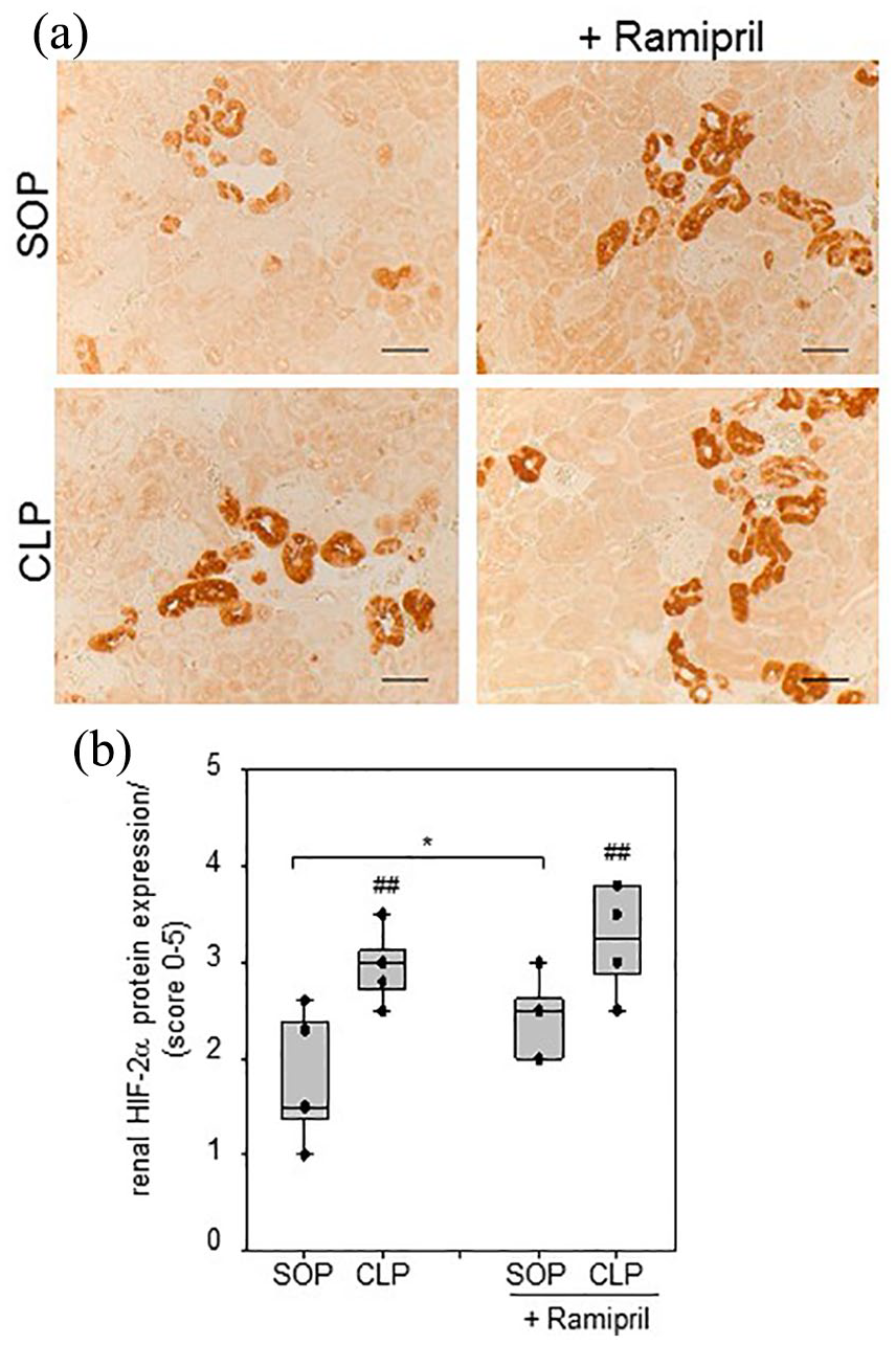
Ramipril elevates the septic accumulation of renal HIF-2α protein
We recently reported that HIF-2α, but not HIF-1α, protein expression was significantly increased in endotoxemic mice.
11
Therefore, we examined whether ramipril pretreatment may affect HIF-2α expression in renal tissue under basal and septic conditions. Immunohistochemistry studies revealed that, in agreement with our previous observation, the protein expression of HIF-2α was significantly elevated in CLP mice compared with the control SOP mice (
Impact of ramipril pretreatment on renal inflammation in septic conditions
There is increasing evidence reporting that angiotensin II (ANG II) is an important factor leading to renal inflammation by increasing the renal expression of pro-inflammatory cytokine as TNF-α.
12
Therefore, we analyzed the effect of ramipril pretreatment on renal inflammation. We investigated the expression of the pro-inflammatory cytokines tnf-α and IL-1β mRNAs in renal tissues. As shown in

Furthermore, we also tested the influence of ramipril on sepsis-induced T-cell infiltration in renal tissue, using immunological detection of CD3+ T lymphocytes. In agreement with our previous observation,
11
sepsis induced a massive accumulation of interstitial T cells in renal tissue (
Influence of ramipril on renal apoptosis and clinical severity
It has been previously shown that of ANG II can induce apoptosis in the kidney.
13
Therefore, we analyzed whether the suppression of ANG II generation will affect sepsis-dependent renal apoptosis. We assessed the levels of the cleaved caspase-3 as an apoptosis marker in kidney tissue by exploring immunohistology (

We further extended our analyses on the influence of ramipril pre-treatment on sepsis by CSS analyses. Not surprisingly, CSS was significantly higher in animals after CLP for up to 24 h compared with SOP controls (
Discussion
Septic conditions cause early renal injury. In intensive care units ANG II has received much attention as a possible therapeutic agent to increase and stabilize the blood pressure of septic patients. 14 In addition, antihypertensive agents such as ANG II receptor blockers or ACE inhibitors are constantly analyzed for their action in septic conditions and whether their application is associated with an increased mortality and renal failure in septic hypertensive patients. 15 Nevertheless, whether ramipril administration before sepsis induction could have a beneficial renal effect in septic conditions has not been investigated much. Thus, in the present study we aimed to shed more light on the influence of ramipril pretreatment on renal function during subsequent sepsis. We chose to perform the studies using a murine model of CLP-induced sepsis, which is thought to be more clinically relevant then sepsis induced by endotoxemia. 5 Ramipril treatment was stopped before induction of sepsis. This approach was used for the following reasons. First, we did not want the ACE-inhibitor to interfere with the development of the septic systemic response; second, we wanted to mimic more closely the clinical situation with continuous ramipril treatment (e.g. for hypertension), which would certainly be immediately terminated if the patient became septic. However, ramipril pretreatment significantly improved renal inflammation, which is unsurprising because we and others have previously shown that ANG II exerts pro-inflammatory activities through both AT1 and AT2-receptors,14,16 –20 renal function and structure, and animal survival was significantly impaired by ramipril pretreatment. The application of ANG II is also shown to induced hypoxia-inducible factor (HIF)-s activation16,17 and we recently demonstrated that suppression of prolyl-hydroxylase (PHD) activity during sepsis, respectively pre-conditional HIF accumulation and stabilization of HIFs protein expression, has a local renoprotective effect. 9 One major drawback of our study is the lack of blood-pressure measurements. Therefore, we do not know whether ramipril pretreatment may have accelerated the hypotension that is typical of sepsis. However, we found that HIF-2α was slightly increased in the group of mice with ramipril pretreatment, although this effect may be due to hypoxic conditions as a result of the reduced blood pressure by ramipril action leading to HIF stabilization in basal conditions. The reduced blood pressure initiates a cascade of responses leading to activation of the RAAS, the main body mechanism for regulating the levels of arterial blood pressure and tissue blood supply. 16 Thus, as a speculation, ramipril pretreatment may have interfered with this adaptive activation of the RAAS in sepsis leading to more severe hypotension and more severe renal injury. Patients receiving ACE inhibitors at baseline should be monitored with special care under septic conditions, because they may develop more severe and early renal damage then patients who did not receive such medical pretreatment. In contrast to our experimental findings, a clinical study in Korea of patients with sepsis demonstrated lower mortality rates associated with prior use of ACE blockers or ANG II receptor blockers. 1 The discrepancy between our experimental results and the clinical study is unclear, but further experiments using our animal model may help gain better insights into potential pathophysiological mechanism(s).
In conclusion, we found in a murine model of experimentally induced sepsis that ramipril pretreatment for 4 days reduced kidney inflammation, did not improve renal function and structure, and even worsened the clinical status of septic mice.
Footnotes
Acknowledgements
We are thankful to Drs I Löffler and M Liebisch and the helpful discussions during the analyses of experiments and to W Palm and U Vetterling for their excellent technical assistance.
Declaration of conflicting interests
The author(s) declared no potential conflicts of interest with respect to the research, authorship, and/or publication of this article.
Funding
The author(s) disclosed receipt of the following financial support for the research, authorship, and/or publication of this article: This study was supported by grants from the German Federal Ministry of Education and Research within the Center for Sepsis Control and Care (grant 01EO1002, Project D1.3 to GW).