
Abstract
Background
Osteopontin is a glycophosphoprotein aberrantly expressed in several tumor types, which exhibits several isoforms generated by post-translational and post-transcriptional mechanisms, including alternative splicing. Among total osteopontin (tOPN), the osteopontin-c (OPN-c) splice variant has been the most explored with an oncogenic role described for a range of tumor types. Especially in ovarian cancer (OC) cells, OPN-c is found overexpressed, presenting both diagnostic and prognostic implications.
Objective
In this review article, we aim to outline OPN-c roles in cancer, particularly in OC, in which it has been reported as a diagnostic biomarker.
Methods
We used PubMed search, and experimental procedures were summarized at the Figure legends.
Results
We identified cytoplasmic, perinuclear, and nuclear OPN-c in OC cells that overexpress this OPN splice variant. Moreover, we report that OPN-c splicing isoform is found highly expressed in endometrioid OC patients’ samples, compared to non-neoplastic ovarian tissues. Also, OPN-c expression levels have been associated with worse overall survival and worse progression-free survival in patients with both endometrioid and serous OC. Furthermore, OPN-c may be involved in a wide range of tumor features evoked by signaling pathways, such as AKT, ERK, and FAK.
Conclusions
Therefore, a better comprehension of OPN-c roles in OC can further contribute to its application as a biomarker as well as a target for putative treatment strategies, especially those aiming to sensitize tumor cells to chemotherapeutic agents currently used in the OC treatment.
Keywords
Introduction
OPN gene structure and known roles
Osteopontin (OPN) is a highly phosphorylated glycoprotein, encoded by the secreted phosphoprotein 1 (SPP1) gene, also known as early T-lymphocyte activation 1 protein (ETA1).1,2 The SPP1 gene is located on chromosome 4q1322.1, containing seven exons. While the exon 1 is untranslated, exons 2–7 contain the coding sequences.3,4 OPN modulates interactions with the extracellular matrix through integrins and CD44 receptors.5,6 OPN has also been proposed as a biomarker for human malignancies, including breast, 7 ovarian, 8 and gastric cancers. 9 Differential expression of OPN has also been observed during viral infections, such as influenza and HIV. 10
OPN isoforms
OPN presents several isoforms, generated by post-translational modifications (PTMs), alternative translation initiation, and alternative splicing. The sum of all these isoforms constitutes total OPN (tOPN), generally referred as OPN. The OPN protein undergoes extensive PTMs, such as phosphorylation, 11 O-linked glycosylation, 12 tyrosine sulfation, 13 transglutamination, 14 and sialylation. 15 These modifications are cell-type specific and depend on pathophysiological mechanisms to modulate OPN functions. Alternative translation of full-length OPN transcript can generate secreted OPN (sOPN) and/or intracellular OPN (iOPN) isoforms. Although most studies focus on sOPN, intracellular roles have been also described for iOPN. 16 Depending on its intracellular or extracellular localization, these two isoforms exhibit different levels in various cell types, leading to distinct biological effects. The translation of sOPN is initiated from a canonical AUG start codon, while the iOPN begins translation from a non-AUG alternative start codon located downstream of the canonical AUG, preventing this isoform from being directed to secretory vesicles.16,17 In cancer cells, tOPN aberrantly activates several signaling pathways, such as phosphoinositide 3-kinase (PI3K)/protein kinase B (PKB/AKT), mitogen-activated protein kinase (MAPK)/extracellular signal-regulated kinase (ERK), and focal adhesion kinase (FAK).18–20 These signaling pathways, in turn, can activate both intracellular and secreted OPN expression in tumor cells, promoting tumor progression and resistance to therapies. 19 The hyperactivation of PI3K/AKT signaling pathway has been shown to be a critical mechanism of drug resistance induced by OPN in non-small-cell lung cancer cells. 20 Additionally, overexpression of OPN and integrin αVβ3 has been demonstrated to contribute to acquired resistance to gefitinib and cisplatin via activation of the PI3K/AKT/FAK signaling pathway.18–20
The alternative splicing of the OPN human primary transcript results in at least five isoforms (OPN-SI), named as OPN-a (the full length gene product), OPN-b (exon 5 is deleted), OPN-c (exon 4 is deleted), OPN-4 (both exons 4 and 5 are deleted), and OPN-5 (arises from an inclusion of intron 3).21–24 OPN-SI have been described as key modulators of cancer hallmarks, due to their differential aberrant expression in tumor cells. 25 The expression of these isoforms has been correlated with tumor development and progression,26,27 metastasis formation,28,29 and response to treatment. 30 The OPN-SI expression is tumor and cell-type specific and may influence tOPN roles in malignancy, besides being widely reported as cancer biomarkers.31–33 Since OPN-SI presents differential levels in tumor in relation to non-tumor tissues, it is necessary to investigate the mechanisms by which OPN-SI may contribute to tumor progression.34–38
Ovarian cancer and total osteopontin
Ovarian carcinoma (OC) is a highly heterogeneous and aggressive disease. It is the most frequent among gynecological tumors and exhibits high mortality rates, mainly due to late diagnosis. 39 Histologically, the most frequent epithelial ovarian carcinomas are serous, clear-cell, endometrioid, and mucinous tumors, although they are treated as a single disease. 40 Despite significant advances in the treatment of OC and initially high responsiveness to platinum-based chemotherapy, therapeutic failures are still observed, contributing to tumor progression and high mortality rates. 41 Therefore, a better comprehension of the expression patterns and functional roles of OPN gene products aberrantly expressed in OC, including those mediating drug resistance, is key to better treatment options for this tumor type.
Total OPN is interestingly found overexpressed in OC and is currently one of the most studied serum biomarkers in this neoplasia. OPN levels in plasma are higher in patients with OC than those presenting ovarian benign tumors or samples of healthy ovarian epithelium. Moreover, OPN may be clinically useful when used complementary to CA125 in detecting and predicting OC, being proposed as a useful tumor biomarker for OC screening tests and as an improvement factor for diagnostic performance of a multimarker OC diagnostic test.42–44 Moreover, OPN levels have been associated with OC progression and metastasis. 45 In the context of OC microenvironment, an RNA single-cell analysis has revealed that OPN levels are found high in exhausted T cell and associated with worse prognosis, providing new clues for OPN as an immunotherapeutic target in OC. 46
OPN-c in ovarian cancer and related signaling pathways
The specific OPN splicing patterns in OC were largely unknown until the studies conducted by our group. Among the OPN-SI, the OPN-c variant has been the most extensively studied across several tumor contexts. 47 In 2011, we were the first to show that OPN-c splicing isoform is found highly expressed in aggressive serous ovarian adenocarcinoma patients, compared to non-neoplastic ovarian tissues. 48 Therefore, in this review, we will focus on discussing this splice variant in more detail in OC.
Total OPN has been widely implicated in the modulation of multiple oncogenic signaling pathways in cancer proliferation and drug resistance.
22
In OC, tOPN has been shown to promote tumor growth in an AKT/HIF1α-dependent manner.
49
Notably, we have previously demonstrated that OC cells overexpressing OPN-c exhibit enhanced proliferative capacity, as shown by functional assays in vitro and in vivo. Interestingly, when OC cells overexpressing OPN-c were exposed to a neutralizing polyclonal anti-OPN-c antibody, their proliferation rates were suppressed, and apoptosis was induced, suggesting that these effects depend on the secreted form of OPN-c.
48
Considering the impact of OPN-c on tumor proliferation, we investigated the phosphorylation levels of the AKT, ERK, and FAK oncogenic kinases in our OC cell model stably expressing OPN-c. Remarkably, we found that phosphorylated ERK and AKT levels are reduced upon OPN-c overexpression (Figure 1(a) and 1(b)), while FAK phosphorylation is increased (Figure 1(c)). This finding initially appears unexpected and may suggest that these parallel pathways could compensate for each other depending on the cellular context. Adding complexity to the interplay between signaling pathways in cancer, a recent study showed that AKT and FAK kinase are differentially expressed depending on cell contractility, extracellular matrix stiffness, and the epithelial–mesenchymal transition (EMT) phenotype.
50
Although these findings were obtained in a model of oral squamous cell carcinoma, they suggest that AKT and FAK requirements and concomitant activation may be context-dependent. AKT, ERK, and FAK signaling pathways are modulated by OPN-c overexpression. Ovarian cancer cells overexpressing OPN-c (A2780/OPN-c+) and the control cells transfected with the empty expression vector (A2780/pCR3.1) were harvested in exponential growth culture for Western blotting analysis, and the phosphorylation levels of AKT (A), ERK (B), and FAK (C) were assessed. GAPDH was used as a constitutive control. The relative protein levels of phospho-AKT (P-AKT), phospho-ERK (P-ERK), and phospho-FAK (P-FAK) were determined based on the total levels of AKT, ERK, and FAK, respectively, and the ratios between phospho (P-) and total AKT, ERK, and FAK were calculated. Western blot band intensities were determined using ImageJ, which were shown below the representative Western blot bands.
Consistent with the differential modulation of signaling pathways in OPN-c overexpressing cancer cells, OPN-c has been implicated in additional features of OC progression. Using in vitro and in vivo approaches, we found that OPN-c promoted migratory and invasive phenotypes in OC cell models. OC cell lines overexpressing OPN-c presented higher VEGF and MMP-2 and MMP-9 transcript levels, when compared to cells overexpressing OPN-a and OPN-b. 48 Later, OPN-c overexpression was reported to induce gene expression patterns involved not only in migration but also in angiogenesis, again including changes in the VEGF gene. Moreover, we functionally demonstrated that conditioned media secreted by OC overexpressing OPN-c stimulated proliferation, migration, and adhesion of human endothelial cells, further evidencing OPN-c roles on promoting angiogenesis. 51 Notably, cells overexpressing OPN-c resulted in extremely rapid tumor growth and formation of larger tumors, when compared to those tumors formed by cells overexpressing OPN-a and OPN-b. We also found that OPN-c overexpression in OC cells increased the number of soft agar colonies formed, whereas OPN-b inhibited it, evidencing a higher metastatic potential of OPN-c in OC cells. Otherwise, OPN-a promoted no significant effect on the number of colonies formed. 48 While our studies do not directly demonstrate how each OPN splice variant influences tumor metastasis, the observed effects for OPN-c strongly suggest that this splice variant likely plays a role in stimulating ovarian cancer metastasis. To definitively clarify the specific contributions of OPN-c to OC metastatic potential, future research using appropriate animal models will be necessary.
Consistently, we found that OPN-c was expressed in higher levels in an OC cell line model resistant to cisplatin, compared to OPN-a and OPN-b isoforms. Silencing OPN-c in these cells affected the gene expression of epithelial–mesenchymal transition (EMT) markers and related cytokines. According to these findings, OPN-c may be involved in the modulation of EMT that contributes to cisplatin resistance in these cells. These transcriptional alterations were directly linked to decreased cell viability in response to cisplatin and doxorubicin chemotherapeutic agents, further highlighting OPN-c as an additional factor in the complex response of cancer cells to drugs. 52 Otherwise, OPN-c-overexpressing cells had higher cell viability but were interestingly more sensitive to cisplatin than control cells. This suggested that OPN-c could be a preferential target for cisplatin’s cytotoxic effects in OC cells, leading to reduced cell survival and viability. These data are in alignment with previous information showing that OPN-c supports prostate cancer cell survival by mediating resistance to docetaxel. 53 In gastric cancer, low OPN-c expression is correlated with poor prognosis. In vitro studies showed that OPN-c overexpression suppressed cell proliferation, adhesion, migration, and invasiveness of gastric cancer cells and increased reactive oxygen species (ROS) production after 5-fluorouracil treatment. 54
Altogether, these data implicate OPN-c in a wide range of ovarian tumor hallmarks, including proliferation, migration, invasion, angiogenesis, metastatic potential, and several biological processes linked to drug resistance, such as cell survival, the EMT phenotype, and the production of EMT-related cytokines. These initial insights into the role of OPN-c in OC cancer offer a basis for exploring this variant and its related signaling pathways as new molecular targets to control OC progression.
OPN-c expression in ovarian cancer: Biological and clinical implications
Reports on OPN-c transcript expression patterns in OC are quite scarce to date. Our analysis of OPN-a, OPN-b, and OPN-c RNA levels in ovarian cancer cell lines and human ovarian tissues (both cancerous and non-cancerous) revealed that OPN-c was uniquely expressed in tumor samples, while OPN-a and OPN-b are expressed in both normal and tumor tissues. Within ovarian tumor cell lines, OPN-a and OPN-b showed higher expression levels compared to an immortalized non-tumoral ovarian cell line. Importantly, OPN-c expression was absent in benign and non-tumoral ovarian tissues, evidencing the diagnostic potential of OPN-c levels in this tumor type. 48 The mechanisms underlying the deregulated expression of OPN-SI in OC remain poorly understood, but some evidence points to aberrant expression of genes encoding splicing factors, such as serine/arginine-rich (SR) and heterogeneous ribonucleoproteins (hnRNsP). 55 It was found that high OPN-c expression levels are associated with a higher hnRNP to SR transcriptional ratio and that OPN exon 4 (which is deleted in OPN-c) and adjacent introns have predicted binding sites for some of these splicing factors. Thus, differential expression of these splicing factors in OC cells might be a mechanism for aberrant OPN-c expression in OC cells. This hypothesis is further reinforced by findings showing that silencing of the splicing regulator SRSF7 impacts the OPN-SI levels and leads to a reduction in the proliferation of kidney cancer cells. 56 Future work aiming at preventing OC progression by downregulating OPN-c might include strategies regulating these splicing factors.
Moreover, we have extracted data for endometrioid OC patient samples from the GEPIA2 public data bank, which revealed that OPN-c isoform is also found to be overexpressed compared to non-tumoral tissues (Figure 2(a)). This data further suggests that OPN-c could be a useful diagnostic biomarker in OC, irrespective of the molecular subtype. Interestingly, early diagnosis blood tests including tOPN and additional serological markers have been increasingly proposed for OC, indicating that OPN seems to be robust and useful in distinguishing OC.
57
Other studies have also reported OPN-c expression and their roles in different cancer types, such as melanoma,
29
prostate,
58
leukemia,
30
breast,
59
and lung cancer
60
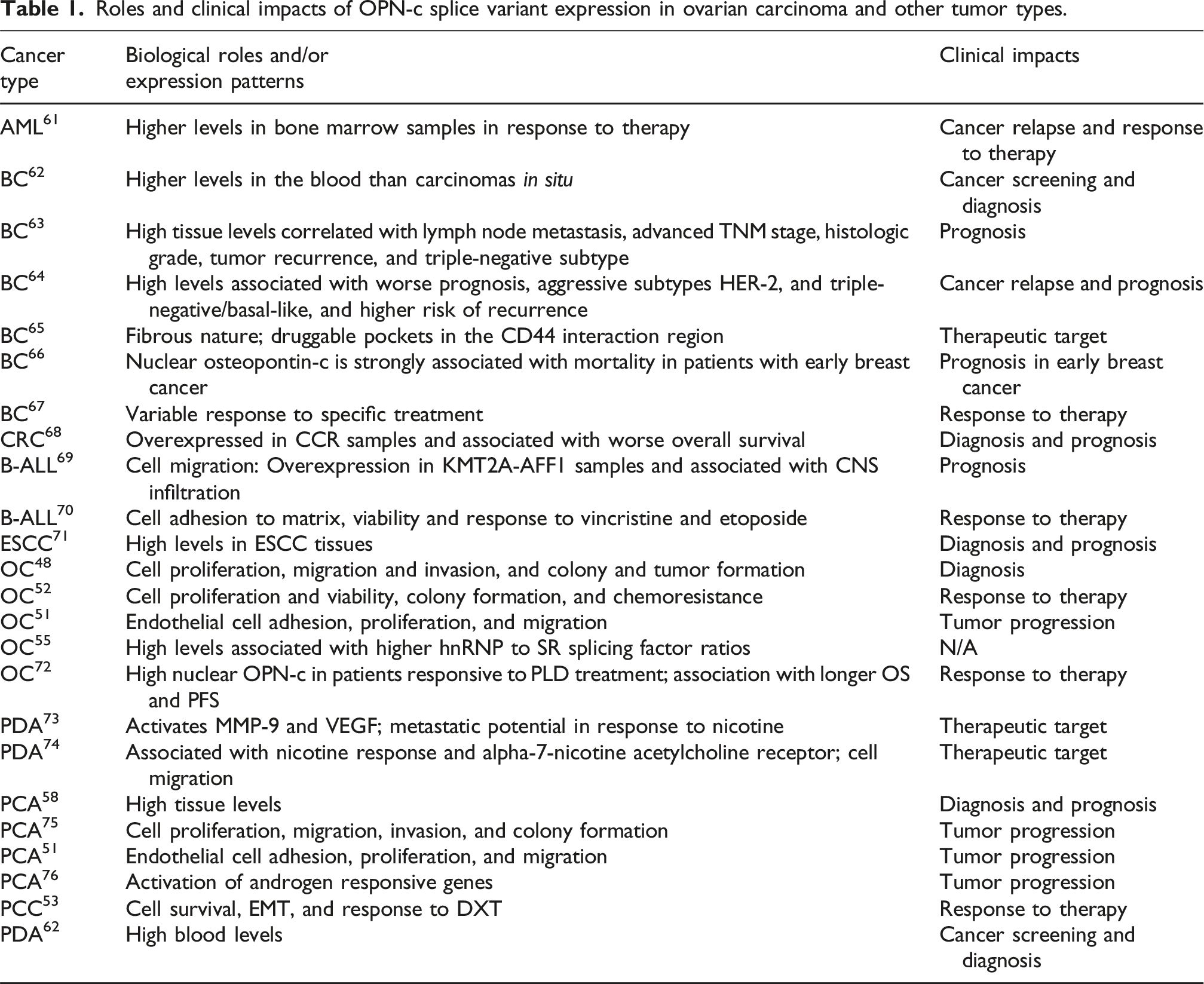
(Table 1). Transcriptional expression of the OPN-c isoform in tissue samples from ovarian cancer (OC) patients: The OPN-c transcript levels were analyzed in tumor (red) and non-tumor (gray) tissue samples (A) from endometrioid OC (n = 514), from the GEPIA2 database originated from the TCGA and GTEx projects. The transcriptional expression value refers to the unit Log2 (TPM+1) referring to the amount of RNA present in a sample. Significant difference presented as an asterisk: *p-value ≤0.01. Overall survival (OS) (B) and progression-free survival (PFS) (C) were associated with high (red) and low (blue) OPN-c expression in endometrioid OC tissue samples. For all Kaplan–Meier curves, the log-rank test was applied, and p-values ≤0.05 were considered significant. In high-grade serous OC subtype, overall survival (OS) (D, G), progression-free survival (PFS) (E, H), and disease-free survival (F, I) were according to high (red) and low (blue). OPN-c expression levels in OC patients treated or not with platinum chemotherapies. The survival analysis was performed using OPN-c mRNA expression data from the TCGA cohort. The survival (v3.5-8) and survminer (v0.4.9) packages were used to determine optimal cutoff points for stratifying patients into different risk subgroups. Data visualization and statistical assessments were carried out using ggplot2 (v3.5.0), including calculations of p-values, hazard ratios (HR), and 95% confidence intervals (95% CI). The log-rank test was used to compare survival curves between the stratified groups. GEPIA: gene expression profiling interactive analysis; GTEx: genotype-tissue expression; TCGA: The Cancer Genome Atlas. Roles and clinical impacts of OPN-c splice variant expression in ovarian carcinoma and other tumor types.
We have found OPN-c was the most upregulated OPN-SI in prostate cancer tissue samples. In this tumor model, high OPN-c levels were strongly associated with prostate cancer occurrence and high Gleason score, providing evidence that OPN-c can improve prostate cancer diagnosis and prognosis. 58 In human esophageal squamous cell carcinomas (ESCCs), OPN-c is significantly upregulated and could also serve as a diagnostic marker, given its association with disease staging and patient survival. 71 Not only does OPN-c expression provide important diagnostic information, but it can also predict clinical evolution in OC. In the same cohort of patients with endometrioid OC, we found that high OPN-c transcriptional levels were associated with decreased overall survival (Figure 2(b)). When it comes to disease-free survival, we have found no significant differences among patients with high and low OPN-c transcript levels (Figure 2(c)). Similar findings have been observed in serous ovarian cancer subtype, in which higher OPNc expression levels were also associated with worse overall survival in patients treated or not with platinum chemotherapies (Figure 2(d) and 2(g)), although with no evident impact to worse progression-free survival (Figure 2(e) and 2(h)) and disease-free survival (Figure 2(f) and 2(i)).
As shown in our studies in OC, the OPN-c isoform has also been reported as a prognostic indicator for invasive breast cancer following papillomatous growth. 77 In human breast tumors, OPN-c expression correlates with lymph node metastasis, 78 advanced TNM stage, histologic grade, and survival rates. 63 OPN-c in breast tumors may be critical for metastasis. 78 Studies on metastatic melanoma samples also show that high OPN-c expression levels are associated with advanced tumor progression and poor prognosis. 26 Our group also evaluated OPN-c expression levels in B-cell acute lymphoblastic leukemia (B-ALL) cases presenting KMT2F-AFF1 and ETV6-RUNX1 gene fusions, where OPN-c expression contributed to poor prognostic features in B-ALL patient subgroups, suggesting it could be a potential biomarker in B-ALL. 69 In prostate cancer, OPN-c expression supports sustained proliferative survival, primarily mediated through PI3K signaling. 75 OPN-c overexpression in the PC-3 prostate cancer cell line enhanced xenograft tumor growth, proliferation, migration, invasion, and soft agar colony formation, as well as MMP2, MMP-9, and vascular endothelial growth factor (VEGF) expression, as has been found in OC cells.48,75 OPN-c also appears to be a biomarker for PCa and modulates androgen receptor (AR) signaling, contributing to PCa progression. 76 Conditioned medium secreted by prostate cells overexpressing OPN-c activated the expression levels of AR-responsive genes through the PI3K pathway. 76 In colorectal cancer, high levels of OPN-c correlate with poor clinicopathological features and shorter progression-free survival (PFS), and they are associated with the BRAFV600E activating mutation, which promotes tumor progression. 68 Moreover, in colon cancer cell lines (HT115 and HCT-8) undergoing 5-fluorouracil (5-FU) treatment, stress signals could be transduced by secreting OPN-c, which promotes cell survival in the tumor microenvironment. Secreted OPN-c isoform activates nuclear calcium signaling and modulates epigenetic markers such as methyl-CpG binding protein 2 (MeCP2), demonstrating the role of OPN-c in adapting tumor cells within a pro-inflammatory microenvironment. 79
Subcellular localization of OPN-c in OC and other tumor types
Total OPN has been widely reported as a matricellular and secreted protein. Indeed, most data concerning its physiological and pathological roles in tumor cells are mainly related to total secreted OPN. Typically, the immunohistochemical (IHC) detection of tOPN reveals a strong signal in the perinuclear area, indicating its passage through the Golgi apparatus and endoplasmic reticulum for secretion. 80
In distinct cell compartments and specific tumor models, its diagnostic or prognostic implications have been explored.71,72,81 Recent IHC analysis of OC tissues found that high nuclear OPN-c expression in patients exposed to PEGylated liposomal doxorubicin (PDL) treatment was associated with longer OS and longer PFS. Thus, nuclear OPN-c expression appears to be a promising marker of therapy response (PLD or other chemotherapy) in OC patients.
72
Additionally, using immunofluorescence assays, we identified predominant perinuclear OPN-c in OC cells overexpressing this OPN splice variant (Figure 3). In these cells, OPN-c is also found in the cytoplasm and faintly labeled in the nucleus. Nuclear staining for OPN-c has been detected for the first time in breast cancer, although it has not been quantified. Later, high nuclear OPN-c staining patterns were strongly associated with mortality in patients with early breast cancer, serving as a poor prognostic indicator for patient survival. It has also been suggested that incorporating OPN-c IHC into standard pathology evaluations could offer prognostic advantages in the early diagnosis of breast cancer.
81
Analysis of OPN-c protein subcellular localization in A2780 ovarian cancer (OC) cells ectopically overexpressing this splice variant. Subcellular localization of OPN-c and F-actin and cellular structures indicated by immunofluorescence and confocal microscopy. Labeling with anti-OPN-c antibody (green), phalloidin FD (red) to detect actin, and DAPI to counterstain the nucleus (blue) in A2780 OC cell clones overexpressing OPN-c (A2780/OPN-c+) (E–H) and control cells, which were stably transfected with the empty expression plasmid vector (A2780/pCR3.1) (A–D). Images were captured by using confocal microscopy at 63x magnification. White arrowheads indicate weak and sparse nuclear staining, full arrows perinuclear staining, and dotted arrows cytoplasmic staining of images of A2780/OPN-c + cells at higher magnification (zoom). Bar: 100 µm. Representative images of three independent experiments.
In parallel, cytoplasmic OPN-c has been detected by IHC analysis in esophageal squamous cell carcinoma (ESSC) and found significantly upregulated compared to normal tissues, supporting its value as a potential diagnostic marker. It was also found that high cytoplasmic OPN-c expression in ESCC samples was significantly associated with pathological T stage and overall tumor stage, but not with histological grade or lymph node metastasis. 71 These findings suggest that the impact of assessing the subcellular localization of OPNc in cancer tissues might be tumor-specific and should be further explored in additional cancer types.
Concluding remarks and future perspectives
This review article provides an in-depth analysis of the recent evidence regarding OPN-c expression and functions in OC, identifying gaps in the literature and suggesting future directions for disease research. OPN-c expression patterns in OC cells and its corresponding signaling pathways are summarized in Figure 4. OPN-c is one of the most studied OPN-SI and its altered expression has been widely reported as an indicator of prognosis, survival, and metastasis rates in different types of tumors. Data from our research group demonstrated that OPN-c expression is implicated in survival, migration, invasion, angiogenesis, and sensitivity to chemotherapeutic drugs in OC cells. Therefore, inhibition of OPN-c could impair/prevent these processes, opening new potential avenues for OC treatment. Strategies to directly inhibit OPN-c include the use of antisense oligonucleotides (ASOs) or RNA interference (RNAi).52,82 ASOs are designed to bind to specific mRNA, blocking its expression. Recent advances have developed ASOs targeting key cancer-related genes, such as HIF-1α and survivin, reducing cell viability and invasion in tumor models, besides enhancing the effects of other treatment approaches. For instance, the AZD8701, a next-generation ASO, selectively reduces FOXP3 expression in regulatory T cells, reversing their immunosuppressive function. A phase I study showed that AZD8701 is clinically viable, with manageable adverse effects and effective tumor delivery.
83
RNAi, which uses small RNA sequences to silence genes, has also made progress with nanoparticles for efficient siRNA delivery. A phase I study of an RNAi-based drug targeting HIF-2α in advanced renal carcinoma showed safety and early antitumor activity, although challenges in RNA delivery, stability, and off-target effects remain.82,84,85 Another therapeutic approach involves inhibiting downstream or upstream signaling pathways activated by OPN-c.
86
Currently, several molecules are used to target the MAPK/ERK/AKT pathways, which are often dysregulated in cancers, thereby modulating cell proliferation, survival, and differentiation.48,87 Additionally, receptor antagonist molecules inhibit key protein interactions by targeting proteins such as PD-1 and VEGF. The combination of these agents with immunotherapy and chemotherapy has shown promise in enhancing cancer treatment outcomes.88,89 Moreover, based on predicted tertiary OPN-c structure on the binding site for CD44, a new druggable pocket has been proposed, which might be used for computational drug design aiming to inhibit those tumor progression features activated for this splice variant.
65
Osteopontin-c expression and signaling in the context of ovarian cancer (OC). In OC cells, OPN-c is a highly expressed protein that can be found in different cellular compartments such as the nucleus, perinuclear region, endoplasmic reticulum, and secretory vesicles. When secreted, OPN-c acts in the extracellular matrix as a signaling molecule, binding to cell membrane receptors such as integrins. We have shown that this binding induces a reduction in the activation of intracellular cascades such as the PI3K/AKT and MAPK/ERK pathways. Conversely, it induces the activation of the FAK signaling pathway. The specific receptors through which OPN-c modulates these signaling pathways have not yet been elucidated. The increased OPN-c expression level in OC cells also induces sensitivity to cisplatin. In turn, the treatment of OC cells with cisplatin reduces the expression levels of OPN-c, suggesting that silencing this splice variant may help sensitizing cisplatin-resistant OC cells to chemotherapy. Created with BioRender.com (accessed on 11 December 2024).
In conclusion, this review outlines the roles of OPN-c in cancer, particularly in ovarian cancer. In this tumor type, OPN-c has both diagnostic and prognostic implications, in addition to playing key roles in modulating cellular responses to chemotherapeutic agents. OPN-c promotes cell proliferation, migration, invasion, angiogenesis, metastatic potential, and cell survival. Moreover, its expression levels correlate with poorer ovarian cancer survival rates, further supporting its use as a prognostic biomarker. Its cytoplasmic, perinuclear, and nuclear localization aligns with the diverse functions performed by this isoform in ovarian cancer and other tumor types. Altogether, these findings implicate OPN-c in a broad range of ovarian tumor hallmarks and biological processes, including epithelial–mesenchymal transition. A better understanding of OPN-c’s roles in ovarian cancer may enhance its utility as both a biomarker and a potential therapeutic target.
Footnotes
ORCID iDs
Author contributions
MCMB: Conceptualization, methodology, formal analysis, investigation, writing—original draft preparation, and visualization. LTC: Methodology, investigation, and formal analysis. ACMSS: Methodology, investigation, and formal analysis. LBF: Methodology, investigation, and writing—original draft preparation. AS: Methodology, investigation, and formal analysis. MB: Methodology, investigation, and formal analysis. GNM: Conceptualization, investigation, resources, formal analysis, writing—original draft preparation, visualization, supervision, project administration, and funding acquisition. ERPG: Conceptualization, investigation, resources, formal analysis, writing—original draft preparation, visualization, supervision, project administration, funding acquisition, and corresponding author. All authors have read and approved the final version of the manuscript.
Funding
The authors disclosed receipt of the following financial support for the research, authorship, and/or publication of this article: This work was funded by Fundação de Amparo à Pesquisa do Estado do Rio de Janeiro (grant numbers: E-26/210.394/2014, E-26/010.002007/2014, E-26/203.204/2015, E-26/202.798/2017, SEI-26003/004812/2021, and N: SEI-260003/001554/2022), Conselho Nacional de Desenvolvimento Científico e Tecnológico (grant numbers: 310591/2014-7,435783/2018-1, 307427/2022-6, and 408314/2021-4), Ministério da Sáude, Universidade Federal Fluminense/Pró-Reitoria de Pesquisa, Inovação, and the L’Oréal-UNESCO-ABC prize for Women in Science and Fundação do Câncer (Programa de Oncobiologia).
Declaration of conflicting interests
The authors declared no potential conflicts of interest with respect to the research, authorship, and/or publication of this article.