
Abstract
Background:
Nasopharyngeal carcinoma (NPC) is a malignant tumor originating from the nasopharynx with high morbidity and mortality in Southeast Asia and south of China. Roundabout guidance receptor 1 (ROBO1) can regulate axonogenesis (axon-like protrusion), which may play an important role in migration. However, the roles of ROBO1 in NPC have not been clarified.
Methods:
A comparative analysis employing the NPC transcriptome (GSE12452) and the axonogenesis-related genes (GO: 0050772) was performed. In total, 124 tissue blocks from patients primarily diagnosed as NPC (1993-2002) were examined using immunohistochemical staining. The connections between clinicopathological variables and protein immunoexpression were analyzed by Pearson’s chi-square test. The Kaplan–Meier method with a log-rank test was employed to plot survival curves. Multivariate analysis was performed using the Cox proportional hazards model to identify independent prognostic biomarker.
Results:
According to transcriptome analysis, we found that ROBO1 is significantly highly expressed in NPC tissues compared with normal tissues. The immunohistochemistry (IHC) staining showed that high expression of ROBO1 was significantly related to primary tumor (T1T2 and T3T4) (P = .024), nodal metastasis status (N0N1 and N2N3) (P = .030), stage (I-II and III-IV) (P = .019), and histological grade (keratinizing, non-keratinizing, and undifferentiated) (P = .065). Importantly, NPC patients with high ROBO1 expression had poorer disease-specific survival (DSS) (P = .0001), distal metastasis-free survival (DMeFS) (P < .0001), and local recurrence-free survival (LRFS) (P = .0001) compared with NPC patients with low ROBO1 expression through the uni-/multivariate and the Kaplan–Meier survival analyses.
Conclusion:
Our report indicates that ROBO1 might be a potential prognostic biomarker for NPC.
Keywords
Introduction
Nasopharyngeal carcinoma (NPC) is a malignant tumor located in the mucous epithelium of the nasopharynx. 1 According to the International Agency for Research on Cancer (IARC) statistics, approximately, 130 000 people worldwide will be diagnosed with NPC in 2020. 2 NPC is considered a rare malignancy globally compared with other cancers, but this type of cancer was more frequently in North Africa, the Middle East, and Asia, particularly South China. 3 Since NPC is unusual, diagnostic markers and treatment strategies for this type of cancer were lacking. Following the treatment guidelines for NPC, radiotherapy (RT) and chemotherapy (CT) are the mainstays of treatment and there are also essential treatments for early-stage NPC patients, and its 5-year survival is around 85%. 4 Unfortunately, due to the specific location of NPC and is often misdiagnosed as other diseases, resulting in more than 70% of patients with NPC presenting with locally advanced stages at diagnosis and even development of distant metastasis. Once recurrence or distant metastasis occurs, the survival rate of patients with NPC is abysmal.5,6 Therefore, comprehensive identification of potential NPC biomarkers may help design more effective and targeted therapeutic strategies.
Cellular migration is a hallmark of distant metastasis in cancer, and it might be the critical factor responsible for inferior prognosis outcomes in patients with NPC. 7 Nevertheless, the great most current therapies for NPC do not show unsatisfactory effectiveness against metastatic. 8 Recently, emerging literature has shown that regulation of axonogenesis may be correlated with tumor metastasis and cancer prognosis in different types of cancers, consisting of gastric cancer, 9 lung cancer, 10 colorectal cancer, 11 and head and neck cancer. 12 However, it remains understood whether axonogenesis-related factors are involved in NPC metastasis. Here, we analyzed the relationship between the positive regulation of axonogenesis Gene Ontology Term (GO: 0003779) and tumorigenesis of NPC in the transcriptome of NPC (GSE12452) expression profiles. We notice that roundabout guidance receptor 1 (ROBO1) is significantly more expressed in NPC patients’ tumors than in non-NPC patients’ tumors.
ROBO1 is the receptor for SLIT1 and SLIT2, and it has been proved to play an essential role in various tumor initiation, development, and tumor progression. 13 In pancreatic cancer, overexpression of ROBO1 significantly decreased cancer cell proliferation through inhibition of cell cycle-associated proteins CCNA2 and CDK2 expression. 14 Furthermore, other studies report that ROBO1 promotes angiogenesis in liver cancer via the regulation of Cdc42 expression. 15 More importantly, recent evidence indicates that an imbalance of ROBO1 protein regulates cell motility and migratory capacity in different cancer types. 13 Zhang et al’s 16 studies indicated that the downregulation of ROBO1 enhances breast cancer metastasis. Inhibition of SLIT2-ROBO1 signaling transaction suppresses the function of epithelial–mesenchymal transition (EMT) and cell migration in gastric cancer. 17 In addition, several studies showed that ROBO1 might be a potential biomarker for diagnostic and prognostic in various cancers, including head and neck squamous cell carcinoma (HNSCC), 18 hepatocellular carcinoma (HCC), 19 and uterine cervical carcinoma. 20 Nevertheless, the correlation between ROBO1 expression, clinical significance, and survival outcomes in NPC patients has not been clarified.
Recent results showed that tumor angiogenesis is inextricably linked to cancer metastasis.21,22 In this study, we evaluated the expression level of ROBO1 in NPC tumor patients’ tissues and non-tumor tissue. Furthermore, we further explored the correlation between the ROBO1 expression and clinicopathological factors and prognosis in NPC patients.
Materials and Methods
Analysis of NPC public transcriptome datasets
In this study, we selected datasets (GSE12452, Affymetrix Human Genome U133 Plus 2.0 Array terrace) from the Gene Expression Omnibus (GEO) database, which has high-throughput resources and an open functional database. The GSE12452 dataset is the data of NPC patients consisting of 31 NPC tissues and 10 normal tissues. We used Nexus Expression 3 software (BioDiscovery, USA) to compare the difference in gene expression in tumor and normal tissue, and focusing the genes related to positive regulation of axonogenesis (GO: 0050772). We analyzed the gene expression and the following criteria (P < .01) were considered statistically significant.
NPC specimens
This study (IRB10302013) was executed after ratification by the institutional review board in Chi Mei Medical Center. The paraffin-embedded tissue blocks were retrieved from 124 patients with NPC, who were not found with distant metastases at initial diagnosis from the years 1993 to 2002. To accomplish a minimum total dose of 7000 cGy radiation, all 124 patients received a daily dose of 180 to 200 cGy and 5 fractions weekly. At least, 3 cycles of cisplatin-containing CT were given for patients with stages II to IV disease. Following the current World Health Organization (WHO) classification, the histological subtypes were reappraised by 2 pathologists. Tumors were staged using the American Joint Committee on Cancer (AJCC) seventh system. Indicative of Epstein–Barr virus (EBV) infection, the expression of EBV-encoded small RNA (EBER) was identified by in situ hybridization (ISH).
Immunohistochemistry staining
The paraffin-embedded NPC tissue sections (3 µm) were deparaffinized and rehydrated based on standard protocols. Immunohistochemical (IHC) staining was following the primary antibody ROBO1 (1:100; Clone: EPR23699-159, Abcam) at 4°C for overnight and the secondary antibody at RT for 30 min. The H-score was used to quantify ROBO1 immunoreactivity and was determined with the following equation: H-score = ΣPi (i + 1), where Pi is the percentage (0%-100%) of stained tumor cells for each intensity, and i is the intensity (0-3+) of staining. To split samples into high and low ROBO1 expression groups, the median H-score was used. Before assessing the staining intensity and area, the slide used Mayer’s hematoxylin to counterstain.
Statistical analysis
Statistical analyses were performed using SPSS. The association between ROBO1 immunoexpression and clinicopathologic characteristics was statistically determined using the chi-square test. From the starting date of RT to the date of an event, the analysis of local recurrence-free survival (LRFS), disease-specific survival (DSS), and distant metastasis-free survival (DMFS) were endpoints. Multivariate analysis using Cox proportional hazards model, and the survival rates were calculated by the Kaplan–Meier method. P-values < .05 were considered to be statistically significant.
Results
ROBO1 is the significant upregulated in NPC patients
We evaluated differentially expressed genes related to axonogenesis positive regulation and tumorigenesis of NPC patients. To determine critical genes involved in the regulation of axonogenesis signaling and NPC pathogenesis, we used GEO database to explore tumorigenesis of NPC in the NPC transcriptome (GSE12452), containing NPC tumor (n = 31) and non-NPC tumor (n = 10) and further compared with classification of positive regulation of axonogenesis (GO: 0050772) in Gene Ontology Term (GO Term) database. Heat map results show that only 2 relevant genes (ROBO1 and AMIGO1) are significantly differentially expressed (P < .05) in the positive regulation of axonogenesis classification and NPC tumorigenesis transcriptome crosslink (Figure 1). Among these 2 genes, ROBO1 was a significant fold change expression (log2 ratio = 1.1499) and significant statistical power (P < .0001) (Table 1); therefore, we focused on the expression of ROBO1 and whether its expression affects the clinical features of NPC patients.

Heat map analysis of differential gene expression in non-NPC tumor (n = 10) and NPC tumor (n = 31) on the GEO database (GSE12452). Particular notice to genes related to positive regulation of axonogenesis (GO: 0050772).
Summary of significant differentially expressed genes related to positive regulation of axonogenesis (GO: 0050772) and associated with tumorigenesis of NPC in the transcriptome of nasopharyngeal carcinoma (GSE12452).

Abbreviations: GO, gene ontology; Ig, immunoglobulin; NPC, nasopharyngeal carcinoma.
The correlation between ROBO1 and clinicopathological variables of NPC patients
To elucidate the clinical significance of ROBO1 in NPC, we incorporate 124 NPC cases that were analyzed. A total of 95 of 124 NPC patients were male, and 29 were female. Ninety-eight NPC patients were < 60 years old, and 26 NPC patients were ⩾ 60 years old. Our clinically relevant characteristics results showed that significant associations between ROBO1 high expression and ROBO1 low expression in NPC patient tumors are closely associated with primary tumor (T1T2 and T3T4) (P = .024), nodal metastasis status (N0N1 and N2N3) (P = .030), stage (I-II and III-IV) (P = .019), and histological grade (keratinizing, non-keratinizing, and undifferentiated) (P = .065) (Table 2). Among them, gender, age, and EBV-encoded small RNA (EBER) have no significant differences in tumors of NPC patients with differential expression of ROBO1. Based on these analyses, we further evaluated ROBO1 expression levels in NPC patients’ tissues by IHC staining and found that the expression of ROBO1 was absent or very low in the normal nasopharyngeal mucosa (Figure 2A). The median H-score of all scored tumor tissues was used to separate samples into weak and strong ROBO1 expression groups. Furthermore, the low stage of NPC patient’s tumor has weak ROBO1 expression (Figure 2B), whereas the high stage of NPC patient’s tumor has strong ROBO1 expression (Figure 2C).
Associations between ROBO1 expression and other important clinicopathological variables.

Abbreviations: EBER, EBV-encoded small RNA; ROBO1, roundabout guidance receptor 1.
Statistically significant.

IHC staining of ROBO1 expression in (A) normal tissue, (B) early-stage (stages I-II) NPC tumor tissue, and (C) advanced (stages III-IV) NPC tumor tissue.
Highly expressed ROBO1 is correlated with poor prognosis outcomes of NPC patients
To confirm the effect of ROBO1 on the survival outcomes of patients with NPC, we used the Kaplan–Meier method to analyze the association between ROBO1 expression and NPC patients’ survival rates. The results showed that the DSS rate (P = .0001) (Figure 3A), distal metastasis-free survival (DMeFS) rates (P < .0001) (Figure 3B), and LRFS rates (P = .0001) (Figure 3C) were significantly lower in the ROBO1 high expression NPC patients group compared with ROBO1 low-expression NPC patients group. To further explore the relationship between the prognostic significance of ROBO1 expression and clinicopathological factors in NPC patients, we performed uni- and multivariate analyses. The univariate analysis found that primary tumor staging (T1T2 and T3T4), lymph node status (N0N1 and N2N3), stage (I-II and III-IV), and ROBO1 expression (high and low expression) were significant correlation with DSS, LRFS, and DMeFS rates (Table 3). However, the clinicopathological variables of gender, age, histological grade (keratinizing, non-keratinizing, and undifferentiated), and EBER (positive and negative) were not significantly different in DSS, LRFS, and DMeFS rates. Moreover, we further performed multivariate survival analyses and revealed that the advanced stage (III-IV) was an indicator of a worse DSS rate (HR: 2.471; 95% CI: 1.244-4.910; P = .010), DMeFS rate (HR: 2.047; 95% CI: 0.985-4.252; P = .055), and LRFS rate (HR: 3.037; 95% CI: 1.170-7.884; P = .022) (Table 4). More importantly, we also found that the expression of ROBO1 may serve as an independent prognostic biomarker of DSS rate (HR: 2.459; 95% CI: 1.439-4.204; P = .001), DMeFS rate (HR: 3.711; 95% CI: 1.945-7.079; P < .001), and LRFS rate (HR: 3.220; 95% CI: 1.572-6.599; P = 0.001) in NPC patients.

Kaplan–Meier survival curves for NPC patients by ROBO1 high or low expression. (A) Disease-specific survival, (B) distant metastasis-free survival, and (C) local recurrence-free survival.
Univariate log-rank analyses.

Abbreviations: DSS, disease-specific survival; DMeFS, distal metastasis-free survival; EBER, EBV-encoded small RNA; LRFS, local recurrence-free survival; ROBO1, roundabout guidance receptor 1.
Statistically significant.
Multivariate survival analyses.
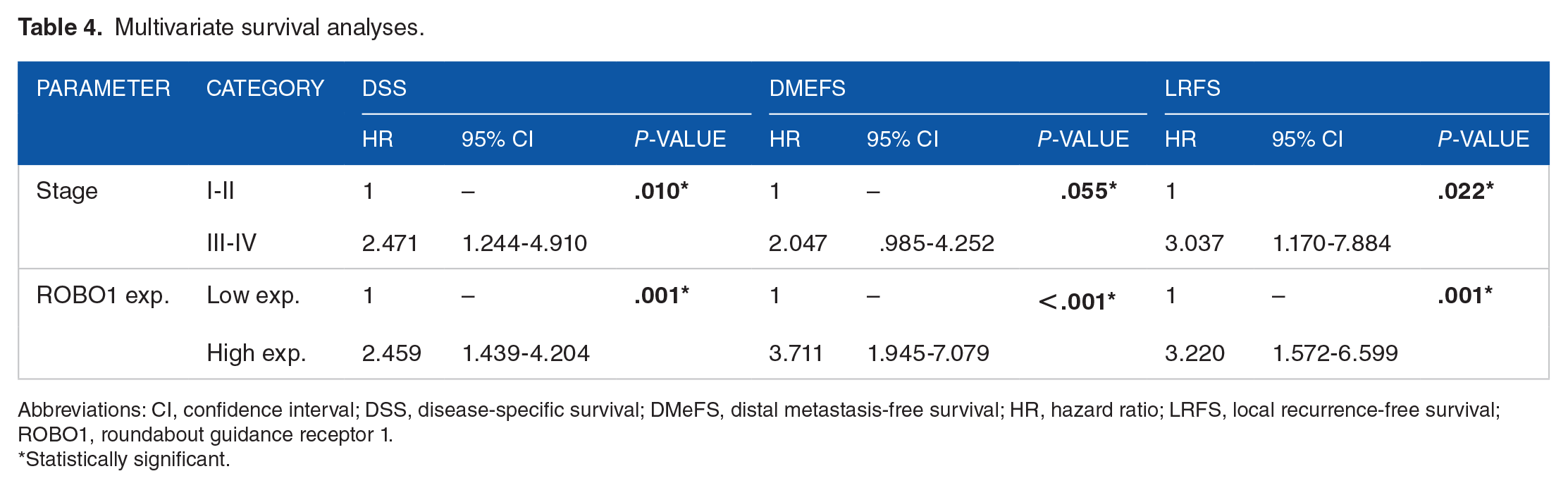
Abbreviations: CI, confidence interval; DSS, disease-specific survival; DMeFS, distal metastasis-free survival; HR, hazard ratio; LRFS, local recurrence-free survival; ROBO1, roundabout guidance receptor 1.
Statistically significant.
Comprehensive analysis of the ROBO1-correlated positive/negative genes and correlated GO biological process enrichment
To elucidate the network of related genes that have interactions with ROBO1 genes, we analyzed the gene expression profiles associated with ROBO1 in NPC patients through the TCGA (The Cancer Genome Atlas) online database. Our analysis demonstrated that the top 200 ROBO1-positively correlated genes and ROBO1-negatively correlated genes in NPC patients, respectively (Supplementary Table S1). Clarifying these critical genes related to ROBO1 expression can help us study the potential regulatory mechanism of ROBO1 in NPC. After elucidating the changes positively and negatively associated with the ROBO1 gene, we further explored the differential expression of biological processes associated with the ROBO1 gene through GO enrichment analysis using the GO resource (http://geneontology.org/). We showed that the top 3 enrichment of biological processes positively related to ROBO1 contains multicellular organism development (GO: 0007275; P = 1.00E–07; FDR = 7.85E–04), developmental process (GO: 0032502; P = 17.36E–08; FDR = 1.15E–03), and regulation of developmental process (GO: 0050793; P = 3.23E–07; FDR = 1.69E–03), the results of other differentially expressed biological processes are shown in Supplementary Table S2. On the other hand, the top 3 enrichment of biological processes negatively related to ROBO1 included the cellular response to zinc ion (GO: 0071294; P = 3.60E-07; FDR = 5.64E–03), stress response to copper ion (GO: 1990169; P = 1.18E-06; FDR = 6.18E–03), and cellular response to cadmium ion (GO: 0071276; P = 3.12E-06; FDR = 6.99E–03) (Supplementary Table S3).
Discussion
NPC is a malignant epithelial tumor with unique geographic and ethnic distribution, which is rare in the United States and Western Europe but endemic in southern China, Southeast Asia, North Africa, and the Middle East.23,24 A large-scale study shows that the occurrence of NPC is inextricably linked to the EBV.25-27 However, although possible EBV infection has been detected in many NPC patients, not everyone infected with EBV will develop NPC. This means that other etiological factors may be involved in the pathogenesis of NPC, such as environmental factors, genetics, tobacco, alcohol, diet (preserved or fermented foods), and personal lifestyle.28,29 According to the WHO histological grade system, NPC is classified into 3 histopathologic types: keratinizing squamous cell carcinoma, non-keratinizing squamous cell carcinoma (differentiated or undifferentiated), and basaloid squamous cell carcinoma. 30 The incidence varies according to different regions; in North America, 25% as a keratinizing squamous cell carcinoma, 12% as differentiated non-keratinizing carcinoma, and 63% as undifferentiated non-keratinizing carcinoma, while in southern Chinese, histological distribution is 2% keratinizing squamous cell carcinoma, 3% differentiated non-keratinizing carcinoma, and 95% undifferentiated non-keratinizing carcinoma. 31 RT and CT are the most common modalities to treat early-stage non-metastatic NPC with favorable clinical and survival outcomes, with 5-year overall survival rates of around 90%. 32 Unfortunately, because NPC is often asymptomatic or can be misdiagnosed with other diseases, approximately, 70% of NPC patients are diagnosed at an advanced stage or even with local spread or metastasis. Once distant metastasis occurs, the overall survival rate of NPC patients is only about 20 months. 33 Although much research has been invested, the pathogenesis of NPC has not been elucidated, thus, finding new biomarkers is crucial to be used for NPC patients.
In this study, we performed cross-linking analysis using the tumorigenesis of NPC in the transcriptome of NPC (GSE12452) and the positive regulation of axonogenesis (GO: 0050772) and revealed that ROBO1 was significantly upregulated in NPC tumor tissues than non-NPC tumor tissues. Moreover, we further found that ROBO1 protein expression was strong express in NPC patients compared with normal tissue by IHC staining. ROBO1 is a receptor for SLIT1 and SLIT2, playing essential roles in cell migration and neuronal development. 34 Several studies have indicated that ROBO1 is involved in cancer progression in different cancer types, including cell proliferation, cell migration, and invasion. 13 For example, Li et al 35 demonstrated that inhibition of ROBO1 expression suppresses breast cancer growth and metastasis via decreased FLNA expression. Moreover, downregulating ROBO1 expression inhibits the EMT process to suppress cell migration and invasion in lung adenocarcinoma cells and HCC cells.36,37 Taken together, these studies report a vital role for ROBO1 in cell growth, migration, invasion, and poor survival outcomes in cancer. To the best of our knowledge, this study is the first to describe the association between ROBO1 and the clinical characteristics of NPC patients.
Our clinicopathological variables indicate that ROBO1 expression is significantly correlated with the primary tumor, nodal metastasis status, stage, and histological grade in NPC patients. Furthermore, we also screened the top 200 positively correlated genes and negatively correlated genes associated with ROBO1 and further ranked the biological process-enriched terms related to the ROBO1 gene by GO dataset. Notably, the enrichment of GO biological processes positively correlated with ROBO1 was found to be related to cellular biological development through our ranking. This finding could allow for more in-depth studies to confirm these results in the future. In addition, we also performed uni- and multivariate analyses, and found that primary tumor, lymph node metastasis, cancer stage, histological grade, and ROBO1 expression have a significant association with DSS, DMeFS, and LRFS in NPC patients by univariate analyses. Similarly, multivariate survival analyses showed that cancer stage and ROBO1 expression were also correlated with DSS, DMeFS, and LRFS in NPC patients. More specifically, NPC patients who exhibited high ROBO1 gene expression had lower DSS rates, DMeFS rates, and LRFS rates than low ROBO1 gene expression in NPC patients.
Of course, our studies have some limitations. First, we only evaluated a single-center retrospective cohort in this study, and further analysis with a larger cohort is warranted to demonstrate our results. Furthermore, this study was unable to evaluate further the associations between ROBO1 and ROBO1-related genes or biological processes in poor survival outcomes in NPC patients. This study provides a scientific basis for diagnostic biomarkers of NPC patients.
Conclusions
In conclusion, to the best of our knowledge, we are the first to find that ROBO1 expression is higher in NPC tumor tissues compared with non-NPC tumor tissue. The retrospective analysis identifies that ROBO1 expression levels were strongly associated with the primary tumor (T), nodal metastasis status (N), stage, and histological grade. Furthermore, in NPC patients, ROBO1 high expression was significantly correlated with worse DSS, DMeFS, and LRFS. The findings of this article indicated that ROBO1 might be valuable as a prognostic factor and potential biomarker for NPC patients.
Supplemental Material
sj-docx-1-onc-10.1177_11795549221113244 – Supplemental material for Roundabout Guidance Receptor 1 Is an Emerging Prognostic Biomarker for Nasopharyngeal Carcinoma
Supplemental material, sj-docx-1-onc-10.1177_11795549221113244 for Roundabout Guidance Receptor 1 Is an Emerging Prognostic Biomarker for Nasopharyngeal Carcinoma by Sung-Wei Lee, Ching-Chieh Yang, Hong-Yue Lai, Hsin-Hwa Tsai, Cheng-Fa Yeh, Yu-Hsuan Kuo, Nai-Wen Kang, Tzu-Ju Chen and Shih-Lun Chang in Clinical Medicine Insights: Oncology
Supplemental Material
sj-docx-2-onc-10.1177_11795549221113244 – Supplemental material for Roundabout Guidance Receptor 1 Is an Emerging Prognostic Biomarker for Nasopharyngeal Carcinoma
Supplemental material, sj-docx-2-onc-10.1177_11795549221113244 for Roundabout Guidance Receptor 1 Is an Emerging Prognostic Biomarker for Nasopharyngeal Carcinoma by Sung-Wei Lee, Ching-Chieh Yang, Hong-Yue Lai, Hsin-Hwa Tsai, Cheng-Fa Yeh, Yu-Hsuan Kuo, Nai-Wen Kang, Tzu-Ju Chen and Shih-Lun Chang in Clinical Medicine Insights: Oncology
Supplemental Material
sj-docx-3-onc-10.1177_11795549221113244 – Supplemental material for Roundabout Guidance Receptor 1 Is an Emerging Prognostic Biomarker for Nasopharyngeal Carcinoma
Supplemental material, sj-docx-3-onc-10.1177_11795549221113244 for Roundabout Guidance Receptor 1 Is an Emerging Prognostic Biomarker for Nasopharyngeal Carcinoma by Sung-Wei Lee, Ching-Chieh Yang, Hong-Yue Lai, Hsin-Hwa Tsai, Cheng-Fa Yeh, Yu-Hsuan Kuo, Nai-Wen Kang, Tzu-Ju Chen and Shih-Lun Chang in Clinical Medicine Insights: Oncology
Footnotes
Acknowledgements
The authors are grateful for the help from Translational Research Laboratory at Department of Medical Research of Chi Mei Medical Centers in the conduction of this study.
Declaration of Conflicting Interests:
The author(s) declared no potential conflicts of interest with respect to the research, authorship, and/or publication of this article.
Funding:
The author(s) received no financial support for the research, authorship, and/or publication of this article.
Author Contributions
Conceptualization: S-WL, S-LC, and H-YL; methodology: S-WL, C-CY, H-YL, H-HT, S-LC, and C-FY; investigation: H-YL, H-HT, C-CY, Y-HK, N-WK, and T-JC; formal analysis: S-WL, H-YL, T-JC, C-CY, C-FY, Y-HK, and H-HT; resources: Y-HK, N-WK, S-LC, and T-JC; validation: C-CY, H-HT, C-FY, S-WL, Y-HK, and N-WK; visualization: C-CY, H-HT, S-LC, C-FY, N-WK, and T-JC; writing—original draft: S-LC and S-WL; writing—review and editing: H-YL; funding acquisition: S-LC; supervision: S-LC. All authors have read and agreed to the published version of the manuscript.
Consent to Participate Statement
As a rule, every participant signed informed consent before being enrolled into the biobank.
Data Availability Statement
The dataset analyzed in the current study (GSE12452) is available in a published transcriptome dataset from the Gene Expression Omnibus (GEO) database (National Center for Biotechnology Information, Bethesda, MD, USA).
Statement of Ethics
This study and its use of tumor samples that were deidentified from the biobank was approved by the Ethics Committee and Institutional Review Board of Chi Mei Medical Center (10302013) and followed the ethical guidelines of the Helsinki Declaration and the regulations of our government.
Supplemental Material
Supplemental material for this article is available online.
References
Supplementary Material
Please find the following supplemental material available below.
For Open Access articles published under a Creative Commons License, all supplemental material carries the same license as the article it is associated with.
For non-Open Access articles published, all supplemental material carries a non-exclusive license, and permission requests for re-use of supplemental material or any part of supplemental material shall be sent directly to the copyright owner as specified in the copyright notice associated with the article.