
Abstract
Background:
Improvement of the management of pancreatic cancer requires a better understanding of the genetic and molecular changes responsible for the development of the disease. The family of p21-activated kinases (PAKs) and especially PAK1 appears to mediate many cellular processes that contribute to the development and progression of pancreatic cancer, but the clinical relevance of PAK1 expression with the disease still remains unclear. Aim of the study was to assess the clinical value and the potential prognostic significance of PAK1 in pancreatic adenocarcinoma.
Methods:
We investigated the relationship between the PAK1 expression and the clinical and histopathologic characteristics of pancreatic cancer patients and the potential significance of PAK1 on survival. We examined tissue samples from 51 patients operated for pancreatic cancer. PAK1 expression was investigated with immunohistochemistry and correlated to clinicopathological parameters.
Results:
PAK1 was detected in all tumor samples and high expression was found in most patients. High PAK1 expression was also associated with younger age and well-differentiated tumors, but no association was found between PAK1 expression and Tumor-Node-Metastasis stage as well as deceased or alive status on follow-up. Moderate to high PAK1 expression favored higher 6-month and 1-year survival and low PAK1 expression 2-year survival but without statistical significance.
Conclusions
Our results indicate that PAK1 could potentially be used as a prognostic marker in pancreatic cancer. Further studies could clarify whether utilization of PAK1 in therapeutic protocols for the treatment of pancreatic cancer will render them more effective.
Introduction
Pancreatic adenocarcinoma is considered as one of the most lethal solid tumors. Worldwide statistics indicate that it represents the fifth most common malignancy and the fourth leading cause of cancer-related mortality. 1 The 5-year survival rate is disappointingly less than 5% and the median survival is only 3 to 6 months, without demonstrating any signs of improvement over the last few decades. 2 This dismal prognosis is the result of both the inability to reliably detect pancreatic carcinoma at an early stage and the lack of successful therapies other than radical surgical excision. Most pancreatic cancer patients miss their chance for a potentially curable resection because symptoms present late during the course of the disease. Even after an extensive, curative-intent excision of the pancreatic cancer, the high rates of liver metastasis and/or local recurrence make treatment efforts almost futile. Better knowledge of the molecular and genetic processes that take place in pancreatic cancer can boost the development of more efficient prognostic markers as well as new therapeutic targets.
In 1994, while searching for proteins that interact with small Rho-like G proteins, Manser et al 3 first discovered the p21-activated kinases (PAKs). The name p21 derives from the common molecular weight of 21 kDa that G proteins share. The PAK family of serine/threonine kinases are effector proteins for the Rho GTPases Cdc42 and Rac and play important roles in a variety of biological activities, including stimulation of cell proliferation, motility, and survival. 4 Deregulation of these cellular processes can easily initiate and promote tumorigenesis. The PAK family consists of six kinases classified into two groups based on sequence, structural homology, and response to activated GTPases. PAKs of both Group I (1-3) and Group II (4-6) share the common features of a N-terminal regulatory domain and a C-terminal kinase domain, 5 but have considerably different regulatory mechanisms.
Group I PAKs show high level of structural homology but variable tissue distribution; PAK1 is found in many organs like the brain, mammary gland, muscle, and spleen, PAK2 is expressed ubiquitously, and PAK3 is found in the nervous system. 3 Their basic characteristics are that they contain an autoinhibitory domain (AID) and they form homodimers. Group I PAK activation is effected by Rho GTPases, which result in disruption of the homodimer forming two monomers, thus allowing autophosphorylation, an action important for the regulation of their function. 6 Group II PAKs share approximately 50% identity in their kinase domains with Group I PAKs, which along with some other overlapping functions among the two groups make specific PAK isoform targeting difficult. 7 PAK4 is highly expressed in embryonic tissues and ubiquitously expressed in low levels in all adult tissues, PAK5 is found in the brain, and PAK6 in the nervous and male reproductive systems. 8 Unlike Group I PAKs, Group II PAKs lack well-defined AID; they are activated by completely different mechanisms and they function as monomers rather than homodimers. 9 PAK1 is the best-documented member among the family of evolutionary conserved family of serine and threonine kinases. It has been identified as an effector molecule for the small Rho GTPases and plays an important role in a variety of cellular functions such as cytoskeletal reorganization, cell motility, apoptosis, and transformation. 10
The most frequent genetic mutation encountered in pancreatic cancer is the K-ras oncogenic mutation. 11 This mutation activates Ras protein which in turn activates a number of signaling pathways—among which Rho GTPases like Cdc42 and Rac—through direct and indirect mechanisms and thus activate PAKs.12,13 PAK activation has an oncogenic effect by increasing proliferation and cell survival and reducing apoptosis. 14 PAK1 has been found to be upregulated in pancreatic cancer as well as pancreatic cancer cell lines.15,16 Overexpression of PAK1 was also found in moderately to well-differentiated pancreatic cancer samples in a BALB/c mouse model of human pancreatic adenocarcinoma xenografts. 17 Despite these findings, the clinical significance of PAK1 in pancreatic cancer is not fully elucidated. This study investigates the relationship between PAK1 expression and various clinical and pathological characteristics of patients operated for pancreatic cancer as well as the possible prognostic role of PAK1 in human pancreatic adenocarcinoma.
Material and Methods
Study subjects
Tissue samples were obtained from surgical specimens of 51 patients operated for pancreatic cancer from 2008 until 2014. Local human ethics committees approved this study. Written informed consent was obtained from each participant. All samples were tested for PAK1 expression and were correlated with the patients’ clinicopathological characteristics age, sex, tumor grade, disease stage, and alive or deceased status. Patients were grouped according to clinicopathological characteristics as ⩽60 and >60 years old, as G1 and G2-G3 tumor grade, as Stage I and Stage II-III disease stage, and as alive or deceased during follow-up. Stage IV patients were excluded from the study.
Immunohistochemistry
PAK1 protein expression was investigated with immunohistochemistry. In brief, surgical specimens were fixed in 10% buffered formalin and embedded in paraffin. Tissue sections of 4 μm thick were obtained from the pancreatic tissue samples, deparaffinized in xylol, and rehydrated in serial ethanol solutions. After blocking of the endogenous peroxidase activity, the sections were incubated with rabbit polyclonal anti-p21 activated kinase antibody (1:300, Sigma). Chromogenic 3,3-diaminbenzidine tetrahydrochloride (DAB) solution was applied to the sections for detection of PAK1 and then counterstained with hematoxylin. Positive and negative controls were used to confirm the proper application in each experiment. Two independent observers graded the staining intensity of each specimen in a semi-quantitative manner. All cases were scored blind to the corresponding clinicopathological data. Intensity grading was categorized as absent (0), weak (1), moderate (2), and high (3).
Statistical analysis
Statistical analysis of the data was performed using the R programming language (R Foundation for Statistical Computing, Vienna, Austria). The chi-square test was used for association testing of PAK1 expression with the study population characteristics. The odds ratios as well as their 95% confidence interval (CI) were calculated to estimate the association of the proteins’ expression with the patients’ characteristics, and multivariate logistic regression analysis was used to determine their independent effects. Survival rates were calculated using the Kaplan-Meier method. The log-rank test was used to assess the statistical significance of the difference between the survival curves. The hazard ratios (HRs) were also calculated and multivariate Cox proportional hazards regression analysis was performed to estimate the independent effect of the expression levels of the proteins and the clinicopathological characteristics on overall survival. All tests performed were two tailed. The statistical significance threshold was set at P values <.05.
Results
Patient demographics as well as grouping according to tumor stage and grade are demonstrated in Table 1. The study included 23 male and 28 female patients with pancreatic cancer. The patients’ mean age was 62.3 ± 9.93 years, with 23 patients being 60 years old or less and 28 patients over the age of 60. Most cases (66.7%) demonstrated moderate or poor histological differentiation and stage grouping according to Tumor-Node-Metastasis (TNM) classification showed that most of the patients (76.5%) were Stages II and III.
Patient clinical and histopathologic characteristics.

PAK1 expression
PAK1 protein expression was detected in all pancreatic cancer specimens. Most cases (45.1%) were found having high PAK1 expression, 37.2% demonstrated low PAK1 expression, and 17.7% moderate expression (Table 2). Representative images showing moderate and high immunohistochemical staining, tumor perineural infiltration as well as negative control are shown in Figure 1A to D.
PAK1 expression according to immunohistochemical staining grading of pancreatic tumor tissue samples.


Immunohistochemistry of PAK1 in pancreatic tissue samples. (A) Moderate PAK1 expression, magnification: ×200; (B) high PAK1 expression, magnification: ×200; (C) perineural infiltration: magnification: ×100; (D) negative control, magnification:×200.
PAK1 Expression and Clinicopathological Characteristics
PAK1 expression was found statistically significant higher in the ⩽60 years age group (P = .0002) and in patients with well-differentiated tumors (P = .0021). On the contrary, no significant difference in PAK expression was found between TNM stage groups (P = .1137) as well as between survivors and deceased patients (P = .9702) (Table 3).
Relationship between PAK1 expression and clinicopathological characteristics.

PAK1 expression and disease-free survival
Survival analysis of the patients operated on for pancreatic cancer using the Kaplan-Meier and log-rank test showed that although only few patients with low PAK1 expression demonstrated 2-year survival, 6-month and 1-year survival was higher in the moderate and high PAK1 expression groups compared with the low PAK1 expression group but without statistical significance (Figure 2). The mean overall survival was 11.95 ± 1.16 months (95% CI: 9.7-14.2). Comparison between the various expression groups revealed statistical difference between the moderate and the high PAK1 expression groups (Table 4).
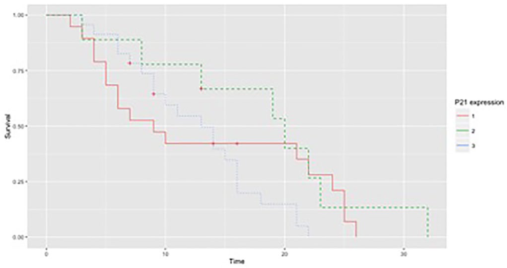
Kaplan-Meier curves for pancreatic cancer patients with low (1), moderate (2), or high (3) p21 expression.
Relationship between patient survival and PAK1 expression.

Discussion
The present study demonstrates that PAK1 is expressed in all pancreatic cancer tissue samples with high expression levels being detected in most cases. High PAK1 expression was found in patients with age ⩽60 years and with well-differentiated tumors and moderate to high rather than low PAK1 expression tends to favor 6-month and 1-year survival. There is accumulating evidence that PAK1 overexpression is a constant finding in gastrointestinal malignancies. Besides studies investigating PAK1 expression levels in pancreatic cancer tissues,15,18-20 similar findings have been detected in other malignancies like urinary bladder, ovary, and breast.21-23 A study by Jagadeeshan et al 15 that analyzed the expression of PAK1 in pancreatic cancer tissue samples found that PAK1 levels are significantly upregulated as compared with adjacent normals, a finding that was also confirmed by Yeo et al. 19 Zhou et al 20 found that 86% of primary pancreatic adenocarcinoma tissue specimens stained positive for PAK1, with one third of the specimens exhibiting moderate to strong intensity. MUC13, an important transmembrane mucin which is involved in PAK1 signaling, was found to be overexpressed in pancreatic cancer which was correlated with increased expression and activation of PAK1. 16 Han et al 18 compared PAK1 expression in primary pancreatic cancer samples with samples from metastatic liver tissues and found that primary tumors have significantly higher PAK1 expression. Immunohistochemical analysis of tissue samples from gastric cancer tissues showed PAK1 is significantly overexpressed. 24 The significance of PAK1 in colorectal and gastroesophageal cancers was investigated in two similar studies, which similarly reported PAK1 overexpression in cancer tissue samples.25,26 All these data, combined with our finding that PAK1 is unanimously expressed in all cancer tissue samples, indicate the importance of PAK1 in the development and progression of pancreatic cancer.
Our study found that two clinicopathological characteristics of the pancreatic cancer patients were associated with high PAK1 expression: younger age group (⩽60 years) and well-differentiated tumors. Both findings are in accordance with results reported in a study by Han et al, 18 which investigated PAK1 expression from cancer tissue samples of 72 pancreatic cancer patients. However, both univariate and multivariate analysis conducted in the same study showed that age is not a prognostic factor for survival of patients with pancreatic cancer. One could assume that higher PAK1 expression in younger age groups is indicative of a more aggressive disease, but this speculation is not supported by the above analysis. In addition, high PAK1 expression does not correlate with younger age groups in studies of non-pancreatic cancers. Study investigating PAK1 expression in gastroesophageal junction adenocarcinomas concluded that age was not an independent prognostic factor for overall survival, even though higher PAK1 levels were found in patients over the age of 60 years. 26 Other reports from gastric and colorectal carcinomas failed to demonstrate any association between age and PAK1 expression levels.24,27 The exact etiologic association between age and PAK1 expression levels is therefore unclear. Further studies, which would include additional features that possibly play a role in pancreatic cancer outcome along with age such as socioeconomic status, smoking, and comorbidities, could prove useful.
Previous report suggesting that well-differentiated pancreatic tumors are associated with high PAK1 expression is confirmed in our study. 18 The above finding is further supported by an experimental study by Makisumi et al. 17 This study used immunized Balb/c mice with human pancreatic adenocarcinoma xenografts and showed strong positive PAK1 immunostaining in moderately and well-differentiated pancreatic tumors compared with poorly differentiated tumors. Association between high PAK1 expression and well-differentiated tumor grade is encountered solely in pancreatic tumors and it is not reported in non-pancreatic tumors. In a study involving 403 premenopausal women with primary breast cancer, high PAK1 staining intensity was strongly correlated to poor tumor differentiation. 28 Similarly, high PAK1 expression was found in tissue samples from gastric, gastroesophageal junction and upper urinary tract cancer samples with moderate and poor differentiation while PAK1 expression was unaffected by tumor grade in colorectal adenocarcinomas.24-26,29 Due to the fact that prognosis in well-differentiated tumors is better compared with poorly differentiated ones, PAK1 overexpression could have more positive prognostic value in pancreatic cancer, contrary to other non-pancreatic cancers in which it is related to poor differentiation and subsequently unfavorable prognosis.
Our study showed that low PAK1 expression had lower 6-month and 1-year survival compared with moderate and high PAK1 expression but without statistical significance. Han et al 18 could clearly demonstrate that high PAK1 expression results in better survival than low expression possibly because two expression groups were used in their study instead of the three groups used in our study which included the moderate expression group. The same authors report that PAK1 expression is lower in metastatic tumors compared with the primary cancer sites, which leads to the conclusion that PAK1 plays an important role mostly during the initial phases of carcinogenesis and less during tumor progression and metastasis formation. On the contrary, data from non-pancreatic tumors show that PAK1 overexpression is related to metastatic disease and poor prognosis.
The unique position of PAKs at the crossroad of various signaling pathways makes them potential prognostic markers as well as attractive therapeutic targets, aiming to increase the effectiveness of cancer treatments. Glaucarubinone, a natural product that was originally used as an antimalarial drug, is being tested for its anticancer activity, mainly because of its ability to inhibit PAK1. Combined with gemcitabine, glaucarubinone reduced proliferation of pancreatic cancer cells in vitro and tumor growth in vivo more than treatment with either glaucorubinone or gemcitabine alone. 30 Another agent under investigation for its effectiveness against pancreatic cancer is FRAX597. This agent is a small-molecule pyridopyrimidinone that targets Group 1 PAKs through binding to their ATP site. 31 FRAX597 inhibited proliferation, survival, and migration of pancreatic cancer cells and when combined with gemcitabine, synergistically inhibited pancreatic cancer proliferation in vitro and inhibited tumor growth in vivo. 19 Other ATP-competitive inhibitors developed for PAK4 include LCH-7749944, which blocks the effect of PAK4 to various targets 32 and PF-3558309, which although demonstrated high PAK4 inhibition could not discriminate between specific PAK isoforms and inhibited both Group 1 and Group 2 PAKs. 33 An alternative approach for PAK inhibition includes the use of allosteric inhibitors like IPA-3 (inhibitor of PAK3), which bind to sites other than the ATP-binding pocket achieving higher specificity 34 and the use of dominant-negative forms or fragments of PAKs, which take the place of PAKs and prevent activation of downstream signaling pathways. 35 The above new data suggest that PAK inhibition may be of clinical value as a target for pancreatic cancer therapy.
Our results demonstrate that there is a strong association between PAK1 and pancreatic cancer since all tissue samples expressed PAK1. High PAK1 expression was detected in most samples and was related to younger age and well histological differentiation. Contrary to other cancers, high to moderate PAK1 expression in pancreatic cancer seems to favor 6-month and 1-year survival, and although no statistical significance was found, this could indicate the possibility of PAK1 having a potential positive prognostic significance. Further investigation will clarify the effectiveness of PAK1 as a prognostic marker for pancreatic cancer as well as its potential value in the management of pancreatic cancer patients.
Footnotes
Funding:
The author(s) received no financial support for the research, authorship, and/or publication of this article.
Declaration of conflicting interests:
The author(s) declared no potential conflicts of interest with respect to the research, authorship, and/or publication of this article.
Author Contributions
NS, ML, AT and CS contributed to the design of the study. ML, EP, CA, AT, AK, CN, and AK contributed to data collection and data analysis. ML and CA contributed to statistical analysis. NS and EP contributed to writing of the study. ML, AT and CS gave final approval for the publication of the study.