
Abstract
Uraemic Cardiomyopathy (UC) is recognised as an intricate and multifactorial disease which portends a significant burden in patients with End-Stage Renal Disease (ESRD). The cardiovascular morbidity and mortality associated with UC is significant and can be associated with the development of arrythmias, cardiac failure and sudden cardiac death (SCD). The pathophysiology of UC involves a complex interplay of traditional implicative factors such as haemodynamic overload and circulating uraemic toxins as well as our evolving understanding of the Chronic Kidney Disease-Mineral Bone Disease pathway. There is an instrumental role for multi-modality imaging in the diagnostic process; including transthoracic echocardiography and cardiac magnetic resonance imaging in identifying the hallmarks of left ventricular hypertrophy and myocardial fibrosis that characterise UC. The appropriate utilisation of the aforementioned diagnostics in the ESRD population may help guide therapeutic approaches, such as pharmacotherapy including beta-blockers and aldosterone-antagonists as well as haemodialysis and renal transplantation. Despite this, there remains limitations in effective therapeutic interventions for UC and ongoing research on a cellular level is vital in establishing further therapies.
Keywords

Introduction
Uraemic Cardiomyopathy (UC) is classically characterised by diastolic dysfunction in association with left ventricular hypertrophy (LVH) and myocardial fibrosis in patients with chronic kidney disease (CKD).
1
Patients with underlying end-stage renal disease (ESRD) and resultant myocardial remodelling often have associated high levels of cardiovascular morbidity and mortality.2,3 Whilst the pathophysiology of UC is traditionally multifactorial, there is emerging research in this area which may help expand therapeutic options for this patient population.4,5 There is growing evidence to suggest that cardiovascular death among these patients are increasingly secondary to LVH and its sequela of congestive cardiac failure (CCF), rather than purely atherosclerotic heart disease, highlighting the mechanistics behind this entity.3,6-8 Appropriate screening for these patients requires a multimodality imaging approach with transthoracic echocardiogram (TTE) and Cardiac Magnetic Resonance Imaging (CMRI) with current interventions centred around appropriate cardiac-specific pharmacotherapy, haemodialysis as well as renal transplantation
3
Epidemiology
The burden of cardiovascular disease (CVD) in patients with ESRD is significant, with mortality from CVD approximately 15 to 30 times higher than the general population. 9 The phenotypic hallmark of LVH which characterises UC is found in over 70% of patients with ESRD. 10 In patients with ESRD on dialysis, sudden cardiac death (SCD), usually indicative of an underlying cardiomyopathy, accounts for roughly 40% of all deaths. 5 There are predictions that the number of patients receiving dialysis for ESRD in the United States (US) will increase from a total of 320,000 in 2003 to 2 million by 2030, illustrating both the current and future burden of disease.3,10
Pathophysiology
The pathogenesis of UC remains complex and multifaceted with many implicative factors. These factors include anaemia, haemodynamic overload, hypertension, alterations in mineral metabolism, endothelial dysfunction, insulin resistance and cardiotonic steroids as well as several circulating uraemic toxins.1,5 See Table 1. However recently there has been an emergence of the pathophysiological role of Chronic Kidney Disease-Mineral and Bone Disorder (CKD-MBD) in UC with hyperphosphatemia, high FGF-23 levels and reduced serum levels of Klotho integral in the process of abnormal myocardial remodelling and resultant cardiac sequelae.1,2,11

UC may manifest as a result of pressure overload, volume overload and a systemic uremic state. 3 Left ventricular (LV) pressure overload may occur as a result of hypertension, arteriosclerosis and aortic stenosis whilst LV volume overload can occur in the setting of haemodynamic overload and anaemia.3,15 LV pressure overload mediates hypertrophy through increasing of LV wall thickness with minimal change in chamber size whereas LV volume overload results in increased chamber size but regular LV wall thickness. 15 Whilst LV hypertrophy (LVH) initially is a compensatory adaptative response, continual LV overload leads to cardiomyocyte death. 3 This acute or chronic loss of cardiomyocytes will eventually lead to systolic heart failure. 16 This process with increased passive stiffness and fibrosis will eventually lead to the development of diastolic dysfunction, a characteristic hallmark of UC. 16 It is also increasingly being recognised that the degree of myocardial fibrosis correlates strongly with the development of arrythmias and sudden cardiac death. 17
The uraemic state also contributes to LVH through the accumulation of substances such as endothelin, parathyroid hormone (PTH), tumour necrosis factor alpha (TNF- α), interleukin 1 alpha (IL- 1α) and IL-6.18,19 Another example of such hypertrophic substances are endogenous cardiotonic steroids classed as cardenolides (oubain and digoxin) and bufadienolides (marinobufagenin and proscillaridin A). 20 These substances interact with the α-subunit of the Na, K-ATPase transmembrane protein on the surface of cardiomyocytes, and whilst their exact mechanism is unknown, they are thought to be vital in the molecular pathogenesis of UC.3,21-23
Oxidative stress and inflammation, and endothelial dysfunction
Endothelial dysfunction represents a characteristic finding in patients with ESRD who have high rates of heart failure and cardiovascular mortality. 24 Intricately intertwined with this is the role of oxidative stress and inflammation, often seen in CKD. 25 For instance, CKD often results in a persistent inflammatory state, which may result in endothelial dysfunction and worsen atherosclerosis. Such inflammatory effects may also have other detrimental effects, such as vascular calcification, erythropoietin (EPO) resistance, increased hepcidin and decreased iron absorption. 25 In turn, anaemia itself contributes to cardiac dysfunction, evident in the CKD population. 26
Soluble α-Klotho and Vitamin D also help maintain endothelial integrity, with their declining concentrations in ESRD contributing to the dysfunction of the endothelial lining. 27 The subsequent increase in circulating FGF-23 and phosphate further damage the endothelial lining. 27 The underlying renal dysfunction also leads to an excess of inflammatory mediators and uraemic toxins that inhibit the recovery of the damaged endothelial lining. 27 There is evidence to suggest that this impaired endothelial function correlates with abnormal left ventricular structure and function as well as cardiovascular mortality in CKD.27,28 Endothelial dysfunction can be further analysed through microvascular or macrovascular endothelial dysfunction. Macrovascular endothelial dysfunction does not have a significant association with increased LV mass in ESRD. 24 Contrastingly, microvascular endothelial dysfunction has been shown to be associated with LV diastolic dysfunction, RV systolic dysfunction and RV diastolic dysfunction in ESRD. 24 The clinical significance of this is still unknown, with the lack of an established causal relationship between endothelial function and cardiac function, however it illustrates the need for trials of novel endothelial therapies to determine the possibility of regression of LVH and cardiac failure in patients with ESRD. 24
Carnitine deficiency
One postulated contributing factor for uraemic cardiomyopathy is the role of carnitine, which plays an important part in myocardial energy metabolism, given its role in beta-oxidation of fatty acids. Carnitine deficiency is often observed in uraemic patients, particularly those on haemodialysis, given that it can be lost through dialytic membranes. 29 While some studies have shown that cardiac hypertrophy may develop without alteration in myocardial metabolism in renal dysfunction, 30 there is a growing amount of evidence that suggests a positive role for carnitine supplementation in terms of improving cardiac function, exemplified by a case report by Kaneko et al whereby a haemodialysis patient’s cardiac dysfunction improved following the administration of L-carnitine. 31
Insulin resistance
Insulin resistance, which may commonly occur in patients with CKD, represents an independent risk factor for cardiac disease in CKD.31-37 One explanation to the increased prevalence of insulin resistance in CKD is the interruption in the intracellular insulin pathway that occurs because of increased angiotensin II, inflammation, metabolic acidosis and uremic toxins. 37 The phosphoinositide-3 kinase (PI3K)-Akt pathway is of particular interest in the development of LVH, myocardial fibrosis, cellular apoptosis and metabolic dysfunction with insulin resistance conferring maladaptive alterations within this pathway. 38 Akt1 and Akt2 are also predominantly found in the heart, with imbalance of these substances resulting in a cardiac phenotype similar to the uraemic heart.38,39 In insulin-resistant states, an Akt2 defect produces a compensatory hyperinsulinemia and upregulation of Akt1 signalling, producing a cardiac hypertrophy associated with fibrosis. 39 This is particularly important as there are therapies which have been shown to target the Akt pathway. Rapamycin, a mTOR target downstream of Akt, has shown to reduce cardiac hypertrophy and fibrosis in uraemic mice whereas Glitazones have been shown to reduce cardiac hypertrophy in mice.40,41 Thus, through our understanding of insulin resistance and its alterations in the Akt pathway, we can understand its cardiac effects and devise potential therapeutic options.
CKD-MBD
The role of CKD-MBD in UC is now increasingly being recognised following advancements in dialysis and resultant correction of uraemia-related abnormalities.42-44 In order to fully appreciate the role of CKD-MDB in the development of cardiomyopathy, it is important to analyse the role of parathyroid hormone (PTH), phosphate, FGF-23 and Klotho. 5 Current literature states the role of high PTH levels and its correlation with LV mass and degree of diastolic dysfunction, with these effects reversible post treatment of hyperparathyroidism.5,45-47 Vitamin D deficiency, especially in the setting of secondary hyperparathyroidism has been associated with LV dysfunction and increased risk of cardiac events, including CCF. 48 Phosphate plays a central role in the CKD-MDB and phosphate toxicity can contribute to cardiovascular mortality.5,43 Hyperphosphatemia has been directly associated with increased LV mass and diastolic dysfunction.49-51 It can promote LVH, potentially through changes in arterial stiffness or by directly acting on the myocardium.49-52
FGF-23
There is now increasing data to support the contributions of FGF-23 and Klotho to uraemic cardiomyopathy in a coadjutant manner. 11 FGF-23 regulates phosphate and Vitamin D metabolism and binds FGF receptor/Klotho co-receptor complexes, with the net result of stimulating phosphate excretion, inhibiting PTH secretion and decreasing active Vitamin D.11,53 FGF-23 has been shown to directly induce hypertrophic growth of the cardiac myocytes, with this process requiring the presence of FGR-4. 54 FGF-23 can activate FGR-4 in cardiac myocytes, resulting in stimulation of the phospholipase Cγ (PLCγ)/calcineurin / nuclear target of activated T cells (NFAT) signalling pathway and subsequent cardiac hypertrophy in a blood-pressure and αKlotho independent manner.5,11,54 Although significant exploration of the role of FGF-23 and FGR-4 has taken place in rat models, there is still support for its role in humans, if not direct causal evidence. 39 A recent retrospective study has shown that in children with ESRD, FGR4 expression levels have been associated with cardiac hypertrophy, with another study showing that lowering serum FGF-23 levels in CKD patients is associated with reduced cardiovascular events and mortality.55,56 There is also increasing evidence that FGF-23 stimulates cardiac fibrosis through the TGF-β and β-catenin pathways via activation of FGR-4, which has been demonstrated in mice models.19,57 Thus, it is evident that increased FGF-23 levels contribute to UC, with myocardial FGR4 representing a promising therapeutic target.5,11
Klotho
Klotho acts as a co-receptor for FGF-23 and helps mediate its’ role in the regulation of phosphate haemostasis. 11 Klotho exists in a deficient state in CKD. 5 The Klotho gene encodes the αKlotho protein which is expressed in tissues and organs including the kidneys and parathyroid glands. 58 Although the mechanism by which αKlotho acts is poorly understood and the myocardium does not express αKlotho, it is known that FGF23 does not seem to induce myocardial hypertrophy in the presence of normal levels of soluble αKlotho.59-62 There is some evidence of a mechanism of αKlotho inhibiting TRPC 6, a calcium permeable cation channel that helps modulate cardiac remodelling and induces cardiac hypertrophy through the calcineurin/NFAT pathway.11,63 Xie et al also demonstrated in a mice model, that soluble Klotho protects against UC independent of FGF-23 and phosphate.60,61 In patients with ESRD, as the kidney is the main source of systemic αKlotho, it may represent the primary alteration in CKD-MBD. 5
Thus, the pathogenesis of UC and its complexities are evident. There is a significant role of CKD-MBD in UC with the evolving understanding of elevated FGF-23 and αKlotho deficiency in particular pivotal to the development of appropriate targeted therapy in the future.
Diagnostics
As UC is a heterogeneous disease process, the accurate diagnosis incorporates the use of both imaging and non-imaging modalities. The non-imaging diagnostic tools include laboratory investigations and electrocardiography (ECG), whereas imaging tools involve a multi-modality approach with non-invasive investigations such as transthoracic echocardiography (TTE), cardiac CT and cardiac magnetic resonance imaging (CMRI). Furthermore, invasive diagnostics, whilst rarely performed, involve myocardial biopsy with histological assessment. These tools continue to evolve in the 21st century, providing additional prognostic information and guidance on selected therapeutics.
Electrocardiography
Bedside ECG is the most readily available test for rapid assessment of presence of LVH in this patient demographic. UC is known to ubiquitously manifest with signs of LVH, as fulfilled by Sokolov-Lyon criteria on ECG. 64 In early stages of the disease process, LVH manifests as an eccentric hypertrophy with a subsequent concentric hypertrophy and progressive worsening of CKD. 64 Other important ECG findings in UC include the presence of Q waves, dynamic ST segment changes, prolonged QRS duration, tachycardia and left and right atrial enlargement as shown by Shafi et al. 65 These findings may assist in prompt diagnosis and quantification of burden of UC, allowing for directed pharmacotherapy.
Electrocardiography may also have a role in SCD and mortality stratification in patients with ESRD on dialysis. 66 There is association between the ECG parameters of QT interval, spatial QRS-T angle, signal averaged ECG, heart rate variability and T-wave alternans with increased rates of mortality and SCD in patients on haemodialysis, however it is unclear if these are independent risk factors or whether they predispose to structural cardiac disease such as LVH that may predispose patients to SCD. 66
Echocardiography
Cardiac remodelling is a fundamental process that occurs in UC, affecting all chambers through intricate mechanisms involving pressure and volume overload. TTE is a non-invasive and readily accessible imaging modality that provides detailed geometric and functional assessment of the cardiac structures throughout systole and diastole. 67 Its utility also lies in that we know echocardiographic cardiovascular disease is already present in a high proportion of patients starting ESRD therapy and are independent mortality factors. 68 In a trial of approximately 600 patients on HD without symptomatic cardiac disease or cardiac dilatation, there was progression of LVH, with LVMI (LV Mass Index) 114 g/m2 at baseline increasing to 128 g/m2 at 96 weeks. 69 LVH also portends significant all-cause and CV mortality with (HR 2.9) and (HR 2.7) respectively. 70 In addition to presence of LVH as determined by LVMI, there is also evidence of direct impairment in LV systolic function, and this correlates with cardiovascular mortality as shown by Kramann et al. 71 However there has been conflicting data on whether patients undergoing renal replacement therapy have progression of LVH and LV systolic dysfunction, with Shi et al demonstrating the maintenance of stable LV structure and systolic function in a cohort of 40 patients on peritoneal dialysis (PD). 72 Furthermore, peak global longitudinal strain is also an independent risk factor for cardiovascular mortality and circumferential early diastolic strain rate is an independent risk factor for all-cause mortality. 71 Other studies have shown similar findings, as assessed by 2-dimensional speckle tracking echocardiography (2D-STE), demonstrating impaired LV longitudinal strain in patients with CKD when compared to controls (P < .001), highlighting significant subclinical systolic dysfunction. 72 These findings highlight the importance of echocardiography and speckle-tracking echocardiography for sensitive assessment of LV systolic function in this population group for diagnosis and prognostication.
The severity of UC also parallels grades of diastolic dysfunction (elevated E/e’, P < .001), highlighting a direct correlation between renal failure, volume overload and impaired relaxation. 73 LA indices are a surrogate for diastolic impairment in many cardiac pathologies. Patel et al. have shown that an elevated LA volume indexed (LAVI) correlates with poor survival in patients on haemodialysis, and that LAVI and LVF portend similar correlations with death on multivariate analysis. 74 In addition, Zapolski et al have shown evidence of reverse atrial remodelling in patients post renal transplantation with significant reductions in LAVI from a mean of 34.63 to 27.57 after 3 years. 75 These findings highlight the significant association of left atrial dysfunction and prognosis in these patients. In essence, a correction of the underlying pathology and subsequent haemodynamics results in reversal of LA myocyte dysfunction.
However, despite its immense utility, there are limitations to TTE in UC. A meta-analysis of 73 studies by Badve et al in 2016, which investigated whether LVM could be used as a surrogate end point for all-cause and cardiovascular mortality in CKD, particularly in the context of pharmacologic or nonpharmacologic interventions, suggested that there was no consistent association between intervention-induced LVM change and mortality. 76 Therefore, while TTE has a role in assessment of cardiac function, clinicians should be cognisant of its limitations within such contexts.
Cardiac MRI
The role of cardiac MRI in quantification of myocardial fibrosis in UC is a growing field of expertise, specifically as the extent of fibrosis is a strong biomarker for cardiovascular death. 67 This fibrotic process can be demonstrated non-invasively on CMR by T1 mapping; a technique that quantifies the relaxation time of protons on inversion recovery prepared images (T1 times) by using analytical expression of image-based signal intensities. 77 T1 relaxation times increase with interstitial expansion due to oedema, infarction, infiltration and fibrosis. 10 Patients on haemodialysis have been shown to have higher global T1 (ms) (1171 vs 1154), LV mass indexed (g/m2) (69.8 vs 55), LVH (%) (42.4 vs 3.6) and a lower peak GLS (%) (−17.7 vs 21.8) when compared to healthy controls. 78 In the group of patients on haemodialysis, the peak GLS significantly correlated with LV mass indices (R = 0.426) as well as galectin-3, a biomarker of cardiac fibrosis. 78 This highlights the presence of findings consistent with myocardial fibrosis on CMRI and a potential relationship with structural and functional abnormalities. 78 In perspective, Charytan et al showed 12% increase in myocardial fibrosis in stage 3 to 4 CKD patients and a 77% increase in stage 5 CKD when compared to healthy controls. 79 In addition to myocardial fibrosis, myocardial oedema as demonstrated by dynamic changes in T1 and T2 mapping values with volume-removal on HD can help differentiate between UC and other hypertrophic phenotypes. 80 Kotecha et al demonstrated both native T1 and T2 values reduce significantly post HD with these changes suggestive of reduction in myocardial water content rather than regression of LVH. 81 These findings elucidate the importance of this underlying fibrotic process and myocardial oedema as a key mechanism in the pathophysiology of UC.
Myocardial biopsy
Myocardial biopsy is an invasive diagnostic modality that may be of use in undifferentiated pathologies or for guidance of appropriate treatment. Studies on myocardial biopsy show that many subjects with ESRD have myocardial appearances resembling the dilated phase of hypertrophic cardiomyopathy (HCM) with severe myocyte hypertrophy and myocyte disarray. 82 In severe cases, there is evidence of diffuse myocardial fibrosis (DMF) and replacement fibrosis with significant increases in volume of extracellular matrix. 83 Whilst the identification of myocardial fibrosis has historically been done with myocardial biopsy, its role is limited by sampling error in addition to significant morbidity and mortality associated with the procedure. 17 Hence myocardial biopsy could be cautiously considered in conjunction with cardiac MRI to assist quantification of fibrosis and in the diagnosis of UC where imaging modalities alone are indefinite.
Management
The management of UC is multi-faceted and involves a comprehensive approach in a multi-disciplinary (cardiologist, nephrologist, dialysis team) setting with a complete understanding of the underlying pathophysiological disease states (see Table 2). Specifically, overactivity of the underlying sympathetic nervous system (SNS) and renin-angiotensin activating system (RAAS) require directed treatment approach. 84
Treatment of uraemic cardiomyopathy.
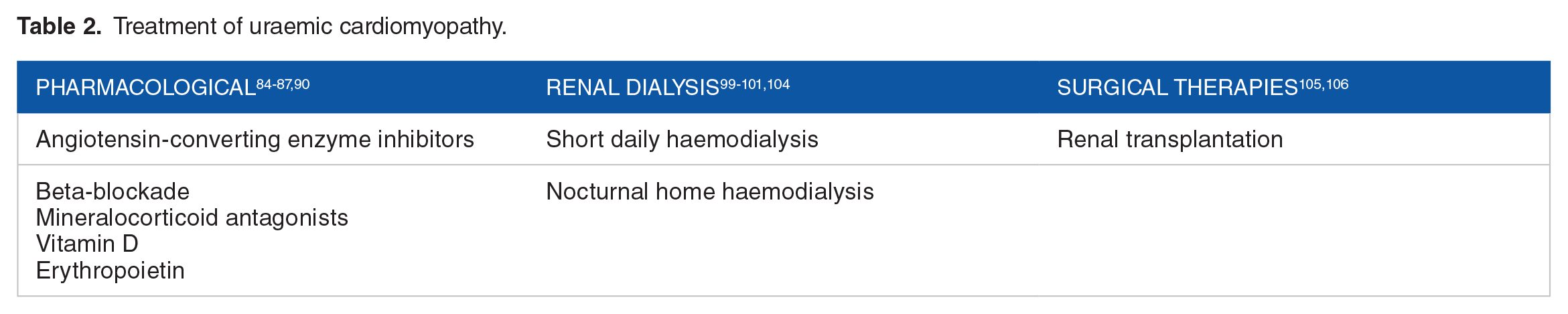
Pharmacological
There are several derived pharmacological treatment approaches for management and reversal of this disease process, which includes beta-blockade, angiotensin receptor blockers, mineralocorticoid antagonists and HMG-CoA reductase inhibitors. 84
Carvedilol has been shown to improve symptoms, LV End-Diastolic Pressure (LVEDP), LV End-Systolic Volume (LVESV) (74-62 ml/m2) and LVEF (26.3%-34.8%) along with a reduction in all-cause cause mortality and hospital admissions in patients on haemodialysis with dilated cardiomyopathy. 85
The role of renin-angiotensin system (RAS) blockade in patients with CKD with and without diabetic nephropathy is well known. RAS blockade was associated with a decreased risk of heart failure in patients with diabetic nephropathy (0.78, 95%CI 0.66-0.92, P = .03) and without diabetic nephropathy (0.74, 95%CI 0.58-0.95, P = .02) when compared to placebo in addition to a reduction in the risk of myocardial infarction and total CV outcomes. 86 Spironolactone, when used in patients with stage II-III CKD has been shown to reduce LV mass (−14 ± 13 g vs +3 ± 11 g) and arterial stiffness when compared to placebo. 87 However, within the haemodialysis population, there appears to be a more limited benefit with Spironolactone; for instance, a study by Hammer et al suggested that in comparison to placebo, Spironolactone did not improve left ventricular mass index in the haemodialysis population, 88 and this was also supported by a study by Charytan et al which compared Spironolactone to placebo in the haemodialysis population, and they too did not find an improvement in cardiovascular function or structure in the Spironolactone group. 89 There has also been significant concern with Angiotensin Converting Enzyme-Inhibitors (ACE-I), Angiotensin Receptor Blockers (ARB) and Spironolactone causing hyperkalaemia, which is not limited to the population with ESRD. 1
The SHARP trial has shown that the addition of simvastatin plus ezetimibe in patients with CKD has resulted in a reduction of first major atherosclerotic events. 90 However, it is worth noting that the 4D and AURORA investigators found that in the haemodialysis population, statins, in comparison to placebo, did not reduce the risk of cardiovascular events in the AURORA study and did not improve the composite primary end point of cardiovascular death, nonfatal myocardial infarction, and stroke in the 4D study.91,92 On the other hand, in the renal transplant population, the ALERT study, which compared Fluvastatin to placebo, suggested an improvement in cardiac deaths and non-fatal myocardial infarction in those treated with Fluvastatin; however, it is worth nothing that Fluvastatin did not generally reduce rates of mortality or coronary intervention procedures. 93 Therefore, the beneficial role of statins may be dependent on the context of the population managed.
The role of Vitamin D is also increasingly being explored, with a study by McGonigle et al demonstrating that the addition of Vitamin D in patients on HD resulted in a significant reduction in PTH, increase in LV fibre fraction shortening and an increase in mean velocity of LV fibre shortening. These findings highlight the importance of targeted pharmacotherapy in this population group. 94
The role of erythropoietin in treatment of anaemia in CKD and its effect of mortality has been a controversial area but is best exemplified by several studies. The CHOIR investigation explored whether EPO treatment to a higher haemoglobin (Hb) target of 13.5 grams (g) per decilitre (dL) compared to Hb target of 11.3 g/dL would be beneficial, and found that this higher target was not associated with additional improvement in quality of life and in fact was associated with increased harm. 95 The CREATE investigators investigated the role of EPO in CKD stage 3 and 4 in early and complete correction of anaemia versus partial correction of anaemia, and found that cardiovascular events were not reduced in those with early and complete correction. 96 This was supported by the TREAT investigators, who found that the use of darbepoetin alfa in CKD patients not undergoing dialysis with co-morbid type 2 diabetes and moderate anaemia to a target of 13 g/dL, compared to placebo (albeit with rescue darbepoetin alfa when the Hb was less than 9 g/dL), resulted in an increased risk of stroke and did not confer a survival benefit. 97 Therefore, while correction of anaemia with EPO may be required in such populations, clinicians need to be cognisant of its potential adverse effects.
Intertwined with the administration of EPO in CKD patients, the PIVOTAL investigators explored the role of high dose versus lose dose intravenous iron supplementation in CKD patients recently initiated on haemodialysis with ferritin concentrations less than 400 μg/L and a transferrin saturation of less than 30% who were receiving an EPO agent. They found that the high dose supplemental group had fewer major adverse cardiovascular events and lower risk of death. 98 Interestingly, this high dose regimen group also required fewer blood transfusions and lower doses of EPO, which as mentioned previously could potentially be associated with adverse effects.
Dialysis
An important treatment for UC in the setting of ESRD is haemodialysis (HD). There is evidence to suggest it is associated with a reduction in LVH, and reversal of systolic dysfunction (LVEF change (%) 31–50).99,100 Short daily HD (~2 hours daily) in comparison to conventional HD has also been shown to reduce LVH (LVMI change: 120.1 ± 60.4 g/m(2)) and antihypertensive use. 101 Therefore, frequent dialysis with close maintenance of euvolemia largely potentiates reverse cardiac remodelling. Similarly, nocturnal home HD has also shown to reduce LVMI by 22% compared to a 6% progression on conventional HD in a cohort of patients with ESRD associated cardiomyopathy. 102 However, despite many studies that have shown HD can potentiate and reverse some of the clinical sequelae of UC, there is conflicting data from other studies. In 2015, Foley et al demonstrated in a population of 596 HD patients with no symptomatic cardiac disease or dilatation that there was progressive concentric LVH and hyperkinesis. 72 This suggests the interplay of other factors such as hypertension that may influence the role of HD on UC. There is data to suggest that PD may portend increased mortality in patients with UC which increases with the length of follow up and has increased requirement of anti-hypertensives.3,103 Important complications to monitor for with HD include dialysis-induced myocardial stunning, resulting in further hypotension, which may compound the underlying cardiomyopathy. 104
Renal transplantation
Renal transplantation can partially reverse the underlying disease state and confers a significant survival advantage compared to the other treatment modalities described. Dzemidzić et al have shown that renal transplantation dramatically reduces the proportion of patients with LVH from 67% to 37% with correlations in improved creatinine clearance and in reduction of the serum creatinine values; as well as values of parathyroid hormone. 105 Additionally, Wali et al have shown that 70% of patients with a baseline LVEF <40% prior to renal transplantation, recovered at follow-up at 6 months with LVEF >50% on ventriculography-gated blood pool. Normalization of LVEF was associated with improvement in NYHA class and was the only significant factor associated with reduced hazard for CCF hospitalization and death (RR 0.90). 106 Native T1 measurements, a method of assessment of myocardial fibrosis in CMRI without gadolinium contrast, was explored by Contti et al where it was found that 6 months following renal transplantation, the native myocardial T1 time decreased, suggestive of regression of reactive fibrosis. 107 These findings demonstrate significant reverse cardiac remodelling as a result of renal transplantation in this high-risk population group.
Conclusion
UC remains a complex and multifaceted disease with high associated morbidity and mortality. Amongst existing therapies, haemodialysis and kidney transplantation are instrumental in halting disease progression. 3 The role of non-invasive and invasive diagnostic tools alongside our growing understanding of the pathophysiology of UC remains vital in illuminating the path for future medical and surgical therapies. Thus, a structured approach which utilises existing therapies in addition to ongoing research on a cellular level may represent the best opportunity of reducing the burden of UC.
Footnotes
Funding:
The author(s) received no financial support for the research, authorship and/or publication of this article.
Declaration of Conflicting Interests:
The author(s) declared no potential conflicts of interest with respect to the research, authorship and/or publication of this article.
Author Contributions
All authors contributed significantly to the final manuscript.
CREDIT Author Statement
• Conceptualisation
• Visualisation
• Writing original draft
• Project administration
• Writing, Reviewing and Editing
• Formal analysis
• Formal analysis
• Writing original draft
• Resources
• Writing, Reviewing and Editing
• Supervision