
Abstract
Objectives
The aim of this study was to evaluate, in vitro, the load and type of failure of the sutured ventral abdominal fascia of cats with different sizes of suture material made of polydioxanone (PDX) (2-0, 3-0, 4-0, 5-0 USP).
Methods
A total of 32 samples of the ventral abdominal wall from 16 cadaveric cats were harvested using an hourglass-shaped template. The samples were sectioned longitudinally along the linea alba and then sutured together in a continuous pattern using four different randomly assigned sizes of pdx suture material (2-0, 3-0, 4-0, 5-0 USP). A universal testing machine was used for linear distraction of the samples. The tensile strength and type of failure were recorded and analysed. Three types of failure were defined: suture material failure (S), suture line failure (T1) and failure of the abdominal wall further away from the linea alba (T2).
Results
The frequency of suture material failure decreased with increasing suture size. Suture size 5-0 failed due to a S failure in 6/8 samples, PDX 4-0 failed in 2/8 samples and PDX 3-0 failed in only 1/8 samples. However, PDX 2-0 failed due to only T1 or T2 failures, with both failures being almost equally represented. No statistically significant differences in the load to failure between PDX 2-0, 3-0 and 4-0 were noted (P >0.05). The risk of suture failure increased with decreasing suture size diameter.
Conclusions and relevance
PDX 2-0 and 3-0 can be used without reservation for the closure of ventral midline coeliotomy in cats. Although there was no statistically significant difference between PDX 2-0, 3-0 and 4-0, PDX 4-0 showed a higher probability for suture breakage and should be used only after careful consideration of the patient while clinical evaluation is pending. Pdx 5-0 cannot be recommended as a safe suture size for this type of surgical closure.
Introduction
Effective wound closure is a fundamental aspect of nearly every surgical procedure. The most common technique for closing wounds is the use of suture material that sufficiently holds the wound margins together for adequate healing of the tissue. However, every penetration with the needle causes additional trauma to the soft tissue and the presence of foreign material increases the risk of infection.1,2
Selection of the size of suture material is based on the strength of the tissue that is to be closed, whereby the focus is placed on choosing the smallest diameter of suture material that is required to hold the tissue together. 3 Inappropriately large diameters should be avoided because of the unreasonable extra tissue damage, histological changes in the tissue architecture and additional excess of foreign material.1,4
In human medicine, there are many studies that help to create recommendations for the closure of surgical incisions.5,6 For instance, working groups such as the European/American Hernia Society are collecting study data to provide recommendations to minimise the incidence of incisional hernia as a major complication after abdominal wall incisions. 7 Furthermore, they still recommend using a slow absorbable monofilament suture in a continuous fashion. 7 Besides saving time in comparison with simple interrupted stitches, 8 continuous suture patterns allow distributed tension along the entire closure. 9
In contrast to the human literature, veterinary studies, especially biomechanical studies regarding ventral midline coeliotomy closure, are rare and primarily focus on dogs.1,10 In the past, recommendations about closing the abdominal wall were either based on clinical case reports and surgeons’ experiences or extrapolated from human medical data.8,11
To the authors’ knowledge, Rodriguez et al 12 were the first to investigate biomechanical considerations in feline ventral abdominal wall closure techniques. Among other findings, they discovered that the post-umbilical (POU) area of the ventral abdominal wall is biomechanically weaker than the regions more cranially. For this in vitro experiment, they tested all sutured samples by using a single size of suture material (3-0 USP). In addition to a very recent canine study, 1 the impact of various suture material diameters on the feline abdominal wall has not yet been considered.
The aim of the present study was to provide further biomechanical data on feline ventral midline wound strength using different sizes of polydioxanone (PDX) suture material by evaluating the load of failure on the one hand and the types of failure on the other.
Materials and methods
Tissue collection, sample preparation and testing
A total of 32 samples from the ventral abdominal wall were tested biomechanically. These samples were collected from 16 cats (two samples per cat) euthanased for reasons unrelated to the purpose of this study. Informed verbal consent was obtained from the owners of all animals. The breed, age, body weight (BW) and sex of the cats were documented.
Within 1 h after euthanasia, tissue samples were collected. Cadavers were placed in dorsal recumbency and the abdominal hair was clipped. A skin incision was made between the xiphoid process and the cranial edge of the pubis. After removal of the subcutis, the ventral abdominal wall was thoroughly checked for abnormalities or lesions of its integrity. Any structural changes led to exclusion from the study.
Before collecting the samples, we designed a reusable hourglass-shaped template for consistency and practicality. This design, similar to one used by Rodriguez et al 12 , featured bilateral holding sections and a central measuring part. The guide measured 100 mm in length and had a maximum width of 40 mm at its holding portions (see Figure 1a).
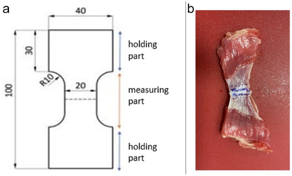
(a) Schematic view of the template, measures in mm; the dotted line corresponds to the linea alba. (b) Sample before suturing; note the marked linea alba (bordered with double straight line) and predetermined suture perforation places (dots)
By using the template, the tissue samples were cut into identical sizes, whereby one cadaver provided two ventral abdominal wall samples.
For protection, the samples were covered with an isotonic saline (0.9% NaCl)-soaked gauze and placed in cylindrical containers made of synthetic material. The containers were labelled with the identification number and collection date. The specimens were stored frozen at −18°C.
Before testing, the samples were thawed to room temperature (Figure 1b). At the Faculty of Mechanical Engineering of the Poznan University of Technology (Poland), the samples were halved along the linea alba and closed with a simple continuous suture in accordance with the biomechanical study of Rodriguez et al 12 by one board-certified surgeon (PS). The suture was secured with a starting square knot of five throws and an ending square knot of six throws.
The samples were randomly assigned a suture size by blindly pulling a suture material out of a prepared box. The study used four different sizes of suture material made of PDX (MonoPlus; B. Braun SE): 2-0; 3-0; 4-0; and 5-0 USP. Every size of suture material was represented eight times throughout the samples.
Immediately after suturing, uniaxial tensile tests were carried out on the universal testing machine Inspekt 20k (Hegewald & Peschke MPT) using a 1 kN loadcell type S2M HBM (Hottinger Brüel & Kjaer). To ensure stable tissue maintenance and adequate friction in the jaws of the testing machine, sandpaper pads were used (Figure 2). All samples were pretensioned with an initial force of 0.5 N. Once the sample and the machine were set up, the test was initiated with a travel rate of 20 mm/min and a data transfer rate of 100 Hz. An automatic stop condition was set up to drop 80% of the maximum force, which corresponded to the rupture of either the tissue or the sutures. During the tests, we recorded time (in s), tensile force (in N) and displacement (in mm) (Figure 3). The tests were performed using LabMaster testing software. Samples were locked using dedicated HP154 mechanical grips (Hegewald & Peschke).
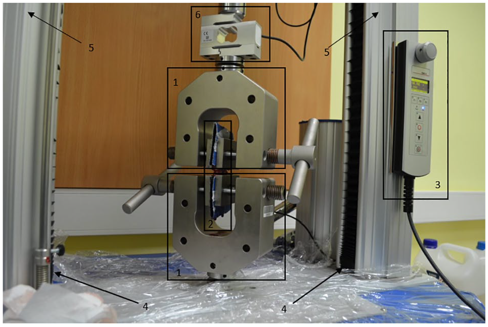
Overview of the universal testing machine, Inspekt 20k (Hegewald & Peschke MPT): 1 = upper and lower holders, 2 = tested sample, 3 = control panel, 4 = ball-screw feed drive, 5 = casing, 6 = load cell
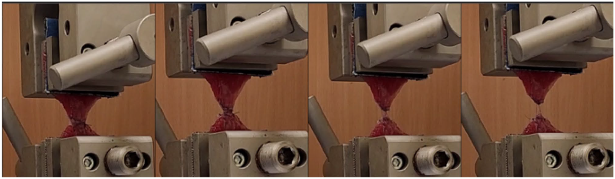
A series of images of a representative sample shows progressive elongation until failure
Statistical analysis
Normality testing was performed for all variables. Descriptive statistics were performed to determine mean ± standard deviation (SD) for load to failure. Spearman’s rank correlation coefficient was calculated to evaluate the correlation between suture material, BW, sex and age.
Mixed model linear analysis was performed to evaluate the effect of suture material, sex, age and BW on the load to failure (dependent variable). Sex and neuter status were considered to be categorical variables in the statistical model. The effect of suture material on the frequency of suture failure was determined by calculating Spearman’s rank correlation. A significant relationship between dependent and independent variables was determined using an ANOVA. P <0.05 was considered statistically significant.
One-way ANOVA was performed for the final statistical model to compare mean load to failure between groups, followed by a post hoc analysis.
Results
In total, 16 cats were included in the study. The mean BW was 5.04 ± 1.81 kg (range 3.00–10.00). The mean age was 10.56 ± 4.63 years (range 2.0–18.0).
When performing the tests with the 32 samples, there were no significant differences of the load to failure between male and female cats, regardless of whether they were neutered or not (P = 0.300). In addition, age (P = 0.105) and BW (P = 0.478) played no significant effect.
In the mixed model, no effect of age, BW and sex was observed on the load to failure. When these variables were removed from the statistical model, suture material was the only significant factor affecting the load to failure. A post-hoc analysis revealed no significant differences between the 2-0, 3-0 and 4-0 groups.
Load to failure was in the range of 29.72–106.62 N (mean 65.98 ± 18.60). SD values are shown in Table 1 and other data are summarised in Figure 4.
Load to failure (Fmax) values for sutures
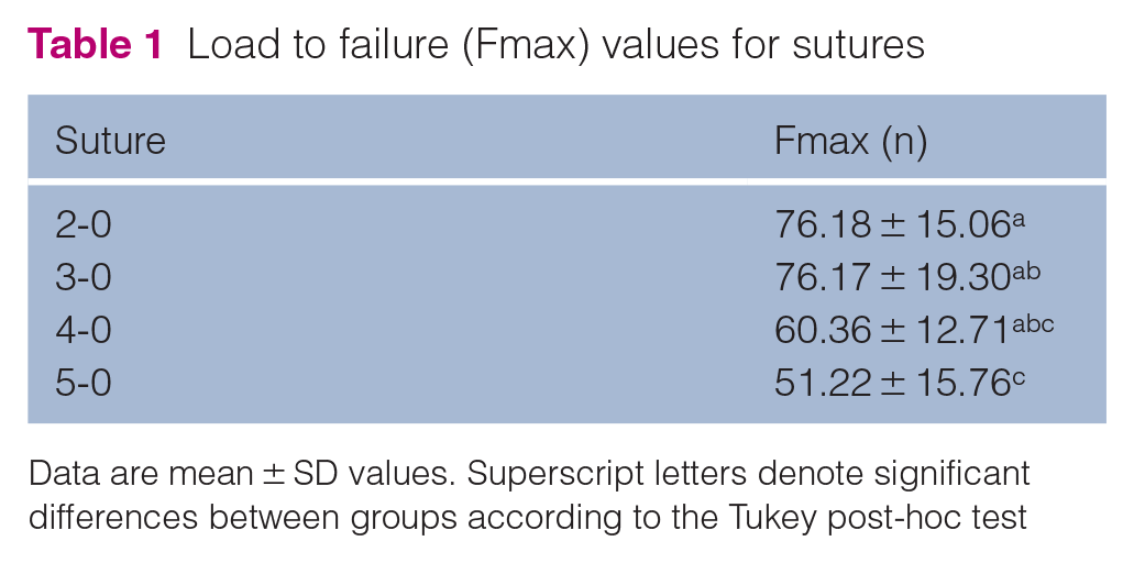
Data are mean ± SD values. Superscript letters denote significant differences between groups according to the Tukey post-hoc test
A significant drop of force was observed for the 5-0 suture vs the 2-0 and 3-0 sutures (by 32.77% and 32.76%, respectively). No differences were recorded between the 4-0 and 5-0 suture groups.
Consequently, one can summarise that the load to failure is comparable for the suture sizes 2-0, 3-0 and 4-0 (P >0.05) (Figure 4).
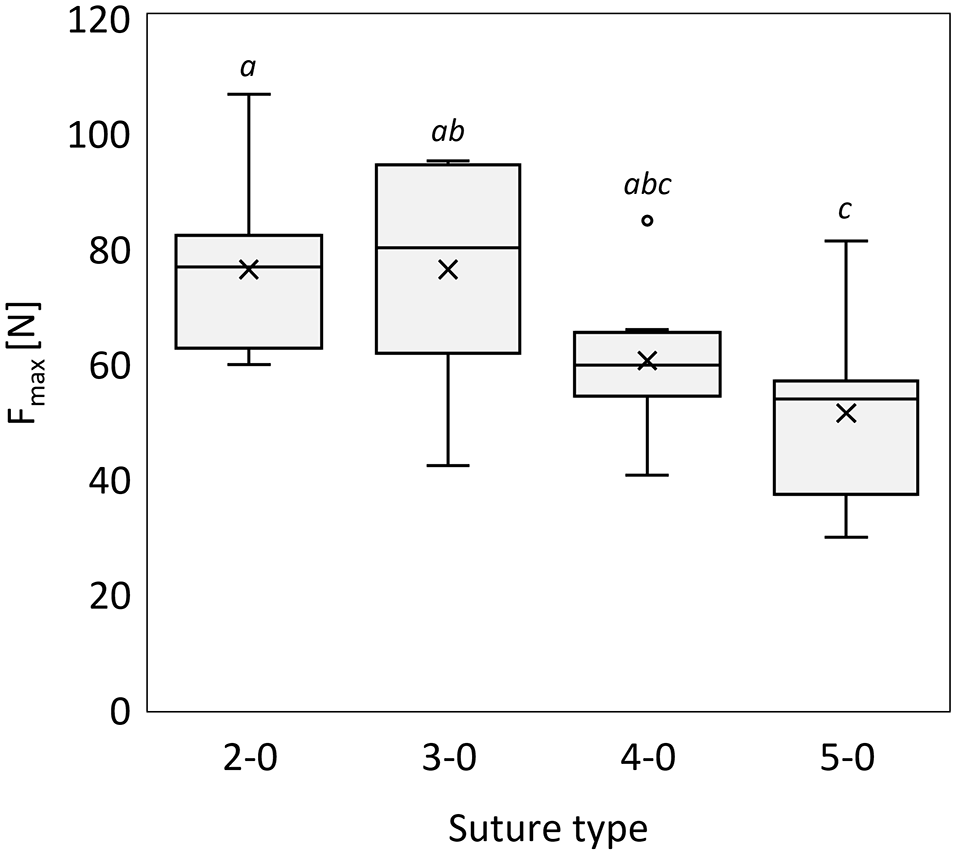
Box-and-whisker plot showing load to failure (Fmax) for each suture type group. Each box represents the middle 50% of observed values. The bottom of the box is the first quartile (25th percentile) and the top of the box is the third quartile (75th percentile). The line in the middle of the box is the median (50th percentile) and ‘×’ is the mean. Error bars (whiskers) indicate the lowest and highest values, excluding outliers. Superscript letters denote significant differences between groups according to the Tukey post-hoc test
Three different types of failure were classified: suture material failure (S); suture line failure, ie, linear tears in the muscle or fascia perpendicular to the linea alba at the site of suture material penetration(s) (T1); and tears in the muscle or fascia distant from the linea alba or celiotomy site (T2) (definitions based on the study by Rodriguez et al 12 ). Among the 32 samples, failure of suture occurred a total of nine times. A T1 failure was counted 15 times and a T2 failure eight times. Samples sutured with 2.0 suture demonstrated T1 and T2 failures (four samples each) but no S failure (Figure 5). The frequency of suture material failure increased with decreasing suture size (one sample for 3.0 [12.5%], two for 4.0 [25%] and six for 5.0 [75%]). T2 failure was equally frequent for 3.0 and 4.0 sutures and was not observed when samples stitched with 5.0 sutures were tested.
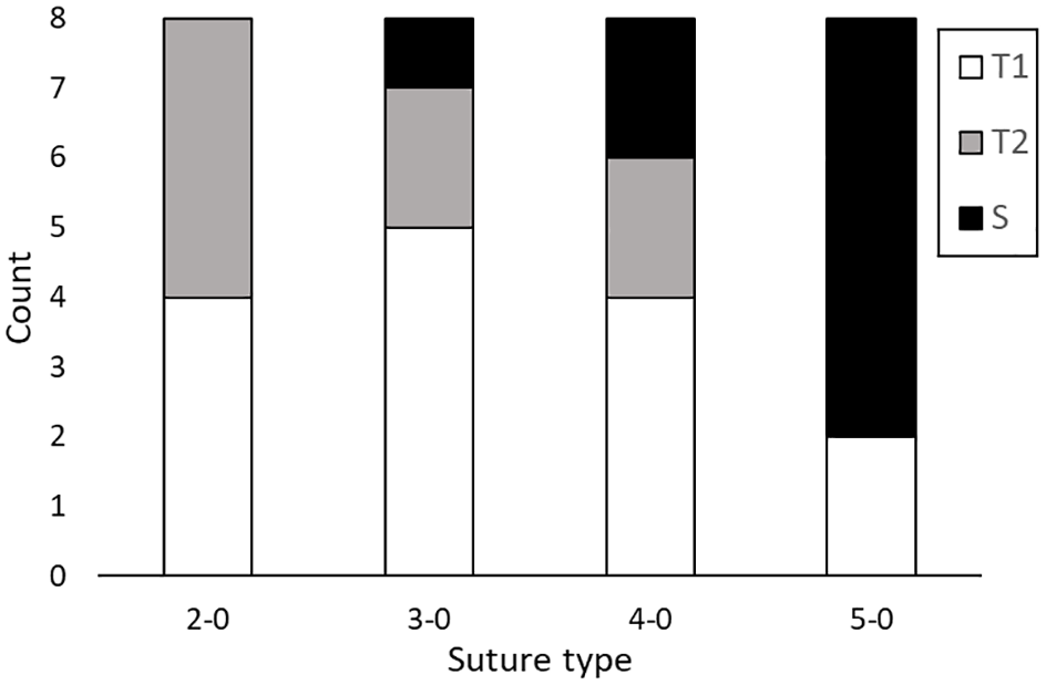
Type of failure recorded for different suture types
The correlation between frequency of suture material failure and suture material was strong (r = 1.000 and P <0.001, Spearman’s rank correlation), indicating that failure by suture is more frequent when the suture diameter decreases.
Discussion
This study suggests that the biomechanical properties of PDX 2-0, 3-0 and 4-0 sutures used for suturing the ventral abdominal wall of cats are comparable. However, the risk of suture rupture increases with decreasing suture diameter.
Samples closed with PDX 2-0 showed no suture failure (S), PDX 3-0 showed 1 (12.5%) suture failure and PDX 4-0 showed 2 (25%) suture failures out of eight samples per group. A different study used only PDX 3-0 for abdominal wall closure followed by tensile testing. 12 From 14 sutured samples closed with a simple continuous pattern – the same as in the present study – no suture (0%) failed. It is worth mentioning, though, that in the mentioned publication, the mean load of failure (N) during linear distraction was 37.93 ± 5.27 N, whereas in the present study, the mean value was higher (65.98 ± 18.60 N). In our work, only 1/8 samples sutured with PDX 3-0 failed by suture rupture at 84.82 N. Early tissue tearing could be the reason for the lack of suture failure in the mentioned study. However, there is a clear discrepancy between the two mean loads of failure.
One possible explanation for this discrepancy may be the condition of the samples at the time of testing. A biomechanical and histological study in dogs revealed reduced tensile strength in muscle tissue when comparing fresh post-mortem tissue with tissue subjected to a standardised freezing/thawing process. 13 Histologically, the frozen specimens exhibited more pronounced muscle fibre damage compared with the fresh specimens. In contrast to this work the above-mentioned study froze euthanased cats in toto at −80°C and harvested the samples immediately before suturing and linear distraction. 12 In addition, the paper by Rodriguez et al 12 does not reveal the exact dimensions of their hourglass-shaped template. Because they were able to collect three samples per cat, it is possible that the sutured part of the linea alba was shorter than that used in the current study (20 mm). These differences in the method of preparation of the samples before testing could lead to discrepant biomechanical results. However, cadaver-based biomechanical studies and their in vitro characteristics do not necessarily correspond to the physiological conditions of a living organ.
Our research used 16 cadaveric cats with two samples per cat. Tissue failure occurred in 23/32 (71.9%) samples, where most of the samples failed at the suture–tissue interface (T1: 15/32, 46.9%). Each suture line causes small tissue perforations and can therefore be seen as a line of weak points. Whether T1 failure would also be the most common type of failure when tested in vivo or whether this failure occurs as a consequence of the repeated freezing process causing changes to the tissues’ histological architecture remains unknown. One study performed tensile testing of the abdominal wall in dogs without a sutured linea alba and showed – without exception – muscular failure distant to the linea alba (17/17 samples, 100%). 10 In a similar study, Rodriguez et al 12 showed the same ratio in feline abdominal walls (6/6 samples, 100%).
One limitation of the ex vivo model presented in this study is that it is impossible to calculate or estimate all biological factors of tissue-strength behaviour. 14 On the other hand, there are further mechanical parameters that can be measured and play a huge role in the biomechanical properties of the abdominal wall. In particular, the individual thickness of the linea alba is important. In humans, horses, dogs and cats, studies have confirmed that a thicker linea alba is biomechanically superior to a thinner one.10,12,15,16
Furthermore, Rodriguez et al 12 compared different parts (pre-umbilical [PU], umbilical [U] and POU) of the feline linea alba and showed that the anatomical architecture changes and is thinner in the more caudal aspects. As a result of this investigation, they concluded that the POU region is biomechanically weaker than the PU and U parts of the linea alba. However, as a limitation due to varying thicknesses of the linea alba depending on their anatomical origin, the present study used two samples per cat but did not differentiate between the different locations. We also experienced that obtaining three samples from one cadaver that were still in correspondence to our template would have been almost impossible. In addition, we did not focus on measuring the thickness of the linea alba.
The major limitation of our study is the small number of samples, which was limited by cadaver availability. As a consequence, an a priori power analysis of ANOVA was not performed. This creates a possibility for Type II error in the non-significant comparisons (sex, neuter status, BW). For the character of a biomechanical study, a larger number would be desirable, although the current study was able to demonstrate statistical relevance for suture type. Comparable biomechanical studies also demonstrate similar available resources.1,12
In addition, our study did not integrate a uniform control group, which would represent a clear added value.
Recommendations for coeliotomy closure in cats are rare, often extrapolated from other species and mainly based on older surgical habits that have become established over the years.8,11 A recent textbook of feline surgery recommends that suture material sizes of 2-0 and 3-0 provide enough support for the incision to heal properly. 17 Based on our research, we can confirm this recommendation.
That aside, based on the recent literature, a smaller diameter of sutures is recommended to minimise side effects resulting from suture material.1,2 Despite the relatively small number of samples in our study and a higher probability of suture breakage as the diameter decreases, PDX 4-0 can be used taking into account the patient's specific conditions. Undoubtedly, this recommendation requires clinical evaluation. Regularly checking the integrity of the sutured abdominal walls using cautious palpation and sonography would be a first feasible way to detect abnormalities in proper wound healing. One can simultaneously assess the extent of inflammatory response based on the specific size of the chosen suture material.
Conclusions
PDX 2-0 and 3-0 can be used without reservation for the closure of ventral midline coeliotomy in cats. Although there is no statistically significant difference between PDX 2-0, 3-0 and 4-0, PDX 4-0 shows a higher probability for suture breakage and should be used only after careful consideration of the patient while clinical evaluation is pending. PDX 5-0 cannot be recommended as a safe suture size for this type of surgical closure.
Footnotes
Conflict of interest
The authors declared no potential conflicts of interest with respect to the research, authorship, and/or publication of this article.
Funding
This study was financially supported by the AniCura Research Fund. Suture material for this study was donated by Aesculap B Braun. This research was partially funded by the Polish Ministry of Science and Education as a part of annual research subsidy (projects 0612/SBAD/3605 and 0614/SBAD/1579).
Ethical approval
The work described in this manuscript involved the use of non-experimental (owned or unowned) animals. Established internationally recognised high standards (‘best practice’) of veterinary clinical care for the individual patient were always followed and/or this work involved the use of cadavers. Ethical approval from a committee was therefore not specifically required for publication in JFMS. Although not required, where ethical approval was still obtained, it is stated in the manuscript.
Informed consent
Informed consent (verbal or written) was obtained from the owner or legal custodian of all animal(s) described in this work (experimental or non-experimental animals, including cadavers, tissues and samples) for all procedure(s) undertaken (prospective or retrospective studies). No animals or people are identifiable within this publication, and therefore additional informed consent for publication was not required.