
Abstract
Objectives
Leptospirosis is a re-emergent zoonotic bacterial disease associated with renal and hepatic injury. In free-roaming cats in some regions, a high prevalence of Leptospira antibodies has been identified, and pathogenic leptospires have been detected in renal tissue, indicating that they may play a role in Leptospira epidemiology. The objective of this study was to determine the prevalence of Leptospira seroreactivity and urinary shedding of Leptospira DNA in free-roaming cats from northern California and southern Texas. A secondary objective was to compare the results of a point-of-care (POC) assay, designed to detect Leptospira antibodies, with the results of the microscopic agglutination test (MAT) when applied to serum samples from feral cats.
Methods
Specimens were obtained from free-roaming cats from northern California (n = 52; 2020) and southern Texas (n = 75; 2017). Leptospira quantitative PCR was performed on blood and urine specimens from Californian cats. Serum samples from Californian and Texan cats were subjected to MAT to categorize them as Leptospira antibody-positive or antibody-negative. The performance of the POC assay was assessed using the MAT as the gold standard.
Results
Leptospira DNA was not detected in the blood or urine of any cats tested. The results of the MAT were positive in 17.3% (n = 9) of Californian cats and 10.7% (n = 8) of Texan cats (P = 0.3). The median MAT titer was 1:100 (range 1:100–1:200) in Californian cats and 1:200 (range 1:100–1:800) in Texan cats. The POC assay was negative in all specimens.
Conclusions and relevance
Free-roaming cats in California and Texas are exposed to Leptospira species and may have the potential to act as sentinel hosts. No cats had evidence of current infection, as determined using PCR on blood and urine specimens. The POC test did not reliably detect anti-Leptospira antibodies in these cats. The role of cats in the maintenance or shedding of pathogenic leptospires requires further investigation.
Introduction
Leptospirosis is a widespread zoonotic, infectious disease caused by infection with spirochetes of the genus Leptospira, of which the species Leptospira interrogans and Leptospira kirschneri are responsible for the majority of disease. 1 Individual Leptospira genotypes have adapted to specific reservoir hosts, such as rodents, which become subclinically infected and maintain a carrier state. 2 Infections of incidental hosts with pathogenic leptospires can lead to clinical disease.
Leptospirosis in dogs is often characterized by the acute onset of fever, signs of hepatic or renal damage, pancreatitis, pulmonary hemorrhage, vasculitis and uveitis. 3 Although relatively uncommon, cats can also develop leptospirosis.4,5 Estimates of seroprevalence in cats have been as high as 35%.6–12 Outdoor, free-roaming cats might be at high risk of exposure because they hunt rodents, the primary reservoir host species for Leptospira species worldwide. 13 Studies have demonstrated the presence of leptospiral DNA in renal tissue and urine in cats, including 42% of cats on Christmas Island, Western Australia. 14 Pathogenic leptospires have also been isolated in culture from the urine of cats from southern Chile. 15 However, alternative studies have shown a lower prevalence of Leptospira exposure and infection,11,12 suggesting that the importance of cats as a reservoir host may vary based on geographic location.
The diagnosis of leptospirosis in dogs requires consistent clinical signs combined with demonstration of a four-fold rise in antibody titers, as measured by the microscopic agglutination test (MAT), or detection of Leptospira DNA in blood or urine specimens using PCR.16,17 Point-of-care (POC) diagnostic assays are available for the detection of Leptospira antibodies in dogs, but their use in cats has not been investigated. 18
The objective of this study was to improve our understanding of the potential role of free-roaming cats in the epidemiology of leptospirosis by determining the prevalence of leptospiral antibodies and DNA in free-roaming cat populations residing in two regions of the USA where leptospirosis is regularly diagnosed in dogs.19–21.The secondary aim of this study was to determine the performance of a canine POC assay for the detection of Leptospira antibodies in free-roaming cat sera, using the MAT as the gold standard for comparison purposes.
Materials and methods
Cat specimens
Whole blood, serum and urine specimens were collected by venipuncture or cystocentesis from free-roaming, unowned cats in Klamath and Hoopa counties, California, in October of 2020. These cats were deemed sufficiently healthy to permit anesthesia for spay-and-neuter surgery, although their precise age was unknown. Specimens were transported at 4°C to the laboratory at the University of California, Davis, within 24 h. An abbreviated serum biochemistry panel that included electrolytes, urea and creatinine concentrations was performed on specimens when serum volume was sufficient (Dobas c501/6000 analyzer; Roche). Urine specific gravity (USG) was recorded using a refractometer. Banked serum specimens were obtained from free-roaming cats that had been euthanized for management purposes at a large shelter in the Rio Grande Valley region of Texas throughout 2017. Cats in this population were euthanized for various reported reasons, such as significant upper respiratory tract infections, quality-of-life concerns from suspected systemic disease, behavioral concerns and other concurrent infections (such as feline leukemia virus infection and dermatophytosis). Specimens were held at −20°C for 3 years before being transported overnight on ice to the University of California, Davis.
Quantitative PCR design and validation
A homologous region on the 16S rRNA gene of Leptospira was targeted for quantitative PCR (qPCR) assay design to ensure broad species detection. Vector NTI software (Thermo Fisher Scientific) was used to confirm the alignment of multiple Leptospira sequences. The assay was predicted to detect L interrogans, L kirschneri, Leptospira fainei, Leptospira santarosai, Leptospira weilii, Leptospira noguchii, Leptospira inadai, Leptospira borgpetersenii, Leptospira meyeri, and Leptospira manara. Two specific primers and one internal, fluorescence-labeled probe were designed with Primer Express software 3 (Thermo Fisher Scientific) on the 50–250 base-pair range of GenBank accession number EF536997.1. To monitor the amplification of the target sequences, a TaqMan major groove binder [MGB] probe that incorporated a 5′ reporter dye (carboxyfluorescein [FAM]) and a 3′ non-fluorescent quencher (NFQ) were used.
Validation of the assay was performed by running DNA from a known Leptospira-positive sample in triplicate, 10-fold serial dilutions. Cq values were graphed against the dilution factor. The slope and y-intercept of the resulting trendline were used to calculate the efficiency (E = 10(−1/slope) − 1) and sensitivity, respectively. The assay was within the acceptable range for both criteria, 92% efficient and sensitive enough to detect as few as 10 copies of target DNA per qPCR reaction. An off-the-shelf assay targeting eukaryotic 18S rRNA (Hs99999901-s1; Thermo Fisher Scientific) was used as an internal housekeeping gene to ensure successful DNA extraction and absence of inhibition.
Leptospira sample processing and DNA extraction
Whole-blood and urine specimens from Californian cats were submitted to the University of California, Davis Real-Time PCR Research and Diagnostics Core Facility for Leptospira PCR. A total of 200 µl of EDTA whole blood and 200 µl of pelleted urine were extracted on a QIACube HT system (Qiagen), according to the manufacturer’s recommendations. Urine was centrifuged at 3220 × g for 5 mins, supernatant in excess of 500 µl was removed and the resulting cell pellet was resuspended by vigorous pipetting. A total volume of approximately 70 µl of total nucleic acid was eluted in diethyl pyrocarbonate-treated water and used for qPCR.
qPCR assay
Each qPCR contained primers at a concentration of 400 nM (80 nM for the MGB probe) and commercially available PCR master mix (TaqMan Universal PCR Master Mix; Thermo Fisher Scientific) containing 10 mM Tris-HCl (pH 8.3), 50 mM KCl, 5 mM MgCl2, 2.5 mM deoxynucleotide triphosphates, 0.625 U AmpliTaq Gold DNA polymerase per reaction, 0.25 U AmpErase uracil N-glycosylase (Applied Biosystems) per reaction and 5 µl diluted sample total nucleic acid (1:5 dilution for blood, 1:1 for urine) in a final volume of 12 µl. The samples, in addition to positive and negative PCR controls, were placed in a 384-well plate. Amplification was performed under the following conditions on a QuantStudio Q7 Pro PCR system (Applied Biosystems): 2 mins at 50°C and 10 mins at 95°C, followed by 40 cycles of 95°C for 15 s and 60°C for 1 min. Fluorescent signals were collected during the annealing temperature, and the quantitative cycle (Cq) was calculated and exported with a threshold of 0.1 and a baseline of 3–10.
Leptospira MAT
Serum specimens from all cats were submitted to the California Animal Health and Food Safety Laboratory (Davis, California) for MAT serology. Serum was reacted with live L interrogans strains (serogroup/serovar/strain; Australis/Bratislava/Jez Bratislava; Canicola/Canicola/H Utrecht IV; Grippotyphosa/Grippotyphosa/Andaman; Sejröe/Hardjo/Hardjoprajitno; Icterohaemorrhagiae/Copenhageni/M 20; Pomona/Pomona/Pomona). The serum dilution resulting in ⩾50% leptospiral agglutination as observed under dark-field microscopy, was reported. If <50% agglutination was observed at the lowest dilution (1:100), the result was recorded as negative.
POC immunoassay
A commercial canine POC Leptospira antibody test (SNAP Lepto Test; IDEXX Laboratories) was performed on cat sera where a sufficient volume was available for testing. The assay was performed according to the manufacturer’s instructions by a single investigator (JFS). The components of the test kit were allowed to equilibrate to room temperature. Three drops of serum were mixed with four drops of manufacturer-supplied conjugate using the supplied dropper and inverted several times to mix. The specimen was then applied to the test cassette. The cassette was incubated at room temperature for 10 mins, and the results were recorded as positive, negative or invalid, as described by the manufacturer.
Statistical analysis
Descriptive statistics were performed to describe the proportion of cats that were Leptospira antibody- or PCR-positive from each geographic location, and 95% confidence intervals (CIs) were calculated using the Wilson–Brown method. A χ2 test was used to compare proportions of positive cats in each geographic location in this study and to historical prevalence studies. The positive and negative percentage agreements were calculated with 95% CIs for the POC assay vs the MAT results. Biochemical parameters were tested for normality using a D’Agostino–Pearson test. Normally distributed data were compared using an unpaired two-tailed t-test. A P value <0.05 was considered to be statistically significant. Data were analyzed using commercial statistical software (GraphPad Prism).
Results
Cats
Specimens were obtained from 52 adult cats in California and 75 cats in Texas (Figure 1). Serum urea nitrogen and creatinine concentrations were within the reference intervals (RIs) for all 34 Californian cats in which testing was performed (Table 1). The median creatinine concentration was 0.7 mg/dl (range 0.4–1.2; RI 1.1–2.2) and the median serum urea nitrogen concentration was 20 mg/dl (range 14–31; RI 18–33). The median USG for 47 Californian cats was 1.044 (range 1.032–1.050).

Geographic distribution of study collection sites in Klamath and Hoopa, California (CA), in October of 2020, and sera from free-roaming cats in Rio Grande Valley, Texas (TX), throughout 2017
Serum biochemical and urine parameters for Californian cats stratified by microagglutination test (MAT) results

Data are presented as mean ± SD or n (%)
Leptospira PCR and MAT results
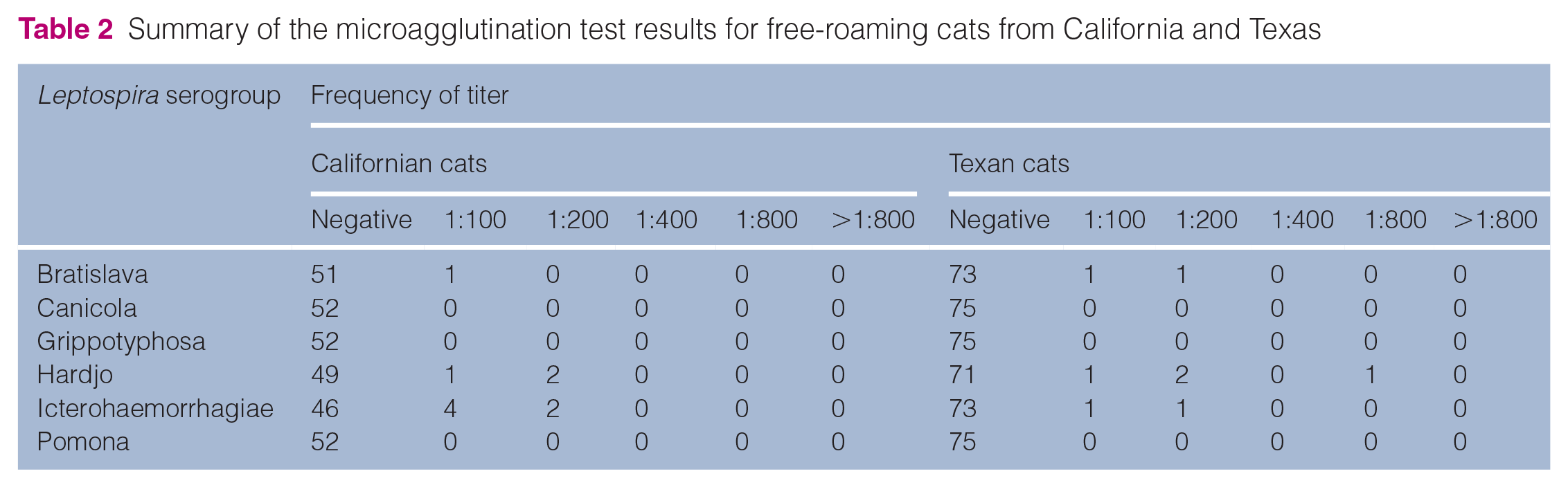
Leptospira DNA was not detected using qPCR of blood or urine from any of the 52 cats from California. The MAT was positive in 9/52 (17.3%; 95% CI 8.0–25.7) Californian cats and 8/75 (10.7%; 95% CI 5.0–17.9) Texan cats. There was no difference in the proportion of MAT-positive cats between the two geographic regions (P = 0.3). The median positive MAT titer was 1:100 (range 1:100–1:200) in Californian cats and 1:200 (range 1:100–1:800) in Texan cats (Table 2). The highest MAT titer was to L interrogans serovar Icterohaemorrhagiae in seven cats, L interrogans serovar Hardjo in seven and L interrogans serovar Bratislava in two; one cat had equal seroreactivity to Icterohaemorrhagiae and Bratislava. There was no difference in biochemical parameters between seropositive and seronegative cats (Table 1).
Summary of the microagglutination test results for free-roaming cats from California and Texas
POC immunoassay results
Adequate volume was present to perform the POC assay on sera from 52 cats from California and 54 cats from Texas; this included all 17 seropositive cats (nine from California and eight from Texas). The POC test was negative in all tested specimens. The positive percentage agreement was 0% (95% CI 0–20.4) and the negative percentage agreement was 100% (95% CI 96.0–100).
Discussion
Our study showed that 17% (n = 9/52) and 11% (n = 8/75) of free-roaming cats from California and Texas, respectively, had evidence of Leptospira species exposure, as detected by MAT. However, no cats from California had qPCR-detectable leptospiral infection or urinary shedding. In a study from Montreal, Leptospira seropositivity was more prevalent in cats with kidney disease than in those classified as healthy. 10 However, we found that seropositive cats in California did not have any biochemical evidence of compromised renal function. The prevalence of seropositivity was similar to that identified in other leptospirosis-endemic regions throughout the world in limited prior studies. 22 When compared with two previous sero-surveys performed in the USA (Massachusetts 4.8% [n = 3/63]; Iowa 8.6% [n = 12/139]),8,23 the seroprevalence in the Californian cats was higher than in cats from Massachusetts (P <0.05). However, there was no difference in seroprevalence among the Texan cats and those from Iowa and Massachusetts. This may reflect temporal or geospatial differences in transmission, differences in exposure within the free-roaming cat population or differences in serologic test performance among studies. It should also be kept in mind that the absence of detectable antibodies does not rule out exposure or active infection, because of limitations in diagnostic test sensitivity or pathogen evasion of the host immune response. The latter has been documented in dogs acting as reservoir hosts for Leptospira species. 24
Of the five L interrogans serogroups tested, antibodies to serogroups Icterohaemorrhagiae and Hardjo were most commonly identified. In comparison, a medical records review of 67 dogs in northern California from 2001 to 2010 tested at the same laboratory seroreacted with highest titers to serogroups Pomona or Bratislava. 25 Although infecting serovars cannot be inferred from MAT results alone, these findings may reflect different transmission dynamics in dogs than in cats, especially given that the primary serovars previously identified in rats in North America consisted of Icterohaemorrhagiae and Copenhageni, with a seroprevalence of 44.1–65.3%. 2 Alternatively, there may be temporal or regional geographic differences between this study and the previous study in dogs. In addition, seroreactivity may not predict the infecting serovar due to paradoxical serologic cross-reactivity and possible absence of the infecting serovar in the panel; MAT serogroups are not serovar-specific and there are antibodies to many serovars within each serogroup that flagrantly cross-react. 1 Further investigation of differences in serovars and strains infecting cats and dogs would require isolation, serotyping and genotyping efforts. 26
The DNA of pathogenic leptospires was not detected in blood or urine from any of the Californian cats in this study. However, studies from other countries have reported the detection of Leptospira DNA in the urine and renal tissue of free-roaming cats, and pathogenic leptospires have been isolated using cultures from the urine of free-roaming cats in southern Chile. 15 The prevalence of urinary shedding or renal infection specifically has ranged from 0% to 67% in various regions,10,12,14,27–29 with all but one study reporting a PCR prevalence of <42%. Recently, a study in the French West Indies found a PCR prevalence of renal infection of 14.3% (n = 6/42) in free-roaming cats. 30 To our knowledge, our study is the first to assess urinary shedding in free-roaming cats in the USA. Although the absence of detectable DNA in urine does not rule out the possibility of renal colonization, our findings suggest that, even though frequently exposed, free-roaming cats in northern California are not important reservoir hosts for pathogenic leptospires. Further investigation is required to determine if this is also true for felids in southern Texas.
Although reports are limited, clinical leptospirosis has been described in cats globally.4,5 Therefore, POC assays may have value in the diagnosis of leptospirosis in cats. Because the SNAP Lepto assay is designed to detect anti-LipL32 antibodies in a species-independent fashion, without a specific anticanine secondary antibody, it has the potential to detect such antibodies in other host species.18,31,32 However, none of the cats that had Leptospira antibodies detected using MAT had positive POC assay results. These results suggest that this commercial canine Leptospira POC assay cannot be recommended as a reliable off-label assay in cats. However, it is important to note that a majority of the peak titers from the MAT-positive cats ranged between 1:100 and 1:200. Prior positive percentage agreement between the MAT and SNAP Lepto test has been reported to be only 64.9% for dog serum samples, with peak MAT titers between 1:100 and 1:400. 18 Further investigation is required to determine whether a greater percentage agreement between these two tests could be achieved when comparing feline results with higher peak titers.
There are some limitations to this study. Blood and urine specimens were only available for the Californian cats, and kidney tissue was not available from the Texan cats that were euthanized. Some of the specimens from Texas had prolonged storage times, which may have impacted the serology results. In this study, the lowest serum dilution tested with the MAT was 1:100, as per standard laboratory protocol. This differs from other laboratories that included a dilution of 1:50.33,34 Therefore, cats with very low levels of anti-Leptospira antibodies may not have been identified in our study. Complete clinical evaluations were also not performed on all cats in this study, so it was not possible to determine whether the cats were exhibiting any clinical signs of leptospirosis. The MAT panel incorporates serovars known to infect domestic animal species in the USA; therefore, it may not have detected antibodies of antigenically unrelated serovars. Urinary shedding of pathogenic leptospires can be intermittent and occur at a level lower than the limit of assay detection, so negative results do not exclude the possibility of infection. There may also be seasonal variation in exposure based on rainfall, as has been shown in dogs. 35 Even within a geographic region like northern California, there may be local variation in the prevalence of infection, depending on opportunity for exposure (eg, free-roaming cats residing in regions where rodent populations are high). Future studies should include longitudinal examination of free-roaming cat populations in regions with known high rodent populations to determine whether such variations exist and influence the potential of cats to act as reservoir hosts.
Conclusions
Cats were previously thought not to play a major role in the epidemiology of Leptospira species. However, studies have shown that cats can not only be reservoir hosts, but can also be clinically affected.4,5 Owing to their interaction with rodents and other outdoor risk factors, free-roaming cats have the potential to play a more significant role than previously anticipated. Our study suggests that cats in northern California and southern Texas are commonly exposed to Leptospira species but may not be important reservoir hosts. Given the high seroprevalence documented in this study, additional longitudinal studies examining a broader cross-section of the cat population are needed for better understanding of the role of cats in the epidemiology of this important zoonotic disease.
Footnotes
Acknowledgements
The authors would like to acknowledge Ms Lisa Auckland of Texas A&M University for her assistance in procuring specimens for this study. The authors would like to acknowledge IDEXX Laboratories, Portland, ME, for donating the SNAP Lepto assays used in this study.
Author note
This work was presented at the 2022 American College of Veterinary Internal Medicine Forum in Austin, Texas.
Conflict of interest
The authors declared no potential conflicts of interest with respect to the research, authorship, and/or publication of this article.
Funding
This work was supported by a grant from the Center for Companion Animal Health at the University of California, Davis School of Veterinary Medicine.
Ethical approval
The work described in this manuscript involved the use of non-experimental (owned or unowned) animals. Established internationally recognized high standards (‘best practice’) of veterinary clinical care for the individual patient were always followed and/or this work involved the use of cadavers. Ethical approval from a committee was therefore not specifically required for publication in JFMS. Although not required, where ethical approval was still obtained, it is stated in the manuscript.
Informed consent
Informed consent (verbal or written) was obtained from the owner or legal custodian of all animal(s) described in this work (experimental or non-experimental animals, including cadavers) for all procedure(s) undertaken (prospective or retrospective studies). For any animals or people individually identifiable within this publication, informed consent (verbal or written) for their use in the publication was obtained from the people involved.