
Abstract
Objectives
This study aimed to determine the prevalence and clinical presentation of ocular diseases in cats in South Korea.
Methods
Medical records of cats that were presented for ophthalmology services at Seoul National University Veterinary Medical Teaching Hospital between 2009 and 2021 were reviewed. Collected data included patient signalment, clinical signs, diagnosed ophthalmic disorders and affected eyes. Odds ratios were calculated when a variable was over-represented.
Results
This study recorded a total of 358 eyes (180 cats). Domestic shorthair (DSH) was the most common breed (42.2%), followed by Persian (13.9%) and Scottish Fold (8.3%); 14 (35.6%) other breeds were recorded. The median age at the first presentation was 3 years (range 2 months to 17 years); the highest percentage of cats presented at <1 year (21.7%). The most affected ocular structure was the cornea (28.5%), followed by the lens (19.9%) and uvea (15.3%). The most frequently reported disorders were corneal ulceration (13.2%), uveitis (11.9%), incipient cataract (11.0%), keratitis (5.4%), secondary glaucoma (5.2%) and sequestrum (4.2%). The Exotic Shorthair breed was significantly over-represented with regard to entropion and periorbital fat prolapse (P <0.01). The DSH breed was significantly over-represented with regard to eyelid agenesis (P <0.01).
Conclusions and relevance
This study provides prevalence information for feline ophthalmic diseases and could contribute important data for diagnosing, treating and preventing feline ophthalmic diseases in South Korea.
Introduction
Several unique ocular disorders in cats have been encountered with no canine counterpart. 1 Epidemiological data regarding feline disease and breed prevalence are therefore useful in facilitating the diagnosis of ophthalmic diseases in cats.2–7
Epidemiological studies regarding ocular disorders in humans, dogs and other species exist.8–11 Previous studies have reported congenital ocular malformations in dogs and cats, and feline hereditary or breed-related ocular diseases.2,5 Several retrospective studies of cats have been conducted for specific diseases, including feline glaucoma, entropion, corneal sequestrum and cataracts.3,4,6,7,12,13 Large-scale investigations are warranted regarding the most common ophthalmic diseases experienced in individual breeds and in the overall population of cats.
Thus, this study aimed to report the types and frequencies of ophthalmic findings in cats referred by general practitioners to a referral centre and provide epidemiological information for feline ophthalmic diseases. It was anticipated that this study might also offer further insight into the prevalence and clinical features of feline ophthalmic diseases, and possible associations between abnormalities and signalments of cats.
Materials and methods
Medical records review
Medical records of cats presented to the ophthalmology service at the Veterinary Medical Teaching Hospital of Seoul National University from January 2009 to December 2021 were reviewed. Patient signalment, diagnosed ophthalmic disorders and affected eyes were reviewed. Unilateral and bilateral diseases, as well as multiple diagnoses for the same eye, were recorded. The anatomical locations were divided into globe/orbit, adnexa, cornea, uvea, lens, retina, nasolacrimal system and glaucoma. Analyses were conducted in the order of signalment, including breed, age at first presentation, sex, anatomical location of the lesion and ophthalmic disorder diagnosis. A cat could contribute one or both eyes in an anatomical location, and a single eye could present with ⩾1 diagnosis, resulting in categorisation in ⩾1 anatomical locations. Laterality and age at the time of diagnosis were evaluated when two or more disorders were diagnosed. If the evaluation of laterality was limited by enucleation or anteriorly located lesions, those eyes were classified as ‘non-classified’.
Ophthalmic examination
A full ophthalmic examination was performed by two board-certified veterinary ophthalmologists (Asian College of Veterinary Ophthalmologists). Slit-lamp biomicroscopy (SL-D7; Topcon) and binocular indirect ophthalmoscopy (Vantage Indirect Ophthalmoscope; Keeler) were performed. When indicated, mydriasis was induced to examine the posterior segment of the eye with 1% tropicamide. A Schirmer tear test I (Schering-Plough Animal Health), fluorescein staining (Flu-Glo; Akorn Pharmaceuticals) and intraocular pressure measurements using a rebound tonometer (Tonovet; Icare Finland Oy) were performed in both eyes of all patients.
Statistical analyses
Descriptive statistical analyses regarding breed, sex, age at first presentation, anatomical location, diagnosis and whether diseases were unilateral or bilateral were conducted using commercial software (Excel 2016; Microsoft). Breed, age and sex were described as a percentage of the total number of cats, while disease prevalence was reported as a percentage of the total number of diagnosed diseases. To compare breed distribution and sex between cats, a paired Student’s t-test was used. Odds ratios (ORs) were calculated when a variable was over-represented in the study population vs the reference population. SPSS was used for analyses (version 26.0; IBM). A P value ⩽0.05 was considered to be statistically significant.
Results
Overall, 358 eyes (180 cats) were presented and examined. Seventeen breeds were represented (Figure 1); there were 53 brachycephalic cats (Persian, Scottish Fold, Exotic Shorthair [ESH] and British Shorthair [BSH]). The most common 10 breeds accounted for 90.6% of the cats examined and there was a statistically significant difference (P <0.001) in the distribution of breeds. The median age at the first presentation was 3 years (range 2 months to 17 years). The highest percentage of patients diagnosed with ophthalmic diseases were <1 year old (n = 39; 21.7%) and decreased from 0 to 5 years of age (Figure 2). The study population comprised 85 (47.2%) females and 95 (52.8%) males; there was a statistically significant difference (P <0.001) in the sex distribution.

Breed distribution of feline ophthalmic patients in this study

Distribution of feline ophthalmic patients according to age at first presentation
Based on anatomical location, 554 different ocular findings were observed, and they were located in the cornea, lens, uvea, adnexa, retina, nasolacrimal system and globe/orbit in descending order (Figure 3). Glaucoma was identified in 29 eyes (Figure 3). The analysis was performed in descending order from the highest to the lowest frequency of occurrences in anatomical locations.
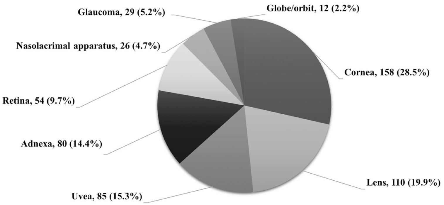
Prevalence rates of ophthalmic disorders according to the anatomical location in cats. Data are shown as number of disorders out of 554 (%)
There were 48 unique diagnoses. The 10 most prevalent disorders among all breeds were, in increasing order of prevalence, corneal ulcerations, uveitis, incipient cataract, keratitis, secondary glaucoma, sequestrum, retinal degeneration, entropion, conjunctivitis and micropuncta (Figure 4).

Number of disorders and prevalence rates (%) of the 10 most commonly diagnosed ophthalmic diseases in this study
Corneal disorders
The prevalence rate of corneal disorders was 28.5%, including corneal ulceration, keratitis, sequestrum, corneal perforation, corneal deposit, acute bullous keratopathy and feline eosinophilic keratitis, in descending order (Figure 5, Table 1). Laterality and age at the time of diagnosis of corneal disorders are provided in Table 2.

Representative images of corneal disorders in cats: (a) corneal ulceration; (b) corneal keratitis; (c) corneal sequestrum; (d) corneal perforation; (e) corneal deposit (arrows); (f) acute bullous keratopathy; and (g) feline eosinophilic keratitis
Total and subtotal prevalence rates of 554 ophthalmic disorders in 180 cats in this study
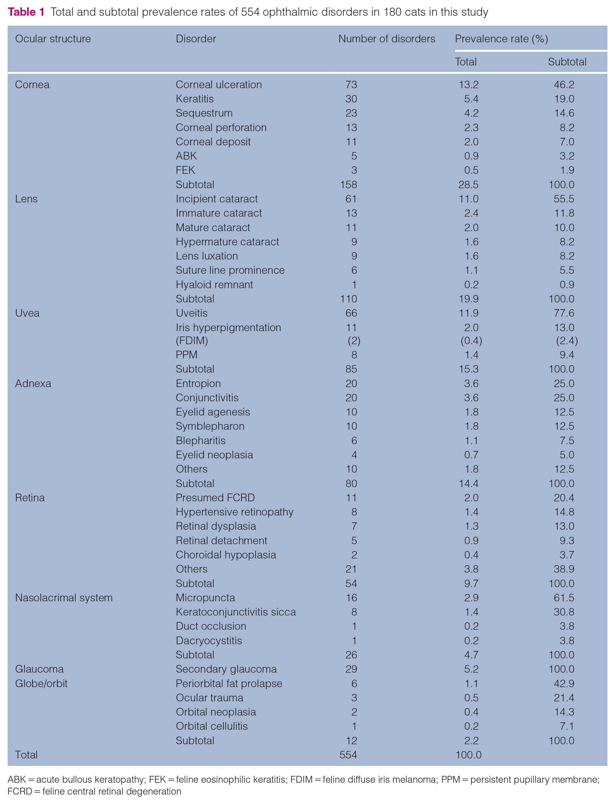
ABK = acute bullous keratopathy; FEK = feline eosinophilic keratitis; FDIM = feline diffuse iris melanoma; PPM = persistent pupillary membrane; FCRD = feline central retinal degeneration
Laterality and age at the time of diagnosis of 554 ophthalmic disorders in 180 cats in this study

Data are n (%) unless otherwise specified
ABK = acute bullous keratopathy; FEK = feline eosinophilic keratitis; PPM = persistent pupillary membrane; FCRD = feline central retinal degeneration
Corneal ulceration (73 eyes) was associated with sequestrum (19.2%), corneal deposit (13.7%), entropion (5.4%), eyelid agenesis (5.4%) and eosinophilic keratitis (2.7%). Brachycephalic breed cats were most frequently affected (n = 13; 68.4%) with sequestrum. Three of five cats with acute bullous keratopathy had a history of systemically administered prednisolone, while 2/5 had a history of glaucoma.
Lens disorders
The prevalence rate of lens disorders was 19.9%, and cataracts were identified in 17.0%, including incipient, immature, mature and hypermature cataracts, in descending order. Further ocular findings classified as cataracts included a suture line prominence and a hyaloid remnant. Lens luxation or subluxation was identified in 1.6% (Figure 6, Table 1). Laterality and age at the time of diagnosis of lens disorders are represented in Table 2. Two breeds with lens luxation or subluxation were represented: the domestic shorthair (DSH; 83.3%) and BSH (16.7%).

Representative images of lens disorders in cats: (a) incipient cataract; (b) immature cataract; (c) mature cataract; (d) hypermature cataract; and (e) lens luxation
Uveal disorders
The prevalence rate of uveal disorders was 15.3%, including uveitis, iris hyperpigmentation and persistent pupillary membrane, in descending order (Figure 7, Table 1). Laterality and age at the time of diagnosis of uveal disorders are provided in Table 2. Two of the 11 iris hyperpigmentations were histopathologically confirmed and identified as feline diffuse iris melanoma. Total and subtotal prevalence rates according to the classification of causes of anterior uveitis are provided in Table 3.
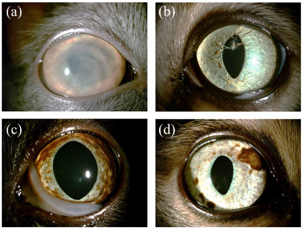
Representative images of uveal disorders in cats: (a) uveitis; (b) persistent pupillary membrane; (c) iris hyperpigmentation; and (d) iris hyperpigmentation identified as diffuse iris melanoma
Total and subtotal prevalence rates according to the classification of causes of 66 cases of anterior uveitis

Adnexal disorders
The prevalence rate of adnexa disorders was 14.4%, including entropion, conjunctivitis, eyelid agenesis, symblepharon, blepharitis, eyelid neoplasia and others, in descending order (Figure 8, Table 1). Details on laterality and age at the time of diagnosis of adnexal disorders are provided in Table 2.
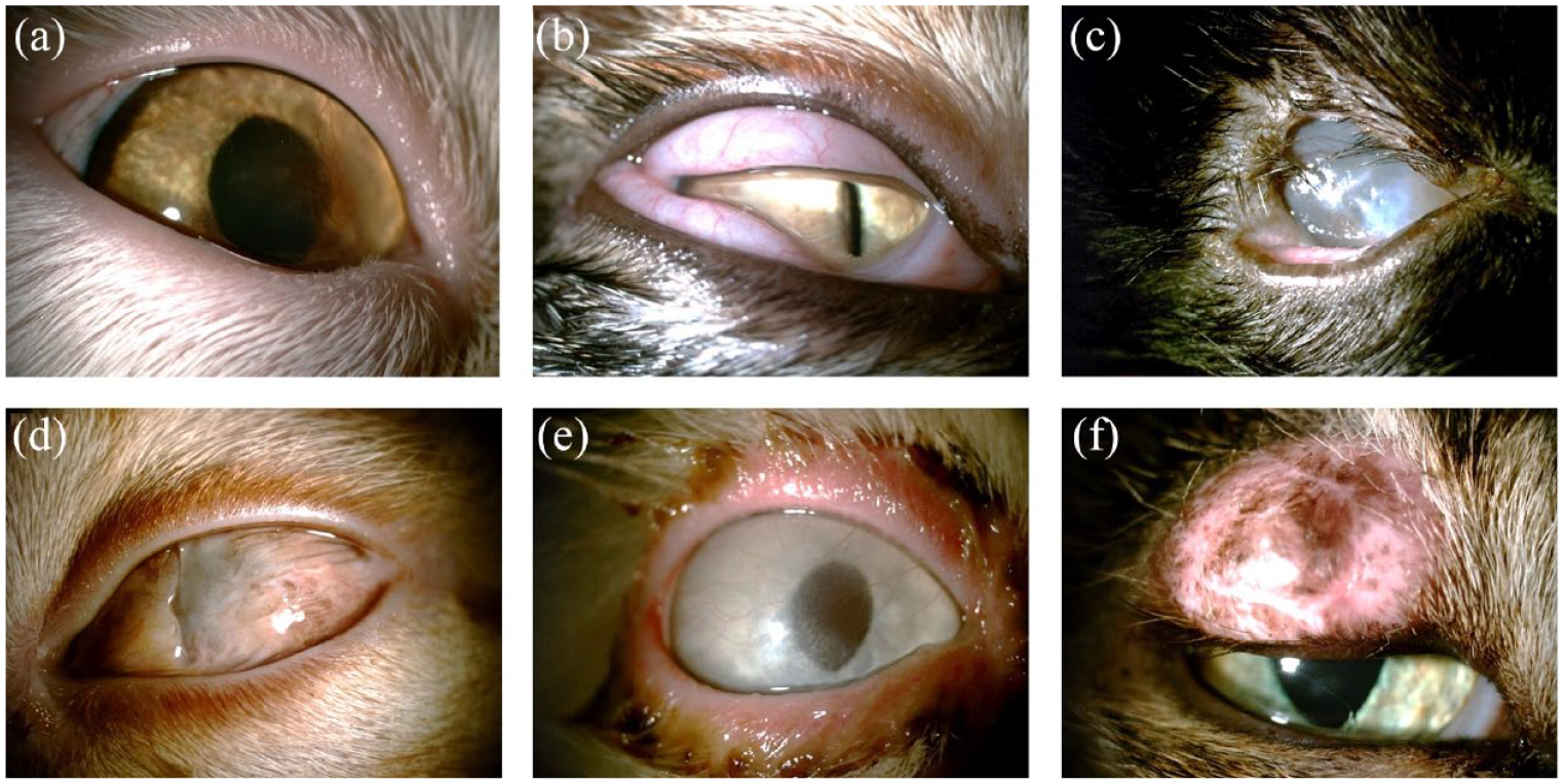
Representative images of adnexal disorders in cats: (a) entropion; (b) conjunctivitis; (c) eyelid agenesis; (d) symblepharon; (e) blepharitis; and (f) eyelid neoplasia
This study diagnosed ocular diseases as primary entropion, which develops at an early age due to facial conformation, and secondary entropion, resulting from chronic painful ocular diseases or in older cats with eyelid laxity or enophthalmos from reduced orbital tissue volume. Primary entropion was found in 15 eyes (2.7%). Brachycephalic cats were most frequently affected (12 eyes; 80.0%). Two breeds were over-represented – Persian (33.3%) and ESH (33.3%) – and a significant association was observed between primary entropion and ESH (n = 3/9; OR 16.6 [P <0.01]). Primary entropion was found medially and temporally in the lower eyelid of 13 (86.7%) and two (13.3%) eyes, respectively. In five eyes (0.9%), secondary entropion was found medially and temporally in the lower eyelid in two (40%) and three (60%) eyes, respectively.
The only affected breed with eyelid agenesis was the DSH. The temporal aspect of the superior lid was affected by eyelid agenesis in all 10 eyes. Two breeds were presented with symblepharon: DSH (85.7%) and Munchkin (14.3%). Eyelid neoplasia was found in four eyes: mast cell tumour (n = 3) and unknown (n = 1).
Retinal disorders
The prevalence rate of retinal disorders was 9.7%, including presumed feline central retinal degeneration (FCRD), hypertensive retinopathy, retinal dysplasia, retinal detachment and choroidal hypoplasia in descending order (Figure 9, Table 1). Other retinal diseases included retinal degeneration of unknown causes, such as post-inflammatory change. Details on laterality and age at the time of diagnosis of retinal disorders are provided in Table 2.

Representative images of retinal disorders in cats: (a) retinal dysplasia; (b) retinal detachment with hypertensive retinopathy; (c) generalised progressive retinal atrophy; and (d) other retinal degeneration
There was no clinical evidence of visual impairment in any cats with presumed FCRD. Of the 11 eyes, five were from street cats, and the remaining six were not evaluated. Two breeds presented with hypertensive retinopathy: Persian (50%) and DSH (50%). Four eyes (80%) with retinal detachment had hypertensive retinopathy. The most affected breed with retinal dysplasia was DSH (75%).
Nasolacrimal disorders
The prevalence rate of nasolacrimal disorders was 4.7%, including micropuncta with epiphora, keratoconjunctivitis sicca, nasolacrimal duct occlusion and dacryocystitis, in descending order (Figure 10, Table 1). Details on laterality and age at the time of diagnosis of nasolacrimal disorders are provided in Table 2. In this study, a micropunctum was defined as a smaller-than-normal punctum with epiphora.

Representative images of nasolacrimal disorders in cats: (a) epiphora and (b) keratoconjunctivitis sicca
Glaucoma
The overall prevalence of glaucoma was 5.2% (Figure 11, Table 1). Laterality and age at the time of diagnosis of glaucoma are represented in Table 2.

Representative images of glaucoma in cats: (a) secondary glaucoma with a Haab’s line (arrow) and (b) secondary glaucoma with buphthalmos
Glaucoma was determined to be secondary based on ocular lesions and primary based on the elevation of intraocular pressure without significant intraocular abnormalities other than those related to glaucoma. All eyes were classified as secondary glaucoma. The aetiologies of secondary glaucoma included anterior uveitis (n = 21; 3.8%), lens luxation (n = 4; 0.7%), uveal/orbital neoplasia (n = 3; 0.5%), including melanoma and lymphosarcoma, and aqueous humour misdirection (n = 1; 0.2%).
Orbital diseases and disorders
The prevalence rate of orbital diseases and disorders was 2.2%, including periorbital fat prolapse, ocular trauma, orbital neoplasia and orbital cellulitis, in descending order (Figure 12, Table 1). Details on laterality and age at the time of diagnosis of orbital disorders are provided in Table 2.

Representative images of orbital disorders in cats: (a) periorbital fat prolapse (asterisk); (b) ocular trauma (arrow); (c) orbital neoplasia (circle); and (d) orbital cellulitis with panuveitis and panophthalmitis
Two breeds had periorbital fat prolapse – ESH (66.7%) and BSH (33.3%) – and a significant association was found between them (n = 2/3 [OR 57.0; P <0.01]).
Discussion
There are few reports on the prevalence of feline ophthalmic diseases and epidemiological analyses in cats. This study investigated the frequency of ophthalmic diseases within a ophthalmic feline referral population in descending order, from the highest to the lowest frequency of occurrences in each anatomical location.
The highest number of patients had ophthalmic diseases at <1 year of age. The majority of cats lived strictly indoors owing to the highly urbanised environment. 14 In a recent study in Korea, only 6.5% of cats had outdoor access; 14 therefore, inherited or congenital diseases could outnumber secondary diseases. 9
The high prevalence of corneal disorders was consistent with a previous study, 15 which stated that persistent or recurrent corneal diseases in cats are a common reason for referral to a veterinary ophthalmologist. Corneal ulceration was the most prevalent ocular disorder (n = 73/554; 13.2%). Ulcerative keratitis was associated with corneal sequestrum, entropion, eyelid agenesis and eosinophilic keratitis in this study population, similar to a previous study. 16 In addition, in the present study, the corneal deposit was also associated with ulcerative keratitis. Primary infectious agents, particularly feline herpesvirus 1, are a much more common cause of corneal ulceration in cats.1,16,17 However, as this study did not systematically explore infectious diseases with PCR in all cats, herpetic origin could not be concluded as a disease entity.
Corneal sequestrum was the sixth most common ocular disease in this study, with a prevalence of 4.1% (n = 23/554). Although genetic or breed-related origins have also been suggested, this corneal disease may be acquired.1,18 In this study, brachycephalic breeds were most often affected (68.4%), which is similar to the rate of 54.7–78.4% reported in previous studies.5,6,18,19 Age at the time of diagnosis varied widely (0.8–11 years), with no specific age preference.
Incipient cataracts – the most common lens disease – was the third most common disorder, with a prevalence rate of 11.0%. Incipient cataracts were present in 61/101 eyes (60.4%), which is higher than the rate of 42.8% reported in a previous study in cats with cataracts. 7 However, similar to the previous study, prevalence decreased as cataracts matured. 7
Hyperpigmentation of the feline iris is a common clinical sign and can be caused by melanosis, diffuse iris melanoma and anterior uveitis. In this study, histopathological diagnosis was achieved in only two eyes that were enucleated due to secondary glaucoma.
Among adnexal diseases, the most common disease was entropion. Primary entropion is an inward turning of the eyelid that leads to trichiasis and develops at an early age owing to facial conformation.3,20 Primary entropion is common in Persians and other brachycephalic breeds, and it usually affects the medial aspect of the lower eyelid.1,20–22 Entropion in ESHs has also been reported,3,5,22 and the present study showed a significant association between ESHs and primary entropion. The prevalence rate of primary entropion (2.7%) was similar to that reported in a previous study (2.2%). 5
Eyelid agenesis has been previously reported in DSH,23,24 Burmese 25 and Persian 26 breeds. In the present study, all affected cats with eyelid agenesis were DSHs. Consistent with published data,1,20 the temporal aspect of the superior lid only was affected (100%) in this study, and lesions were usually bilateral (80.0%). The median age at diagnosis was 8 months, and the age of all affected cats was <1 year in this study.
In this study, the retina was the fifth most affected anatomical location. Furthermore, presumed FCRD accounted for 2.2% (n = 12/554) of the population. The prevalence of FCRD has dramatically reduced since the recognition of the role of taurine in its pathogenesis, and administration of taurine may prevent the progression of the retinal disease and improve rod function.17,27 In the present study, all cats with presumed FCRD were affected bilaterally, which was also the case in a previous study by Bellhorn et al. 28 The cats with presumed FCRD in the present study had no visual impairment.
Of the nasolacrimal diseases, the most common was micropuncta with epiphora. Micropuncta is a smaller-than-normal punctum, of which diagnosis is made by biomicroscopic examination of the punctum and nasolacrimal flushing. 17 However, feline lacrimal puncta are more circular and smaller than those of dogs, and more difficult to cannulate. 29 Therefore, cats diagnosed with epiphora and smaller puncta were classified as having micropuncta with epiphora. Nasolacrimal disorders are uncommon in cats and are usually characterised by epiphora. 30 Furthermore, in brachycephalic breeds, conformational features such as medial entropion, altered nasolacrimal course, a shallow lacrimal lake and tight apposition of the eyelid against the eye may contribute to epiphora. 1 In this study, epiphora and medial canthal entropion were found simultaneously in six eyes, four of them in brachycephalic breeds.
Although glaucoma was identified in only 29 eyes, secondary glaucoma was the fifth most common disease (prevalence rate 5.2%). Glaucoma in cats is less common than that in dogs. 1 In this study, all cases of feline glaucoma appeared to be secondary, consistent with previous studies, which reported that 87–95% of feline glaucoma cases were associated with other ocular or systemic disease processes, including uveitis, neoplasia, trauma and intraocular haemorrhage.12,13 The present study showed that anterior uveitis was the most common cause of feline secondary glaucoma.
The least commonly affected anatomical location was the orbit, including periorbital fat prolapse. In a previous study, orbital fat prolapse was reported bilaterally in Persians. 31 In the present study, all cats were affected bilaterally and ESHs were significantly associated with periorbital fat prolapse.
A limitation of this study was its retrospective nature, as the data available were limited to clinical records. As a systemic examination was not performed for all cats, analysis of anterior uveitis was also limited. Therefore, the aetiology of endogenous uveitis was only classified into idiopathic, infectious, immune-mediated, metabolic and neoplastic. The lack of diagnostic tests for FCRD, including serum taurine levels or histopathological examination, was a drawback; therefore, the suspected lesions with FCRD were named ‘presumed FCRD’. In addition, the low number of cats in each subpopulation might have affected the statistical power of the analyses.
Conclusions
This retrospective study described the prevalence, epidemiology and clinical presentation of feline ophthalmic diseases presented over a 13-year period at a referral centre in South Korea. The most common breed was DSH, and the most affected group was those aged <1 year. The cornea, lens and uvea were the most affected ocular structures presenting disorders. These findings suggest that the most prevalent disorders in this ophthalmic referral cat population were corneal ulceration, uveitis, incipient cataract, keratitis, secondary glaucoma and sequestrum. Breed over-representation was observed in entropion, eyelid agenesis and periorbital fat prolapse. This study provides epidemiological information for feline ophthalmic diseases, which could facilitate the diagnosis, treatment and prevention of feline eye diseases.
Footnotes
Conflict of Interest
The authors declared no potential conflicts of interest with respect to the research, authorship, and/or publication of this article.
Funding
This study was supported by the BK21 FOUR Future Veterinary Medicine Leading Education and Research Center, Research Institute for Veterinary Science (RIVS), College of Veterinary Medicine, Seoul National University, Seoul 08826, Republic of Korea.
Ethical approval
The work described in this manuscript involved the use of non-experimental (owned or unowned) animals. Established internationally recognised high standards (‘best practice’) of veterinary clinical care for the individual patient were always followed and/or this work involved the use of cadavers. Ethical approval from a committee was therefore not specifically required for publication in JFMS. Although not required, where ethical approval was still obtained, it is stated in the manuscript.
Informed consent
Informed consent (verbal or written) was obtained from the owner or legal custodian of all animal(s) described in this work (experimental or non-experimental animals, including cadavers) for all procedure(s) undertaken (prospective or retrospective studies). No animals or people are identifiable within this publication, and therefore additional informed consent for publication was not required.