
Abstract
Objectives
Thelaziosis caused by the nematode Thelazia callipaeda (Spirurida, Thelaziidae) affects the eyes of domestic and wild carnivores, lagomorphs and even humans. The role of cats in spreading thelaziosis remains unclear. The present study assesses the current status of feline T callipaeda infection in Spain and presents the results of the first prophylactic trial conducted in this species.
Methods
We retrospectively analysed the occurrence of feline thelaziosis (study 1), examined its prevalence based on cross-sectional sampling of endemic areas (study 2), and assessed the therapeutic/prophylactic benefits of a spot-on (moxidectin) and therapeutic efficacy of an eye drop (ivermectin) formulation (study 3).
Results
In study 1, 69 Thelazia-infected cats were examined. Autochthonous cases were detected in 18/26 municipalities surveyed in Spain, which corresponds to 88.4% (n = 61/69) of the total number of cases. In study 2, 74 cats (20 from La Vera region and 54 from Orense province) were examined from 2011 to 2013. Ten of these cats (13.5%) were infected with T callipaeda. The infection prevalence was 40% (n = 8/20) in La Vera and 3.7% (n = 2/54) in Orense. The therapeutic efficacy of ivermectin was assessed over a summer (2011) in 12 cats, while that of moxidectin was examined over a 17-month period in seven cats. Four of these seven cats were subsequently included in an all-year-round prophylaxis trial. Treatment efficacy ranged from 91.7% (ivermectin) on day 28 after treatment to 100% (moxidectin) on day 14. Moxidectin prevented reinfections in cats.
Conclusions and relevance
Our data confirm the endemic status of T callipaeda infection in Spain, revealing its high prevalence in cats living in known endemic areas. Moxidectin was effective in treating and preventing infection. We strongly recommend the inclusion of thelaziosis in the differential diagnosis of pets and humans presenting with ocular manifestations, along with adequate preventive measures.
Introduction
Thelaziosis is an emerging arthropod-borne disease caused by Thelazia callipaeda (Spirurida, Thelaziidae), a nematode that, at both adult and larval stages, infects the eyes of domestic (ie, dogs and cats) and wild (eg, red foxes, wolves, beech martens, wild cats) carnivores,1,2 lagomorphs (rabbits, brown hares)1,3 and even humans.4–7 T callipaeda was known as ‘the oriental eyeworm’, as it was thought to be confined to Asian countries, including India, Thailand, China and Japan. 8 However, following the first reports of T callipaeda infection in dogs in Italy,2,9 the distribution of this ‘oriental eyeworm’ has since extended to larger parts of Europe, with enzootic areas in southern Europe. 10
In Spain, the first cases of canine thelaziosis were detected in 2010.11,12 Since initial reports of this infection in dogs, its prevalence has been increasing dramatically in many regions of mainland Spain. Today, Spain is endemic for canine thelaziosis as reports exist of autochthonous cases in 86 municipalities belonging to 19 provinces. 13
Sporadic cases have been described in other animal species (eg, red foxes or grey wolves)14–16 and even in humans.4–7
Clinical cases of feline thelaziosis have been detected in endemic areas for dogs in the majority of cases.17–21 Nevertheless, some authors consider that the epidemiological role of cats in the spread of thelaziosis could be only seemingly less important than the role of dogs.20,22 To treat this eyeworm infection, the combination of milbemycin oxime/praziquantel has proved effective in reducing infection rates in naturally infected dogs and cats. 23 In addition, a moxidectin 1.0% w/v plus imidacloprid 10% w/v spot-on formulation for cats has recently been shown to be highly effective for treating feline thelaziosis. 24 To date, however, no prophylactic efficacy studies have been carried out in this species.
The present study sought to improve knowledge of the treatment and prevention of feline thelaziosis. To this end, we examined the current status of T callipaeda infection in cats in Spain and assessed the therapeutic and prophylactic efficacy of a moxidectin-based spot-on formulation and the therapeutic efficacy of an ivermectin-based eye drop formulation. In addition, we reviewed the literature to identify reports of feline thelaziosis in other countries.
Materials and methods
Study design
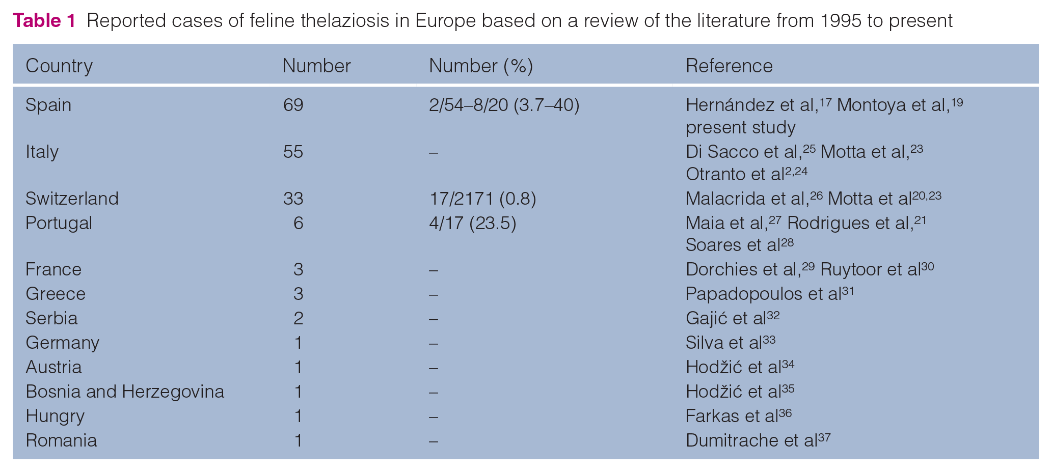
Established internationally recognised high standards (‘best practice’) of individual veterinary clinical patient care were followed in accordance with international guidelines for the Care and Use of Experimental Animals and Spanish Legislation (RD 53/2013). Three studies were conducted. Study 1 involved a survey of clinical cases of feline thelaziosis based on data from clinical records collected from veterinary clinics across Spain. Study 2 was a prevalence study based on cross-sectional sampling of endemic areas. Study 3 was a therapeutic and prophylactic trial to evaluate the clinical efficacy and safety of two antiparasitic formulations. The formulations tested were a spot-on formulation of 100 mg/ml imidacloprid plus 25 mg/ml moxidectin, and an eye-drop formulation (6 µg) of ivermectin 10 mg/ml diluted 10% in propylene glycol. In addition, the literature was reviewed to identify cases of feline thelaziosis in other countries (Table 1).
Reported cases of feline thelaziosis in Europe based on a review of the literature from 1995 to present
Whenever possible, a data collection sheet was completed for each cat in all three studies by the authors or participating veterinarians. Data recorded were date, geographical location, travel history, sex, age, weight, presence of T callipaeda parasites, number of Thelazia specimens, ocular signs and use of preventive measures against T callipaeda infection.
Participating cats were examined after the administration of anaesthetic eye drops (1 mg/ml tetracaine hydrochloride and 4 mg/ml oxibuprocaine hydrochloride). T callipaeda eyeworms were collected from the conjunctival sac of infected cats using sterile cotton swabs or by flushing with physiological saline solution (Figure 1). Nematodes were identified at our laboratory using morphological keys available in the literature. 38
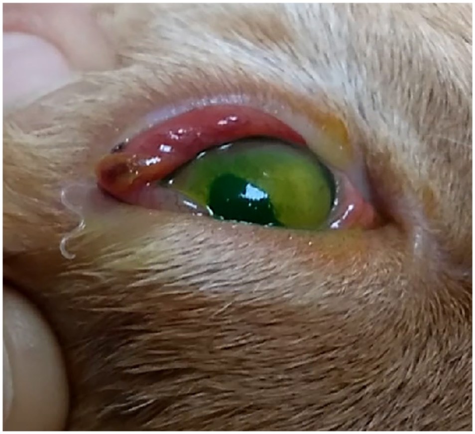
Feline thelaziosis due to Thelazia callipaeda in a domestic cat
Amplification of the mitochondrial cytochrome c oxidase subunit 1 gene was not performed as haplotype 1 is the only one detected in domestic and wild animals in Europe, 39 and this haplotype has already been identified in domestic dogs, domestic cats, red foxes, grey wolves and humans in Spain.5,12,16,17,19
Study area
Cats were enrolled from three different sites (Figure 2).
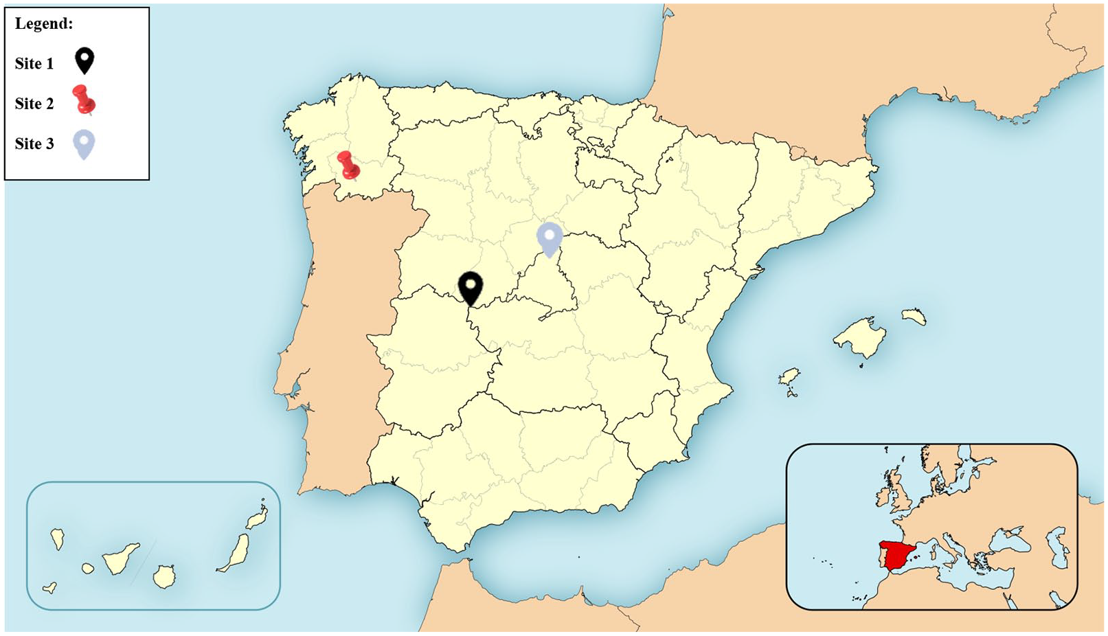
Locations of the three study sites. Site 1: La Vera region and Candeleda municipality (central-western Spain); site 2: Orense province (north-western Spain); site 3: Miraflores de la Sierra municipality (north-west Madrid Community)
Sampling site 1
La Vera region (northern Cáceres Province, 40°9’41"N, 5°23’13"W) and Candeleda municipality (Avila province, 40°9’11", 5°14’29"W) in central-western Spain; altitude 472 m and 424 m, respectively, above sea level (masl). At this site, reported prevalences of T callipaeda in dogs were 40%, 68% and 61% in 2011, 12 201840 and 2020, 13 respectively.
Sampling site 2
Orense (42°22’37"N, 8°02’05"W) in north-western Spain; altitude 914 masl. Here, the prevalence of the infection in dogs is unknown, but our research group reported 135 autochthonous cases in the entire province between 2010 and 2018. 13
Sampling site 3
Miraflores de la Sierra municipality (north-west Madrid Community); Sierra de Guadarrama (40°48’54’’N, 3°46’15’’W); altitude 1147 masl. Reported prevalences of T callipaeda in dogs at this site were 33% and 30% in 201840 and 2020, 13 respectively.
All sampling sites
All areas are characterised by mountains with valleys and river streams and vegetation mainly consisting of oak woods. 41 The mean temperature recorded from May to October ranged from 16°C to 27°C, and average annual rainfall was 550 mm at site 1, 811 mm at site 2 and 428 mm at site 3.42–44
Enrolment and follow-up
Study 1: survey of clinical cases
Veterinarians working in clinics throughout Spain (n = 23) participated in this study by filling in the data sheet described above for any cases of feline T callipaeda infection detected from October 2010 to March 2020.
Study 1: statistical analysis
Data from all cats were subjected to statistical analysis. The variables recorded were: date, geographical location, sex, age, number of Thelazia specimens, ocular signs, use of preventive measures against T callipaeda infection and travel history. The population sample was described in terms of distributions of continuous and categorical variables through their means, SD, median, interquartile ranges and percentages. Associations between the number of T callipaeda specimens present and the other epidemiological variables (autochthonous cases, sex, living outdoors, positive cases obtained by sampling in study 2, ocular signs and use of preventive measures against T callipaeda infection) were assessed using the Wilcoxon test, except for ocular signs for which we used the Kruskal–Wallis test.
Study 2: prevalence study in endemic areas
Cats were recruited from two endemic areas of Spain where cases of thelaziosis in dogs have been previously reported. The sampling sites were: site 1, where a population of 20 owned cats living outdoors was examined in situ in 2011; and site 2, where a population of 54 cats housed in a shelter and living outdoors was examined in situ in 2013 (bioclimatic data indicated above).
Study 2: statistical analysis
The variables recorded in the descriptive study were: geographical location, travel history, weight, sex, age, presence of parasites, ocular signs and use of preventive measures against T callipaeda infection. Correlations between T callipaeda eye infection and epidemiological variables (geographic location, weight, sex and age) were analysed by Fisher’s exact test for categorical variables (geographical location and gender) and Student’s t-test for numerical variables (weight and age).
Study 3: therapeutic and prophylactic trial
In study 3, two populations of cats with thelaziosis were recruited from two sites in the Iberian Peninsula: site 1 and site 3 (bioclimatic data indicated above). Cases were enrolled through local veterinary practitioners. All animals were domestic cats and had access to the outdoors.
At site 1 (La Vera), the therapeutic efficacy of an eye drop formulation (6 µg) of ivermectin 10 mg/ml diluted 10% in propylene glycol was assessed over the summer of 2011 in 12 cats.
At site 3 (Miraflores de la Sierra), the therapeutic efficacy of a spot-on formulation of 100 mg/ml of imidacloprid plus 25 mg/ml moxidectin was assessed over a 17-month period (November 2016 to April 2018) in seven cats. Four of these seven cats were also included in a 1-year-long prophylaxis trial.
During the therapeutic/prophylactic trial each cat was examined and searched for T callipaeda infection.
Inclusion criteria were: (1) age at least 9 weeks; (2) body weight >1 kg; (3) living permanently outdoors; and (4) (a) for inclusion in the therapeutic trial, cats had to be infected with a minimum of one worm and a maximum of 50 at enrolment; (b) for inclusion in the prophylaxis trial, cats had to have taken part in the therapeutic trial and tested negative for T callipaeda infection in at least two subsequent visits.
Exclusion criteria were: (1) pregnant and/or lactating females; (2) cats showing a poor health state or chronic diseases; (3) cats whose owners decided to withdraw them from the study; or (4) deviations produced in the protocol.
Cats enrolled in the therapeutic trial were treated at visit 1 (V1) and checked after 14 ± 4 days (visit 2 [V2]) (only cats treated with moxidectin) and again after 28 ± 4 days for the presence of T callipaeda parasites.
Cats enrolled in the prophylaxis trial were treated on a monthly basis all year round and seen at a total of 14 follow-up visits.
Study 3: statistical analysis
In a descriptive study, we analysed the variables recorded (geographical location, travel history, weight, sex, age, number of parasites, ocular signs and use of preventive measures against T callipaeda infection).
The main outcome measure was treatment efficacy. This was determined by comparing T callipaeda infection prevalences between V1 (day of diagnosis) and the remaining follow-up visits within the same treatment group (intragroup) and between the two treatment groups (intergroup).
Treatment efficacy was calculated as the percentage of cats that did not show the presence of ocular nematodes after treatment according to the following formula:

The prophylactic efficacy of the moxidectin spot-on was calculated as the percentage of cats that did not become re-infected during the trial according to the equation:

To examine the relationship between treatment and the categorical variables (geographical location, sex, ocular signs and use of preventive measures against T callipaeda infection) or numerical variables (weight, age and number of parasites), we used Fisher’s exact test or the Wilcoxon signed-rank test, respectively.
All statistical tests were performed using the Stata v. 15.0 package (Stata Corp). Significance was set at P ⩽0.05.
Clinical staging
In all three studies, each cat was clinically assessed for T callipaeda infection using a clinical score grading ocular signs designed by a veterinary ophthalmologist (MH). In cats in which the nematode was detected, infection was classified as stage 1 (no clinical signs), stage 2 (mild conjunctivitis, ocular discharge or blepharitis) and stage 3 (severe conjunctivitis, keratitis, chemosis and/or purulent ocular discharge) (Figure 3).

Clinical staging of thelaziosis in cats. Stage 1: absence of ocular signs; stage 2: mild conjunctivitis; stage 3: complicated follicular conjunctivitis, chemosis and/or purulent ocular discharge
Literature review
Systematic literature searches were conducted on Scopus and Web of Science as comprehensive databases likely to provide accurate information on Thelazia-related research. The main search heading used was T callipaeda in cats. Searches were limited to European countries and included publications from 1995 to present (Table 1).
Results
Reports of Thelazia callipaeda eye infection in cats in Spain (study 1)
We analysed data recorded in 69 T callipaeda-infected cats from the retrospective analysis (study 1) (n = 59) and the active search in the prevalence study (study 2) (n = 10) in collaboration with the veterinarians and animal owners. Figure 4 provides details of the number of cats testing positive for the eyeworm each year.

Number of cases of Thelazia callipaeda infection recorded in cats in each year. Note that cross-sectional sampling took place in the years 2011 at site 1 and 2013 at site 2
Using this information, we mapped the geographical locations and trends in the cases of feline thelaziosis detected in Spain (Figure 5). The corresponding sites of all 69 cases were 26 municipalities of Spain. Autochthonous cases were detected in 18/26 municipalities surveyed in Spain, which corresponds to 88.4% (n = 61/69) of the total number of cases. Eight of 69 cases had a history of recent travel to/from another part of the peninsula.

Thelazia callipaeda status in cats in Spain based on cumulative data from 2011 to 2020
The 18 municipalities in which autochthonous cases of feline thelaziosis were detected (shown in Figure 5) belong to eight Spanish provinces located in central regions (Avila, Caceres, Madrid, Segovia and Toledo) and the north-west (León, Orense and Pontevedra). While several features of the positive cats (n = 69) were recorded, total numbers may vary owing to some missing data. The ratio between males and females was 20 males (46.5%) to 23 females (53.5%). Mean weight (n = 32) was 3.58 ± 0.85 kg and mean age (n = 34) was 4 ± 3.6 years. One of the main characteristics of the population was that 94.0% (n = 47/50) of the cats had not received preventive treatment against Thelazia infection. Moreover, 98% (n = 48/49) of the cats had access to the outdoors.
In the ocular examination, the mean number of parasites (on 23 cats) identified was 11.48 ± 14.77, with a minimum of one parasite (n = 4) and a maximum of 49 (n = 2). The most frequently observed ocular signs were stage 2 infection (n = 26/44 [59.1%]) followed by stage 1 (n = 13/44 [29.6%]) and finally stage 3 (n = 5/44 [11.4%]). Correlations identified in a bivariate analysis between the number of T callipaeda parasites and the variables sex and sampled vs non-sampled cats from study 2 were significant at P = 0.03 and P = 0.003, respectively (Table 2). The relationship between ocular staging and number of parasites is reported in Figure 6, although, as indicated in Table 2, a significant relationship (P = 0.53) between these two variables could not be demonstrated due to the low number of cases.
Association between the number of Thelazia callipaeda parasites and epidemiological variables recorded

IQR = interquartile range

Relationship between ocular stage and mean number of Thelazia callipaeda parasites
Prevalence study (study 2)
Over the period 2011–2013, of 74 cats (20 from La Vera region and 54 from Orense) examined, 10 (13.5%) were found to be infected with T callipaeda.
Thelazia infection prevalences were 40% (n = 8/20) for site 1 and 3.7% (n = 2/54) for site 2. This indicates a significantly higher risk of infection for cats from site 1 (odds ratio [OR] 17.33, 95% confidence interval [CI] 3.26–92.24; P <0.0001) compared with site 2. The demographic characteristics of these cats from site 1 were mean weight 3.17 ± 0.72 kg and age 1.04 ± 0.50 years. No significant differences were found between these variables at the two sites (P = 0.59 and P = 0.16). Both sexes were represented: seven females (35.0%) and 13 males (65.0%). Five of seven (71.4%) females and 3/13 (23.1%) males were positive, indicating a significantly higher risk for female cats (P = 0.04). These features could not be analysed for site 2 owing to a lack of information (weight, sex and age) from the shelter association. At both sites, cats had access to the outdoors.
Therapeutic/prophylactic trial (study 3)
Data from the cats enrolled in the therapeutic trial are provided in Table 3.
Data recorded in the cats enrolled in the therapeutic study by treatment group

Data are n (%) unless otherwise indicated
IQR = interquartile range; V = visit
Treatment efficacy ranged from a minimum of 91.7% recorded for site 1 (ivermectin eye drops) on day 28 after treatment to 100% recorded for site 3 (moxidectin spot-on) on day 14 (V2) (Figure 7a).

(a) Efficacy data recorded in the different treatment groups. Ivermectin group: eye drop formulation (6 µg) of ivermectin 10 mg/ml diluted 10% in propylene glycol. Moxidectin group: spot-on formulation of 100 mg/ml of imidacloprid plus 25 mg/ml moxidectin. (b) Changes produced in ocular signs in cats infected with Thelazia callipaeda treated with a moxidectin-based spot-on formulation. There were no stage 3 cats in the study group. D = day
At site 1, all the cats had ocular signs, mainly mild conjunctivitis (stage 2) and only one cat presented with keratitis (stage 3). At site 3, of the seven positive cases of feline thelaziosis diagnosed, more than half were asymptomatic (stage 1) (n = 4 [57.1%]) at the time of diagnosis and were presented to the clinic for different reasons (vaccinations, deworming or regular check-ups). Thus, the detection of parasites was an incidental finding in a routine ophthalmological examination. In the other three cases (42.9%), the signs observed were mainly conjunctivitis, ocular discharge, pruritus and blepharitis (stage 2). More severe lesions such as keratitis or corneal ulcers were not seen in any cat from site 3. The mean number of eyeworms detected was 15.1 ± 14.79 and, despite the difference between mean numbers in each group (Table 3), this was not significant (P = 0.09). In relation to breed, all cats were non-pedigree domestic cats. All of the cats had access to the outdoors and, therefore, vector exposure was greater. None of the diagnosed positive cats had a history of having travelled to other areas considered endemic for thelaziosis. All treatments were well tolerated by all of the cats.
The prophylactic efficacy recorded was 100%. The spot-on formulation of 100 mg/ml imidacloprid and 10 mg/ml moxidectin applied monthly all year round (in 14 scheduled visits) prevented infection recurrence in all four cats included (Figure 7a). No adverse effects were observed during follow-up.
Finally, ocular signs improved in the different visits in the moxidectin treatment group, indicating a relationship between the presence of parasites and the severity of the ocular signs. In effect, 85.7% of cats had no clinical signs 28 days after treatment and 100% had no clinical signs on day 56 after treatment (Figure 7b).
Discussion
This is the first epidemiological study to examine the prevalence of T callipaeda infection among cats in Spain and provide useful information on the treatment and prevention of feline thelaziosis.
Here, we report 69 new cases of the infection in cats in 26 municipalities, of which 61 are autochthonous to 18 municipalities of eight Spanish provinces.
The mean number of parasites detected in cats testing positive for the eyeworm was 11.5 ± 14.8, being lower in non-autochthonous cats. This parasite number is higher than the figures reported in other studies carried out in cats. Motta et al 20 reported only one cat harbouring more than 10 eyeworms in the same eye, and Maia et al 27 described that the number of worms per cat ranged from 1 to 14 (4.3 ± 6.5). Such a high number of nematodes may be related to the endemic status of many regions of Spain. Notably, the vast majority of cats (98%) examined here were outdoor cats living in a rural area characterised by a vegetation mainly represented by oaks and by wildlife species richness. This is the typical environment required by the parasite to complete its life cycle. Another important aspect was that 94.0% (n = 47/50) of the cats had not received preventive treatment against Thelazia infection, which is, in fact, a known risk factor in dogs (OR 0.22; P <0.0040). 13 A higher prevalence of the eyeworm was observed in females yet this finding is of negligible epidemiological relevance (considering the few number of cases). The most frequently observed ocular sign was stage 2 infection (mild conjunctivitis) (n = 26/44 [59.1%]), followed by stage 1 (no ocular signs) (n = 16/44 [36.4%]) and stage 3 (complicated conjunctivitis) (n = 5/44 [11.4%]), in agreement with a previous study carried out in 17 Thelazia-positive cats 20 as the study including the largest number of cats so far. In our study, cats with more serious ocular signs had a higher average number of parasites.
The prevalence of T callipaeda detected here was 13.5%: 40% for site 1 and 3.7% for site 2. The populations from both sites of the prevalence study (study 2) were outdoor cats. Unfortunately, epidemiological variables were only known for cats from site 1, precluding our analysis of the whole population. Differences in prevalences between the two sites were significant (OR 17.33, 95% CI 3.26–92.24; P <0.0001). Such a high number of positive cats at site 1 reflects the high endemicity of thelaziosis in this region. 40
Despite cats being considered a suitable primary host for T callipaeda, 39 to our knowledge, only two prevalence studies have been conducted in cats in Europe: one in Switzerland and another in Portugal with reported prevalences of 0.8% (17/2171 examined cats) and 23.5% (4/17 examined cats), respectively.20,27 All other studies have mostly reported isolated single cases, one of which was a cat imported from southern Spain. 33 The rare reports of this infection in cats along with its low prevalence in this species seem attributable to the intensive self-grooming habits and small body mass index of cats, which seems to make them less attractive for Phortica flies. 20 In addition, as cats have nocturnal habits, we propose their reduced exposure to the vector, which will be most active when the temperature is higher during the day. 45 However, it is also true that the prevalence rate of the infection in cats is likely highly underestimated as an in depth ocular examination and lifting of the third eyelid in cats normally requires sedation of the animal.2,20,26 Further, in early infection stages and in those infections caused by a low number of parasites, hosts often do not display clinical signs and the disease may therefore go unnoticed by owners and veterinarians.12,20,26,27 While under-diagnosis could be a cause of lower prevalences in cats, this was not the case in our cross-sectional study.
The typical candidate for feline thelaziosis is a cat that lives mainly outdoors in an area with fruit trees or oaks where there are also dogs (which, as already discussed, are more frequently infected).
Milbemycin and moxidectin are currently registered for use against canine ocular thelaziosis in Europe,23,24,46 and moxidectin has been also recently registered for use against feline thelaziosis. The regular use of moxidectin or milbemycin in endemic areas is highly recommended, especially in dogs.23,24,46–52
In this study, we assessed the therapeutic efficacy of two formulations (ivermectin and moxidectin) and the prophylactic efficacy of the formulation based on moxidectin in cats from areas endemic for canine thelaziosis in Spain. Both products proved effective for the treatment of cats infected with T callipaeda. Efficacy rates ranged from a minimum of 91.7% recorded for site 1 (ivermectin eye drops) on day 28 after treatment, and a maximum of 100% recorded for site 3 on day 14 (V2) (moxidectin + imidacloprid spot-on). The use of moxidectin to treat canine thelaziosis has been previously reported as effective in dogs.46,47,51–53 In cats, the therapeutic efficacy of a moxidectin 1.0% (w/v) and imidacloprid 10% (w/v) spot-on solution against natural infections of T callipaeda was recently analysed in 30 cats in Italy. 24 Efficacy was similar to that observed here and was 93.3% on day 14 and 100% on day 28. In the present study, we confirmed the efficacy of this formulation and report new data on its prophylactic use in cats. Hence, the monthly use of this combination was found to prevent thelaziosis in an endemic area.
In 31 naturally infected cats from Italy and Switzerland, the efficacy of milbemycin oxime/praziquantel tablets was explored. Of the 31 treated cats, 53.3% and 73.3% were free of T callipaeda on day 7 and day 14, respectively. 23 We also observed the efficacy of a combination of 2 mg/kg milbemycin oxime/praziquantel in a single case of a cat from Ourense diagnosed with T callipaeda infection at the Complutense Veterinary Hospital in Madrid. Two weeks after treatment, the animal showed the absence of the parasite. 54 No more studies are available for feline thelaziosis. Because of this, we also report the results of a study in which we tested an eye drop formulation (6 µg) of ivermectin 10 mg/ml diluted 10% in propylene glycol in 12 naturally infected cats in Spain. Of the 12 cats included in the study, 93.3% were free of T callipaeda parasites after 28 days of treatment. The use of ivermectin has been shown effective against canine thelaziosis.53,55–58 However, we do not recommend the off-label use of ocular ivermectin as it may provoke adverse reactions such as pruritus, irritation and redness in some animals. 59
Cats that live in endemic areas or that will be travelling to such areas need to be adequately prophylactically treated for thelaziosis. Products showing therapeutic and prophylactic efficacy against T callipaeda infection in cats need to be tested in large controlled studies. Considering that in endemic areas the prevalence of the infection may be as high as 68% among dogs 40 and 40% in cats, regular preventive measures for dogs and cats is essential to control the infection and minimise possible public health impacts.
To detect subclinical cases, especially in cats that live or have travelled to areas endemic for canine thelaziosis, an exhaustive ophthalmological examination should also be routinely performed. A lack of prophylactic measures in carnivores could be a crucial factor explaining the recent rise in cases of thelaziosis. 23 An increasing awareness of thelaziosis among veterinarians, parasitologists and ophthalmologists has contributed to an increased number of reports in Europe.10,60
Conclusions
The data presented here confirm the endemic status of T callipaeda infection in Spain, revealing high prevalences in cats living in endemic areas. We strongly recommend including thelaziosis in the differential diagnosis of pets and humans presenting with ocular manifestations, along with the use of adequate preventive measures.
Footnotes
Acknowledgements
The authors thank the owners of the cats and the participating veterinarians for their valuable help with the enrolment and follow-up of clinical cases. We are also grateful to Dr Mauro Hernández (ophthalmologist from the Ocuvet Veterinary Clinic) for the ocular staging of the cats and to the staff of all the veterinary clinics for their collaboration, especially Marta Benito (Clínica Veterinaria Candeleda). No specific grant from funding agencies in the public, commercial or not-for-profit sectors was received for this research. The formulations used in the treatment study were provided by Bayer Health Care.
Accepted: 30 January 2021
Conflict of interest
The authors declared no potential conflicts of interest with respect to the research, authorship, and/or publication of this article. The authors have performed this study on the basis of their freedom of research.
Funding
The authors received no financial support for the research, authorship, and/or publication of this article.
Ethical approval
This work involved the use of non-experimental animals only (including owned and non-owned animals and data from prospective or retrospective studies). Established internationally recognised high standards (‘best practice’) of individual veterinary clinical patient care were followed. Ethical approval from a committee was therefore not necessarily required.
Informed consent
Informed consent (either verbal or written) was obtained from the owner or legal custodian of all animal(s) described in this work (either experimental or non-experimental animals) for the procedure(s) undertaken (either prospective or retrospective studies). No animals or humans are identifiable within this publication and therefore additional informed consent for publication was not required.