
Abstract
Objectives
The aim of this multicentre retrospective study was to review the clinical data, outcomes and histopathological features of cats that had been treated for ocular surface dermoids.
Methods
Thirteen cats from various private practices in France with a clinical diagnosis of ocular surface dermoid were included in the study.
Results
The mean age of the study population at the time of diagnosis was 5 months. There were nine males and four females. Three different breeds were domestic shorthair (n = 7), Birman (n = 4) and Havana Brown (n = 2). Two of the four Birmans were related (same sire). The two Havana Browns were also related (same sire). All of the dermoids were unilateral. Five of the dermoids were strictly conjunctival. Four affected both the conjunctiva and the cornea. Three affected both the conjunctiva and the eyelid, and one was strictly corneal. They were located in various positions: temporal (n = 9), inferonasal (n = 1), dorsonasal (n = 1) and dorsotemporal (n = 1). The last dermoid was heterogeneous and involved the nasal, dorsal and temporal quadrants. Concurrent eye diseases were observed in five patients: four cats exhibited associated eyelid agenesis and one cat exhibited persistent iris-to-iris pupillary membranes. Ten dermoids were surgically excised with no recurrences. Surgery was not performed for three cats: one cat died a few days after diagnosis and two cats were lost to follow-up after initial presentation.
Conclusions and relevance
Ocular surface dermoids are a rare condition in cats that can be treated successfully by surgical excision. Although our study reports only a small number of cases, the observation of ocular surface dermoids in two related cats in two different breeds indicates that genetic transmission is likely.
Introduction
A dermoid is a histologically normal cutaneous tissue that develops in an abnormal location during embryonic development.1,2 The composition of dermoids can vary and includes general characteristics of normal skin such as the epidermis, dermis, glandular tissue, fat tissue, hair, hair follicles and blood vessels.1–8 Ocular dermoids can form in a variety of locations, including the eyelids, conjunctiva, nictitating membrane and cornea.3,4 They have been reported in many species, including humans,9–17 dogs,5–8,18–23 cats,24–33 horses,34–37 cattle,38–43 rabbits,44,45 pigs, 46 guinea pigs,47,48 a camel, 49 a rat, 50 a parrot 51 and a deer. 52 In dogs, the most common form is conjunctivo-corneal dermoids at the limbus. 7 In cats, several studies have described lateral canthus dermoids,25–33 and one study has reported a dorsally located corneal dermoid. 24 Although cases of bilateral dermoids have been reported in the literature in dogs,8,18 cats,30,33 cattle,38–41 horses 37 and humans, 9 most dermoids are unilateral.
Dermoids are a developmental abnormality present at birth, although they are sometimes only identified as a result of secondary ocular irritation later in life. Clinical signs associated with an ocular dermoid are manifestations of ocular pain (blepharospasm, epiphora), corneal ulcerations, corneal pigmentation and keratitis.1–4,31
A genetic predisposition has been described in dogs (including Basset Hounds, Dachshunds, Dalmatians, Dobermans, German Shepherds, Golden Retrievers, Saint Bernards and Welsh Corgis),5–8,18–23 cattle (with a high prevalence in Herefords) 38 and cats (Birmans and Burmeses).25,27,30
The purpose of this study was to describe the clinical features, breed prevalence, association with other ocular abnormalities, treatment and histopathological findings of cats diagnosed with ocular dermoids at multiple private practices in France.
Materials and methods
The various private practices involved in the study were VetoOphtalmo at Bois-Guillaume, Pôle Santé Chanturgue Clinique Vétérinaire at Clermont-Ferrand, Clinique VPlus at Saint Germain en Laye, Centre Hospitalier Vétérinaire Saint Martin at Saint Martin Bellevue and the Small Animal Clinic, Université de Toulouse, ENVT at Toulouse. Cases were identified via electronic and picture databases at these institutions. All cases identified as ocular dermoids, in the various private practices involved in this study, were included in this study. The case screening period for the study and the total number of cats examined for ophthalmology during this period was recorded for every clinic. The following data were collected: signalment (breed, sex and age at time of diagnosis), the reason for consultation and the affected eye. The physical characteristics of the dermoids were also recorded: the location (central, nasal, temporal, ventral or dorsal), hairiness (subjectively described as short, intermediate or long), associated clinical signs, concurrent ophthalmic abnormalities, any treatments at the time of diagnosis, surgical procedures, histopathological findings and clinical outcomes.
Results
An overview of the patients included in our study is presented in Table 1. Ten of the 13 recorded patients were referral cases. The remaining three cases were presented as follows: case 7 was presented for emergency consultation at Clinique VPlus (Saint Germain en Laye); and cases 12 and 13 were first-line consultations at the ophthalmology unit of the Small Animal Clinic, Université de Toulouse, ENVT (Toulouse).
Patient overview

Dermoid hairiness: 1 = short; 2 = intermediate; 3 = long
M = male; F = female; DSH = domestic shorthair; OS = left eye; OD = right eye; PPMs = persistent pupillary membranes
Patient data
Thirteen cats were included in the study. Breeds included domestic shorthair (n = 7), Birman (n = 4) and Havana Brown (n = 2). Two of the four Birmans had the same sire but different mothers. Both of the Havana Browns were males and they also had the same sire but different mothers. Nine were males (69.2%) and four were females (30.8%). The cats were aged between 6 weeks and 9 years (mean 5 months) at the time of diagnosis. All but one of the cats were <1 year old.
The reason for the consultation was ‘hair in the eye’ for five of the patients and ‘an odd-looking eye’ in two. A ‘modified eyelid opening’ was reported for two of the patients. Three cats had a diagnosis of ocular dermoid, as determined by the treating veterinarian. The last dermoid was an incidental observation in a cat presenting with non-ocular disease (case 7).
Clinical data
The clinical data (number of cases per structure, case screening period of the study, total number of cats examined during the screening period, mean number of cats examined per year and proportion of overall ophthalmology cases the dermoids represented) are presented in Table 2.
Clinical data of the cats with dermoids that were included in the study

The case screening period of the study was extended from 6 to 12 years. The number of dermoids seen during this period ranged from one to five per clinic. The total number of cats examined per structure ranged between 600 (in 6 years) and 3000 (in 12 years), with a mean number of cats examined per year of between 100 and 250. The proportion of dermoids seen per structure, compared with the total number of cats examined for ophthalmology, was between 1/200 (in 6 years) and 1/3000 cases (in 12 years).
Dermoid characteristics and concurrent ophthalmological abnormalities
Macroscopic presentation of ocular dermoids in all of the cases except for case 12 is presented in Figure 1. All of the dermoids were unilateral (nine OS, four OD). The dermoids were found to occur at various locations, including: conjunctival (n = 5), conjunctivocorneal (n = 4), conjunctivopalpebral (n = 3) and corneal (n = 1). They were located in various positions, including: temporal (n = 9), dorsonasal (n = 1), inferonasal (n = 1) and dorsotemporal (n = 1). One cat had a plurilobulated dermoid with a nasal, temporal and dorsal distribution (case 13).
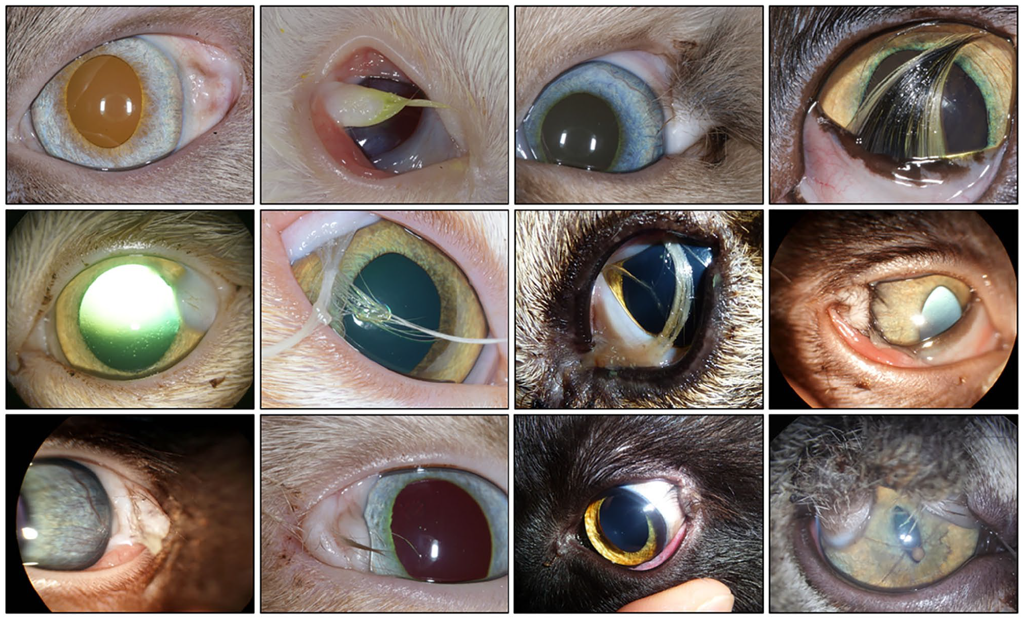
Macroscopic presentation of ocular dermoids in all of the cases except for case 12. Images are shown from left to right (case 1 = top left; case 13 = bottom right; case 12 not included)
The degree of hairiness varied between the patients and was associated with the body hairiness in all cases. The hair was short in three cases, intermediate in five and long in two. The dermoid of case 1 was hairless at the time of presentation because it had been manually depilated by the treating veterinarian prior to referral. The hairiness of case 12 was not documented. Discomfort was reported by the owners and confirmed by clinical examination in 8/13 cases. The associated clinical signs were epiphora, conjunctivitis and keratitis. Five patients had no associated clinical signs.
Concurrent eye anomalies were identified in five patients. Two cats exhibited eyelid agenesis (cases 3 and 13), two cats had an incomplete temporal canthus (cases 1 and 10) and one cat exhibited persistent pupillary membranes (case 13).
Treatment prior to surgery, surgery, outcomes and histopathological findings
At presentation, two cats had topical treatments prior to referral and surgery: one cat had been treated with framycetin and dexamethasone (Fradexam; TVM) and one cat had received retinol (Vitamin A Dulcis; Allergan).
Surgery was performed on 10/13 cats. A conjunctivectomy was performed in eight cases, with a complementary blepharoplasty procedure owing to eyelid agenesis in one case. Canthoplasty was performed in the two cases of incomplete lateral canthus. A superficial keratectomy was performed in two cases. No corneal graft was necessary. The postoperative treatments included topical antibiotics for all cases (neomycin and polymyxin B [Tevemyxine; TVM], 1 drop q8h, 2/10 cases), chloramphenicol (Ophtalon; TVM [1/4 inch strip q8h, 7/10 cases]) or tobramycin (Tobrex; Alcon [1 drop q8h, 1/10 cases]) with or without systemic antibiotics (cefalexin [Therios; Ceva] at 15 mg/kg PO q12h for 5 days, 3/10 cases) and non-steroidal anti-inflammatory drugs in seven cases (meloxicam [Melosus; Axience] at 0.05 mg/kg PO q24h for 3–5 days).The outcome was assessed during a postoperative follow-up – the time after surgery was at the discretion of the surgeon. When only a conjunctivectomy or a superficial keratectomy was performed, follow-up happened between 15 and 30 days. When a blepharoplastic procedure or canthoplasty were necessary, follow-up coincided with the removal of stitches at 15–20 days after surgery. The outcome was considered ‘good’ when the associated clinical signs were completely resolved (no further epiphora, conjunctivitis or keratitis), as well as a complete eyelid function recovery. The outcomes were considered good in all of the operated cases. Surgery was not carried out in three cases. Dermoid was a fortuitous diagnosis for case 7; the initial presentation was for pulmonary oedema, but unfortunately the cat died a few days after admission. For cases 12 and 13, surgery was proposed considering that the two cats had clinical signs. Unfortunately, the owners wanted to wait for the cats to grow and they were lost to follow-up.
Histopathology reports were available for 2/10 excised dermoids (cases 5 and 10). The results revealed hyperplasic keratinising stratified squamous epithelium overlying a normal epidermis and dermis with well-developed hair follicles and adnexal structures such as sebaceous glands (Figure 2). Adipose lobules were located under the dermal area and represented the hypodermis layer. This was accompanied by a moderately hyperplasic corneal epithelium and hypertrophied fibroblasts in the corneal stroma with moderate oedema. No signs of inflammation or neovascularisation were noted.
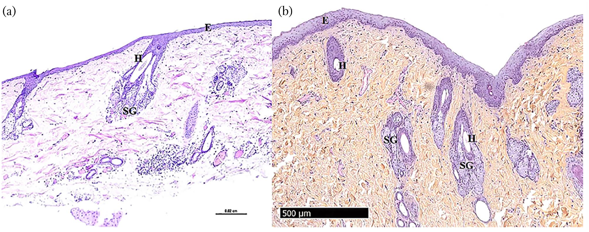
Dermal components of dermoid, including keratinised and pigmented epithelium (E), hair (H) and sebaceous gland (SG). (a) Case 5 (corneal dermoid) and (b) case 10 (conjunctivopalpebral dermoid). Haematoxylin and eosin staining (case 5) and haematoxylin and eosin–safran staining (case 10) at × 4 magnification
Discussion
Ocular dermoids are uncommon in cats. The literature contains four single clinical case descriptions in cats,24,28,30,33 and there have been three clinical case series (in 1979, 1985 and 1992).25–27 In a recent study regarding congenital ocular malformations in dogs and cats (123 cases), ocular dermoids were found in 16 dogs, but no cases were reported in cats, which confirms the rare occurrence of this type of lesion in cats. 5
In our study, the mean proportion of dermoid cases, compared with all the cats seen in ophthalmology in the various private practices, is about one case in 462.
The available epidemiological data confirm the developmental origin of ocular dermoids; however, the mechanisms involved in the pathogenesis of ocular dermoid have not yet been elucidated.1,2 It would appear that an abnormal invagination of ectodermal tissue occurs during gestation. 1 The resulting anomaly comprises ectodermal elements (keratinised epithelium, hair and glandular structures) combined with mesenchymal elements (fibrous tissue, adipose tissue and cartilage).8,19,40,43 Several terms can be used to characterise dermoids. Hamartomas are described as a non-neoplastic overgrowth from faulty development in a normal location, whereas choristomas are histologically normal tissue found at an abnormal location.1–8 This is why ocular dermoids can be considered to be hamartomas when they involve the eyelid, and they can be considered to be choristomas when they involve the eye surface. 1 Balland et al 8 have suggested that the developmental abnormality and the biological mechanism causing choristomas and hamartomas are probably the same.
In our study, the patient signalment was similar to previously reported cases, with only slight differences. Corneal dermoids have been described in the domestic shorthair, Burmese and Birman breeds.24,33 Conjunctival dermoids have been described in domestic shorthair and Burmese breeds.25–28 The results of our study are similar, with an over-representation of domestic shorthairs and Birmans. Interestingly, we reported two cases involving Havana Browns. To our knowledge, this is the first study describing ocular dermoids in the Havana Brown breed. This is a relatively new breed, developed in Great Britain in the 1950s by crossing a Siamese cat with a black shorthair cat. In France, the Havana Brown breed is very uncommon, with only two breeders. Birman, Burmese and Havana Brown breeds all have Siamese ancestors. We hypothesise that Siamese mixed-breeds may be predisposed to ocular dermoids.
Two retrospective studies in humans have revealed a female predisposition.13,14 In dogs, a study has suggested a female predisposition (n = 17/22 [77%]), 21 while another study noted a male predisposition (n = 28/44 [63.6%]). In cats, none of the studies to date have had enough cases to identify a sex predisposition. In the present study, males were over-represented, but the number of cases is too low to draw any definitive conclusions in this regard. Owing to their developmental origin, ocular dermoids are present at birth but frequently not diagnosed until the cat is several weeks of age. 31 In our study, all but one of the cats were less than 1 year of age at initial presentation.
Ocular dermoids have been reported to involve many ocular structures, including the limbus, cornea, conjunctiva, nictitating membrane and eyelid.5–8,18–52 While the condition exists in humans, there is no universal grading for it in veterinary medicine.11,13 In cats, the most commonly reported locations are the conjunctiva and the cornea, with a majority of composite lesions involving both.24–33 Our study describes strictly conjunctival, strictly corneal and composite locations that also involve the eyelid and the canthus, which is comparable to the literature. In cats, the most frequent location of ocular dermoids is the temporal canthus, 1 although one study has described a case of a dorsally located corneal dermoid. 24 The majority of the dermoids in our study were also located at the temporal canthus, but three cases were dorsally located and one was located at the inferonasal bulbar conjunctiva, which, to our knowledge, has not been described before.
Our study reports an equal distribution of right (OD, n = 6) and left (OS, n = 7) eyes, with only unilateral cases. We found two reports of bilateral ocular dermoids in cats; one cat with several abnormalities resembling Goldenhar syndrome, 33 and one cat with a single dermoid that involved multiple locations (sclera, bulbar conjunctiva and upper eyelid) in one eye and a small conjunctival dermoid on the other eye. 30
Concurrent ophthalmic abnormalities have been reported in Burmese cats, including incomplete temporal canthus, prolapse of the nictitating gland and associated nasal dermoids.25,27 Concurrent congenital ocular diseases have been described in a cat with several abnormalities resembling Goldenhar syndrome. The patient presented with bilateral microphthalmia, anterior and posterior segment dysgenesis, and cataracts, in addition to symmetrical corneal dermoids. 33 The concurrent eye diseases found in our study were eyelid agenesis, incomplete lateral canthus and persistent pupillary membranes. The first two are commonly described in association with conjunctival and palpebral dermoids, probably because these dermoids often modify the architecture of the canthus.2,29,31,32 To our knowledge, persistent pupillary membranes have never been described in association with dermoids in cats. However, this has been reported in three dogs.7,8,21 The link between ocular dermoids and persistent pupillary membranes remains unclear, and the presence of both was probably a coincidental finding.
A review of the clinical characteristics of the cases of ocular dermoids in cats reported in the literature is summarised in Table 3.
Clinical characteristics of cases of ocular dermoids reported in the literature
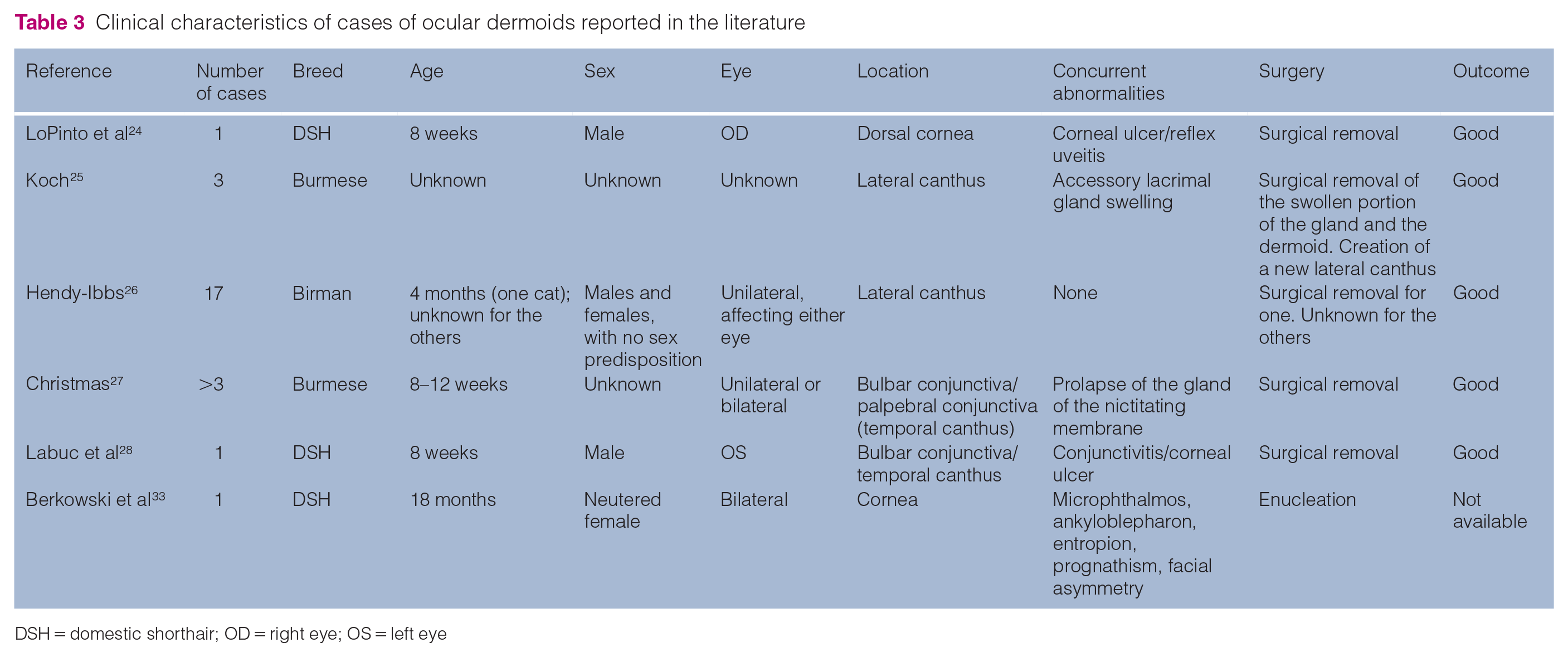
DSH = domestic shorthair; OD = right eye; OS = left eye
It is not clear whether dermoids are hereditary, although a number of studies have found evidence of a hereditary pattern in humans, 17 cats,26,27 dogs 21 and cattle. 38 In cats, epibulbar dermoids in Birman kittens from three separate, but related, litters have been described. 27 Hendy-Ibbs published a report regarding five related Birman cats with epibulbar dermoids, and there appeared to be multifactorial inheritance with a threshold phenomenon. 26 In our study, 2/4 Birman cats had the same sire, as did both of the Havana Browns, strongly suggesting a genetic basis. Unfortunately, no conclusions about the mode of inheritance could be made and further studies are warranted in this regard.
Histological evaluation has revealed similar structures and elements across animal species, with normal epidermis, dermis and subcutaneous tissue. The outer surface has a stratified and squamous epithelium, with keratinisation and various levels of pigmentation.8,19,21 Elements often noted by histopathological assessment include glandular tissue, adipose tissue, hair and hair follicles, and occasionally bone and cartilage.1,8,19,21All of these elements can be found with a different ratio between specimens. One limitation of this study is the small number of histological analyses performed after surgery. As dermoids are clinically diagnosed with an ocular examination, histopathology after curative surgery may not be accepted by the owners. Analysis available for the two cases were performed for the purposes of the present study (ie, not requested by the owners).
Surgery is usually recommended for treating ocular dermoids.21–23,27 It consists of removal of the abnormal parts in the involved area. The surgical procedure depends on the location of the dermoids. The procedure of choice for corneal dermoids is complete excision by a superficial lamellar keratectomy. When a deep keratectomy is required, a reconstruction graft may be necessary.4,21,23 Adnexal dermoids are corrected by conjunctivectomy or various types of blepharoplastic surgery. In the present study, conjunctivectomy or superficial lamellar keratectomy were performed without the need for a reconstruction graft. One case (case 5), which exhibited a strictly corneal dermoid, had corneal surgery, but the lesion was not associated with pain. The decision to remove these relatively benign cases surgically is debatable. One other case needed blepharoplastic procedures to treat the associated eyelid agenesis, and two of the cats needed a canthoplasty. Particular attention should be paid to eyelid reconstruction, in order to preserve the palpebral function and to remove any risk of iatrogenic ocular irritation. 8 Postoperative complications are rare. Dermoid regrowth has been reported to occur when the resection is incomplete.7,8,20–23
Conclusions
The aim of this retrospective study was to describe the clinical presentation of ocular dermoids in cats. Although the number of cases presented in this study is low, our findings are reason to suspect a breed predisposition and a potential hereditary nature of ocular dermoids that warrants further investigation. Surgical excision was curative in our case series.
Footnotes
Acknowledgements
The authors wish to thank the owners and the referring veterinarians of all of the cats involved in this study. We also wish to thank Dr Alexandra Nicolier (DMV, DESV Anatomie Pathologique, Dipl ECVP), Dr Nicolas Pouletty (DMV, DES, Dipl ACVP) and Dr Marie-Capucine Tricaud (DMV, MCMVS, PhD) for the histopathology reports and pictures.
Conflict of interest
The authors declared no potential conflicts of interest with respect to the research, authorship, and/or publication of this article.
Funding
The authors received no financial support for the research, authorship, and/or publication of this article.
Ethical approval
The work described in this manuscript involved the use of non-experimental (owned or unowned) animals. Established internationally recognised high standards (‘best practice’) of veterinary clinical care for the individual patient were always followed and/or this work involved the use of cadavers. Ethical approval from a committee was therefore not specifically required for publication in JFMS. Although not required, where ethical approval was still obtained, it is stated in the manuscript.
Informed consent
Informed consent (verbal or written) was obtained from the owner or legal custodian of all animal(s) described in this work (experimental or non-experimental animals, including cadavers) for all procedure(s) undertaken (prospective or retrospective studies). No animals or people are identifiable within this publication, and therefore additional informed consent for publication was not required.
This paper was handled and processed by the European Editorial Office (ISFM) for publication in JFMS