
Abstract
Clinical presentation:
A 6-month-old female spayed domestic shorthair cat was presented for investigation of acute right hindlimb lameness and paresis.
What is your diagnosis?
Readers are encouraged to review the case history and consider what their diagnostic suspicion is. Also what is the preferred treatment for this injury? And what is the primary cause of this injury?
Keywords
Case history
A 6-month-old female spayed domestic shorthair cat was presented for acute right hindlimb lameness and paresis. The owner reported that they thought the cat had fallen approximately 6 feet (1.8 m) out of a cat tree that morning. Although they did not witness the fall, they heard the cat cry out and observed the cat not moving the right hindlimb.
Upon examination, a non-weight-bearing lameness affecting the right pelvic limb was evident; however, normal motor function was present. A soft, painful swelling was palpable over the right stifle and proximal femur, and the greater trochanter was palpably asymmetrical and suspected to be displaced craniodorsally. All other physical examination findings were within normal limits.
The cat was sedated with dex-medetomidine 0.005 mg/kg and butorphanol 0.2 mg/kg IM. Anterior-posterior and lateral radiographs of the right stifle, and ventrodorsal and lateral radiographs of the pelvis were obtained (Figure 1).

Radiograph taken after presentation
Diagnosis
The cat’s radiograph (Figure 1) shows a chronic right capital physeal fracture with femoral neck resorption. This is consistent with a diagnosis of chronic slipped capital femoral epiphysis (SCFE). On detailed examination of the radiograph, the fracture line extending through the right capital physis is observed, as is medial rotation of the femoral head within the acetabulum in relation to the femur. The right femoral neck is small, rounded and smoothly marginated, with wispy soft tissue to mineral opacities surrounding the neck. Slight cranial displacement of the femur is present. The left coxofemoral joint is normal. Radiographs of the right stifle were within normal limits.
Case management
The preferred treatment for this injury is an excisional arthroplasty via femoral head and neck ostectomy (FHO).1–3 In this case the patient was given pain relief (robenacoxib 6 mg PO q24h) and scheduled for subsequent FHO surgery.
At surgery, a standard craniolateral approach to the hip was performed and exposure of the coxofemoral joint obtained. The femoral head was observed to be separated from the neck of the femur and secured in the acetabulum. The neck of the femur appeared to be resorbing and consisted mostly of fibrous tissue. The remaining femoral head was removed sharply from the acetabulum (Figure 2). The remaining femoral neck was removed using rongeurs and a bone rasp, making a smooth cut aligned between the greater and lesser trochanters. The joint capsule was closed over the empty acetabulum and the rest of the surgical site was closed routinely. Postoperative radiographs were taken to verify removal of all necessary tissue and proper angle of the cut (Figure 3). Additionally the excised femoral head was submitted for histopathology.
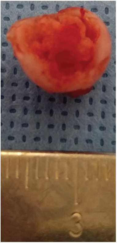
Remaining femoral head immediately after removal

Postoperative radiograph; note the complete removal of the right femoral head and neck, and the smooth surface on the femur
The cat recovered uneventfully and was sent home with pain relief (robenacoxib 6 mg PO q24h and buprenorphine 0.03 mg transmucosally q12h) and instructions to ice the surgical site, perform passive range of motion exercises and restrict to limited activity for 2 weeks.
On follow-up examination 2 weeks postoperatively, the patient was found to be significantly improved, with only mild lameness and near-normal range of motion. The patient was cleared to return to normal activity.
Discussion
The SCFE injury in this cat resulted in a pathologic fracture. SCFE represents a chronic displacement of the capital femoral epiphysis from the rest of the femur occurring through the physis.
Characterization
Lesions are characterized radiographically by widening of the physis with subsequent progressive lytic areas in the head and neck of the femur.4,5 These correspond to the common histopathologic findings of bone necrosis, sclerosis and remodeling. The physeal cartilage is found to contain clumps of irregular chondrocytes and has an absence of normal maturation of the physis and disorganization of the articular cartilage near the physis. 5 The zone of hypertrophy is absent and there is widening of the zone of proliferation. 6 This is in contrast to a traumatic Salter-Harris type I fracture, which would not have radiographic lysis and histopathologically would show normal chondrocytes.5,6 The excised femoral head of this cat was submitted for histopathology and similar lesions of severe physeal dysplasia were found (Figure 4).

(a) Photomicrograph of the femoral head. The physis is severely irregularly thickened and there are numerous irregular chondrocyte clusters. In the center of the epiphysis, irregular cartilage replaces lamellar bone. Hemotoxylin and eosin (H&E) stain; bar = 200 μm. (b) Photomicrograph of the femoral physis. The lamellar bone in the epiphysis is replaced by chondrocyte clusters (asterisk) and abundant eosinophilic or basophilic matrix. H&E stain; bar = 100 μm
SCFE has been compared with avascular necrosis of the femoral head (Legg–Calvé–Perthes disease), which is more commonly seen in dogs. 2 However, the comparison is not accurate as the blood supply to the femoral head of the cat is more robust, including the artery of the teres ligament which provides relatively poor blood supply in the dog. 2 This makes the lesions in the cat more confined to the area distal to the physis, involving little of the femoral head. 2
Signalment
SCFE has been reported in Siamese, Maine Coon and domestic shorthair cats, and is seen occasionally in other breeds such as domestic longhair and Siamese mix cats.1,6 In one study it was found that male cats were more commonly affected, average age of diagnosis was 16 months and being diagnosed as obese was a risk factor. 4 Thirty-eight percent of affected cats were found to have bilateral lesions. 6 Female cats with SCFE were found to have a much younger age of diagnosis (8 months). 6 However, only 13 cats were examined in this study and only two were female so further investigation is likely necessary to understand true risk factors.
Pathogenesis
Although the pathogenesis of SCFE in the cat is poorly understood, it has been suggested that the cause is genetic in nature as it has been found to be statistically more likely in certain breeds and there have been reports of affected littermates.
Treatment and prognosis
The most common treatment for SCFE is FHO, although total hip replacement (THR) has been described for large cats or cats with severe degenerative changes.1,3,7 Rarely, internal fixation of the fracture has been attempted. In one study examining SCFE in 24 Maine Coon cats, 14/24 cats were treated with FHO and 8/24 with THR. 1 However, the numbers are likely skewed towards THR as the study was performed at a major referral hospital and involved Maine Coon cats, which are significantly larger than the average cat. The general population of affected cats likely does not have such ready access to THR.
Patients undergoing FHO have been found to have a good to excellent return to function and it has been reported that cats may be able to return to normal even following bilateral operations.2,3,7
Primary cause of the injury
The timeline of this type of injury is often uncertain even though it is often attributed by owners to a traumatic event. It is possible a fall, as described in this cat, could have caused a fracture of the weakened area. However, this cat’s fall was not observed and it is quite possible that the fracture occurred due to normal climbing activity. The pain of the pathologic fracture may have then caused the normally sure-footed cat to fall, resulting in the perception of a traumatic event.
Conclusions
SCFE is a developmental orthopedic disease affecting young cats. Diagnosis by radiography reveals the damage to the femoral head and neck. Definitive treatment is by surgical FHO and most cats have a good to excellent return to function.
Footnotes
Date accepted: 2 October 2018
Conflict of interest
The authors declared no potential conflicts of interest with respect to the research, authorship, and/or publication of this article.
Funding
The authors received no financial support for the research, authorship, and/or publication of this article.