
Abstract
Objectives
The objectives of this study were to describe the CT characteristics of the adrenal glands in healthy cats, to provide normal reference biometry for adrenal gland size and attenuation values, and to investigate the association with age, sex, laterality and body weight.
Methods
Retrospective evaluation of 30 CT studies of healthy adult cats recruited from September 2013 to July 2015 was performed. Healthy cats >1 year of age were included based on the absence of clinical signs, unremarkable physical examination, normal results of the complete blood count, biochemical profile, feline immunodeficiency virus, feline leukaemia virus and Bartonella species infection tests. The relationship between gland biometry (size and attenuation values) and the age, sex, laterality and body weight of cats were tested by two-way ANOVA. The intraclass correlation coefficient was assessed and mean, SD, range and reference interval provided.
Results
Twenty-seven cats were included. Bilobed, arrowhead and oval adrenal gland shape patterns were recognised, the first being most common. No statistically significant differences were observed between the biometric parameters (length, height and attenuation values) and age, sex, laterality or body weight of the cats. Regarding the width of the adrenal glands, there was a statistically significant effect of sex and laterality. The length (11.6 ± 2.1 mm) and height (6.1 ± 1.3 mm) were the most consistent biometrical parameters to describe adrenal glands.
Conclusions and relevance
Adrenal gland shape, size and attenuation CT data of healthy feline patients are provided in this study, as well as normal reference intervals for morphometric characterisation based on adrenal length and height.
Introduction
Adrenomegaly has been described to be associated with systemic diseases such as acromegaly, 1 hyperadrenocorticism,2,3 hyperthyroidism 4 and hyperaldosteronism. 5 However, interstitial cystitis has been associated with reduced adrenal gland size. 6
Ultrasound (US) is the non-invasive imaging technique of choice for assessing adrenal morphology in cats.7–9 Ultrasonographic characterisation of adrenal glands has been described in healthy cats, both glands being typically ovoid, uniformly hypoechoic, 10–11 mm long and 3.5–4.5 mm in maximum thickness.7–10
Nowadays CT is becoming more available, allowing evaluation of the adrenal glands, as well as the pituitary axis or thyroid glands simultaneously in the same study. Therefore, knowledge of standard adrenal gland characteristics in CT images is essential.
To our knowledge, there are no available data on normal feline adrenal gland CT characteristics. Because of the importance of knowing the normal appearance of an organ in a clinical situation, the purpose of this study was to describe CT characteristics of the adrenal glands in normal feline patients, to provide normal reference biometry for adrenal gland size and attenuation values and to investigate the association with age, sex, laterality and body weight.
Material and methods
Patients
Retrospective evaluation of 30 CT studies of healthy adult cats, >1 year of age, was performed independently by two of the authors (CM and RA) after a training period provided by a board-certified veterinary radiologist (RN) and a senior radiology professor (YE). Patients were previously included in another unrelated prospective study, and recruited from September 2013 to July 2015. 11 Animals were considered healthy based on the absence of clinical signs, unremarkable physical examination, and normal results of the biochemical profile and complete blood count (CBC). Moreover, feline immunodeficiency virus (FIV), feline leukaemia virus (FeLV) and Bartonella species infection, were ruled out by means of a SNAP test and PCR, respectively.
Patient data, including age, sex, breed, and weight, were recorded.
Imaging procedure
Cats were sedated with midazolam (0.2 mg/kg IM [Midazolam; Normon]), butorphanol (0.4 mg/kg IM [Torbugesic; Zoetis]) and ketamine (5 mg/kg IM [Imalgene; Merial]). Anaesthesia was induced with isoflurane 5% dosage 100% O2 at 4 l/min and maintained with isoflurane 1.5–2% in 100% of O2 at 2 l/min (Isoflurane; Abbott Laboratories). All cats underwent whole-body CT scans in dorsal recumbency and images were acquired using a soft tissue algorithm, before and approximately 30 s after manual administration of 600 mg/kg IV Iopamidol (Scanlux 300 mg/ml; Sanochemia Pharmazeutika) in the cephalic vein. Manual hyperventilation was performed before each examination to induce transient apnoea. Scans were performed in a 16-slice helical CT scanner (General Electric Brivo CT 385) with a slice thickness of 0.625 mm, interslice interval of 0.625 mm, collimation pitch of 0.5625:1, 120 kV, 50–90 mA and a matrix of 512 × 512.
All data were transferred to a workstation and analysed using a medical image viewer (Horos version 3.0, 64-bit).
Measurement techniques
The shape, size and attenuation values were determined for each adrenal gland in both non-enhanced and enhanced series, using a window width of 400 and a window level of 40.
The shape of the adrenal glands was evaluated using multiplanar reconstructions, the dorsal plane being the most useful in the majority of cases. When the adrenal gland had different shapes in two different planes (as happened in arrowhead shape), the shape in the dorsal plane was the one considered. Three shape patterns were distinguished: bilobed (oval with a central depression or over the minor axis), oval (without depression) or arrowhead (wedge shape with a rounded caudal tip) (Figure 1). In cases where there was initial disagreement on the adrenal shape, consensus was reached by the authors.

Post-contrast dorsal CT reconstruction images at the level of the adrenal gland displayed in a soft tissue window. Three shape patterns of the adrenal gland were distinguished: (a) arrowhead (wedge shape with a rounded caudal tip); (b) oval (without depression) and bilobed (c) oval with a central depression or (d) a depression over the minor axis
As parameters for evaluation of adrenal gland size, the length was assessed by placing an electronic caliper from the cranial to the caudal border at the maximum diameter using multiplanar reconstructions, sagittal or dorsal planes. Likewise, height and width were also measured in transverse or sagittal and transverse or dorsal planes, respectively. Height was defined as the maximal distance from the ventral to the dorsal border and width was defined as the maximal distance from the medial to the lateral edge, independently of the adrenal gland shape (Figure 2). Each dimension was measured twice for every gland by both radiologists, and the biggest measurement was selected for statistical purposes.
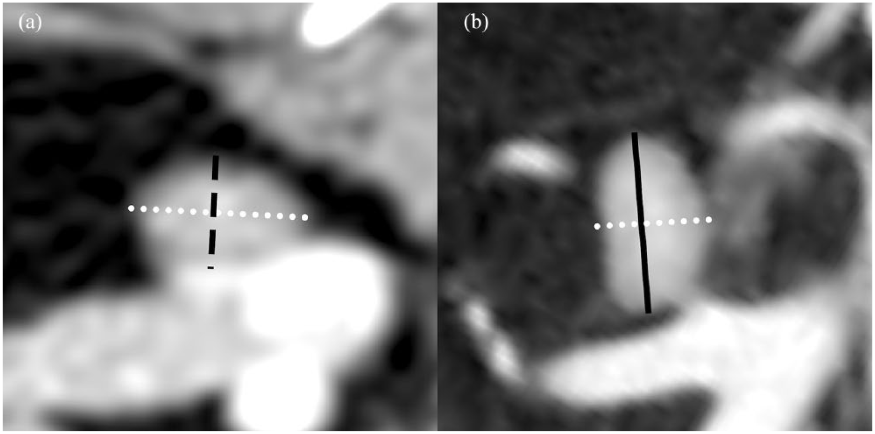
Post-contrast (a) transverse and (b) dorsal CT reconstruction images of the right adrenal gland displayed in a soft tissue window. Maximal length (solid black line), maximal width (dotted white line) and maximal height (black discontinuous line) were assessed in each gland by placing an electronic caliper
Attenuation values of the adrenals were measured in Hounsfield units (HU), using a circular/ovoid region of interest (ROI) on the images of every gland following the same protocol previously used in dogs. 12 Briefly, one measurement was performed for each gland in both the non-enhanced and the enhanced series. The section with the largest cross-sectional area of tissue was selected on one of the images in transverse or dorsal planes. The ROI included one-third to one-half of the area of the adrenal gland in the selected image, excluding the phrenicoabdominal vessels and gland edges, to minimise partial volume effects and to exclude adjacent periadrenal fat (Figure 3). The SD of each gland measurement in both series was also recorded.

Post-contrast (a) dorsal and (b) transverse CT reconstruction images of the right and left adrenal gland, respectively. Images are displayed in a soft tissue window, showing the measurement technique for attenuation values. An ovoid region of interest was placed in the centre of the gland and the software automatically calculated the mean pixel value and SD included in the area
The presence of demarcation between the cortex and medulla in enhanced and non-enhanced series, and the existence of parenchymal mineralisation, were also recorded.
Statistical analysis
Departures from normal distribution of adrenal glands were assessed by the D’Agostino test and inter-observer repeatability by the intraclass correlation coefficient (ICC) that ranges from 0 (no agreement) to 1 (perfect agreement). The relationship between gland biometry (size and attenuation values) and the age, sex, laterality and body weight of cats were tested by a two-way ANOVA. The mean, SD, range and reference interval (RI) were calculated with the ‘rcompanion’ package version 1.13.2 of the statistical software R.
Results
Of the 30 cats initially studied, two animals were excluded because of the presence of respiratory motion artefacts affecting the evaluation of the adrenal glands and another cat was excluded because of age. The cat was 17 years old and despite the blood work-up being normal, we could not rule out other common subclinical diseases at this age; therefore, a total of 27 cats were finally included. The population comprised six entire males (22%), five castrated males (19%), seven entire females (26%) and nine spayed females (33%) with a mean age of 3.8 years (range 1–9 years) and a mean weight of 4.5 kg (range 3–7 kg). Twenty-six cats were domestic shorthairs and one cat was Persian.
Biochemical determinations and CBC were within normal limits. All cats were negative for FIV/FeLV and Bartonella species.
In all 27 cats, both left and right adrenal glands were visualised on the non-enhanced CT images; however, enhanced images were more valuable in identifying the gland boundaries and allowing delimitation with the surrounding abdominal organs, especially between the cranial pole of the right adrenal gland and liver parenchyma, or adjacent vascular structures.
Of the total of 54 adrenal glands, 21 (39%) had a bilobed shape, 16 (30%) oval shape and 17 (31%) arrowhead shape. The right adrenal gland was of bilobed shape in 11 patients (41%) and of an arrowhead shape in 16 (59%), whereas the left adrenal gland was bilobed in 10 cats (37%), oval in 16 (59%) and arrowhead in one (4%). The dorsal plane was found to be the most useful for characterisation of the gland shape, especially for the arrowhead shape.
According to the scale proposed by Bartko, 13 inter-observer agreement was poor for unenhanced attenuation values (ICC = 0.25), fair for width (ICC = 0.5), good for height and enhanced attenuation values (ICC = 0.6 and 0.65, respectively) and excellent for length (ICC = 0.88). The two-way ANOVA test did not reveal statistical dif-ferences for length, height and attenuation values in association with the laterality, age, sex or body weight. Regarding the width of the adrenal glands, there was a statistical significant effect of sex (P = 0.002) and laterality (P = 0.001), with the largest width values being recorded in male cats (males: 6.5 ± 1.3 mm; females: 5.8 ± 1.1 mm) and in right glands (6.6 ± 1.5 mm; left glands: 5.6 ± 1.2). Morphometric and attenuation values were calculated, averaging the results of the two observers to minimise error and improve accuracy. The mean, SD and range for each adrenal gland measurement are summarised in Tables 1 and 2.
CT measurements of the adrenal glands in healthy cats

CT attenuation values of the adrenal glands in healthy cats
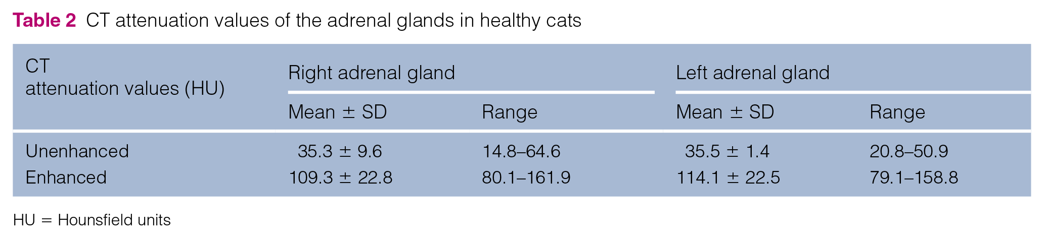
HU = Hounsfield units
According to the previous results, adrenal gland length and height are provided for morphometric CT characterisation of the RIs in healthy cats (Table 3). Adrenal width is considered less reliable owing to its lower reproducibility and wider range of RI values in comparison to the other measurements. The RIs for adrenal gland attenuation in unenhanced and enhanced series are also depicted in Table 3.
CT reference values for length, height and attenuation of adrenal glands in healthy cats

CI = confidence interval; RI = reference interval; HU = Hounsfield units
However, adrenal corticomedullary differentiation was not observed in any of the patients, on either the unenhanced or enhanced series. Only one 5-year-old spayed female cat (1/27 [4%]) presented a unilateral focal punctiform adrenal mineralisation affecting the caudal pole of the left gland.
Discussion
CT was used to measure and characterise the adrenal glands. High body weight and, consequently, accumulation of retroperitoneal fat was found helpful in delimitating the glands.
Three adrenal shapes (arrowhead, oval and bilobed) were found. Altogether, bilobed was the most common shape, with this finding being in agreement with previous studies using US.7,9 However, the left adrenal gland most commonly showed an oval shape, while the right was most commonly arrowhead in shape. This fact may explain the statistically significant effect between width and laterality as the largest width values were recorded in the right adrenal glands and the arrowhead glands were the widest. The arrowhead shape of the right adrenal gland has been widely reported in dogs, 14 but this is the first study to describe a similar pattern in cats.
Normal adrenal CT cross-section measurements, volume and attenuation values have been described in normal dogs.12,15 In addition, CT findings have been described in canine patients with hyperadrenocorticism and adrenal masses.16,17 CT is the gold-standard imaging technique in humans, 9 detecting adrenal abnormalities and characterising them as benign or malignant, by means of the attenuation values, enhancement pattern or wash-out features of adrenal masses. 12
Ultrasonographic morphometric values of adrenal glands in cats have been previously described;4,7 however, this is the first description using CT. The CT measurements obtained in this study are similar to the previously described ultrasonographic references, although they are slightly higher and wider in range in all dimensions. Our findings are in accordance with the literature, whereby morphometric measurements of abdominal organs with US and CT showed considerable differences, revealing that direct comparison of measurements obtained with CT frequently exceeded those obtained with US. 18 A previous study also reported significant differences between post-mortem gross measurements and ultrasonographic measurements in width, indicating underestimation of the dimension. 18 Another hypothesis to explain our higher short-axis measurements in comparison with US, is that in this study the height and width were estimated in its maximum diameter, without regard to its location within the gland (cranial, caudal pole or central), in contrast to US. The authors considered this measurement easier to obtain and compare in CT owing to the variability of the morphology and the relatively small size of the adrenal glands. Consequently, morphometric examinations of the adrenal glands should be performed using the RIs reported in the same imaging modality.
As with US, in CT the length (or long-axis) of the adrenal glands was found to be more uniform and well defined, in comparison with short-axis measurements, which showed higher variability. 9 Our hypothesis to explain the broader range in height and width measurements is the variation of the gross morphology of the adrenal glands, presenting different shape patterns, in comparison with other organs that have a more constant morphology. This fact indicates that adrenal gland shape should be considered when evaluating the morphometric measurements.
It is well understood that morphometric evaluation in imaging diagnosis is highly observer dependent based on skill level. 14 CT measurements of any organ, but particularly the adrenals owing to their small size, depend on the expertise of the observer and the measurement technique for adrenal glands increases with greater training of the observer. At the time the study was completed, one of the observers (CM) was beginning the first year of a radiology residency, and the other observer (RA) was a board-eligible radiologist. Thus, inter-observer differences in our study could be partly due to a difference in experience. However, the mean differences between the two observers’ measurements were minimal for length and height, with the correlation being strong and good between observers, respectively. The width showed moderate inter-observer correlation, indicating lower reproducibility in comparison with the previous measurements, and need of further training of the observers. We speculated that variation in the gross structure of adrenal glands giving a wider range of width measurement might have contributed to this fact. Based on these results, we propose morphometric CT characterisation of the adrenal glands based on length and height; adrenal width is considered less reliable owing to its lower reproducibility and wider range of RI values in comparison with the other measurements.
Consistent with previous studies in the US, 9 in the present study there was no effect of body weight or age on adrenal gland CT measurements. A significant effect was found between width and sex. As described previously, castrated male cats showed larger adrenal glands than intact males and spayed females. 9 A previous study described an increased aldosterone-to-renin activity ratio in healthy neutered cats compared with intact cats and speculated that reduced negative feedback from absent gonadal steroids due to castration may cause enlargement of the adrenal glands in neutered cats. 9 However, this finding has low clinical significance given the small differences in adrenal size compared with the rest of the population.
The attenuation values obtained here represent the mean values of all pixels included within the ROI. In our experience, transverse and dorsal planes were preferred for measuring the attenuation values, allowing us to define the largest ROI while excluding the gland boundaries and the phrenicoabdominal vessels. The left and right gland attenuation values measured in non-enhanced images were similar, and even though the right adrenal glands had a wider range of attenuation values, this difference was not statistically significant. Even despite protocol standardisation, minimal differences in contrast phase (timing, velocity of contrast administration, cardio-vascular conditions of the patient) during the scanning could be responsible for the wider range in the enhanced attenuation values. Similar values and intervals have been reported in dogs, using the same technique. 12
Even though the same technique was used in both measurements, the correlation between the observers was fair and good for the non-enhanced and enhanced attenuation values, respectively; the reason for this difference was unidentified, but could be due to poor delimitation of gland boundaries in the non-enhanced series. For this reason, owing to the lower reproducibility of the non-enhanced attenuation values, the proposed normal values should be interpreted carefully.
Intact male cats showed lower enhanced attenuation values than the rest of the population, the reason for this finding is unknown. However, even though this difference was statistically present, it was not clinically significant.
The feline adrenal gland is mainly described ultrasonographically as uniformly hypoechoic; however, high-frequency transducers sometimes allow differentiation between the cortex and medulla. 19 In the present study, the corticomedullary differentiation was evaluated in both enhanced and non-enhanced series, without evidence of clear demarcation in any patient.
Adrenal gland mineralisation is a common incidental finding in older cats without clinical signs associated with adrenal pathology,20,21 and has been histologically reported in 25–30% of cats in the veterinary literature.21,22 Calcification occurred predominately in the zona reticularis of the adrenal cortex but can affect the entire cortex and even extend into the medulla. 23 The cause and pathogenesis of adrenal calcification is unknown. 24 In this study, only one cat presented with focal adrenal mineralisation of the left gland. The young age of our population might have influenced this finding.
There were a few limitations in this study. First, the retrospective nature of the study. Second, the overall sample size was small and creating authoritative RIs requires a larger population of cats. Specific endocrine testing to definitively rule out underlying adrenal disease was not performed. For future studies, comparing normal healthy cats with cats with confirmed adrenal pathology would allow sensitivity and specificity, as well as false-positive and false-negative rates, to be calculated for our proposed measurements.
Conclusions
Adrenal gland shape, size and attenuation CT data of healthy feline patients are provided in this study, and normal RIs for morphometric characterisation based on adrenal length and height are proffered.
Footnotes
Acknowledgements
The authors would like to thank José Ríos for his contribution with the statistical analysis and are grateful to the staff of the Hospital Clínic Veterinari of the Universitat Autònoma of Barcelona, and to students and hospital clients for their contributions to this project.
Conflict of interest
The authors declared no potential conflicts of interest with respect to the research, authorship, and/or publication of this article.
Funding
The authors received no financial support for the research, authorship, and/or publication of this article.