
Abstract
Case series summary
Pantarsal arthrodesis (PTA) was performed in seven tarsi of six cats, using orthogonal (dorsal and medial) veterinary cuttable plates (VCPs) without postoperative external coaptation. Short-term outcomes, arthrodesis progression and complications were assessed using a retrospective review of case notes (veterinary examination) and radiographs. Long-term outcomes were assessed via owner questionnaire (Feline Musculoskeletal Pain Index [FMPI]). Mean angle of PTA was 136° (range 116–166°). Intraoperative complications were recorded in two cases, both involving failure of the drill bit during drilling for calcaneotibial screws. Postoperative complications were encountered in a case of bilateral single-session PTA. These included gastrocnemius myotendinopathy on the right, and long-term protrusion of a screw head from the skin on the left. Both complications were resolved surgically, through resection of the implicated gastrocnemius tendon of insertion and removal of the plate, respectively. FMPI assessment was performed for all six cats a mean of 8.8 months (range 6–16 months) following surgery. Mean score for the first part (assessing ability to perform normal activities) was 92.2% (range 80.9–97.1%). Mean score for the second part (owner perception of pain) was 95.8% (range 87.5–100%). Mean overall score (mean score for parts 1 and 2 combined) was 92.3% (range 81.6–97.4%). PTA may be performed in cats using orthogonal VCPs to treat severe tarsal injuries. It may be prudent to avoid single-session bilateral PTA in cats.
Relevance and novel information
This case series documents a novel technique as an alternative for PTA in cats with talocrural injuries. Long-term outcome and complications presented in this case series are evaluated and discussed.
Introduction
Pantarsal arthrodesis (PTA) is a salvage procedure involving fusion of the talocrural, proximal and distal intertarsal, and tarsometatarsal joints. 1 Indications in dogs include intractable tarsal pain, severe tarsal fractures, shearing injuries and tarsal joint instability. 2 There is limited information regarding the indications for PTA in cats, although traumatic injuries were the most common indication in one study. 3 The English-language literature addressing surgical techniques and outcome following feline PTA is limited to 32 cats.3–9
Fixation methods, including combinations of pins and lag screws, with or without orthopaedic wire or bone plates, external skeletal fixation and medial, dorsal, lateral or plantar bone plating, have been described in vivo or under mechanical testing for PTA in dogs.10–14 Fixation methods reported for PTA in cats include the use of a type II external skeletal fixator, 15 circular external skeletal fixator8,9 and dorsal and medial plating.3–5,7,15 A percutaneous technique involving dorsal plating for PTA in cats has been described. 3
Dorsal plating for PTA involves positioning the plate on the compression side of the joint, such that it is subject to increased risk of cyclic load to failure. 15 Plantar plating is preferable in terms of the biomechanics, as the plate is primarily subjected to tension, and this technique has been reported in four dogs. 14 Plantar plate placement is complicated by the regional neurovascular structures, 16 and has remained unpopular. Various plates for dorsal application have been reported, including locking compression plate and dynamic compression plate (DCP),4,7 2.7/2.0 mm hybrid DCP, 5 2.0/2.7 mm stacked veterinary cuttable plates (VCPs), 6 2.4 mm locking reconstruction plate (Unilock; Synthes) 15 and a pre-contoured feline dorsal PTA plate (Feline Dorsal Pantarsal Arthrodesis Plate; Veterinary Instrumentation). 3 A 2.0/2.7 hybrid PTA plate with a 120° bend for medial or lateral application is also available (2.0/2.7 Feline Pantarsal plate Right/Left 120°; Veterinary Instrumentation), although we are unaware of any published reports using this implant.
Medial plating is preferred for tibial fracture repair because the approach is simple on this side. 17 Additionally, medial plating of tibial fractures may improve biomechanics in comparison with dorsal plating as the implant is edge-loaded, increasing the area moment of inertia and resistance to bending forces, 14 which may be applicable to PTAs. Application of a medial plate for PTA has been tested in vitro, 12 and is reported in dogs and in cats using 2.0 mm or 2.4 mm locking reconstruction plates (Unilock; Synthes).11,15
The use of VCPs in small animal fractures was first described by Brüse et al in 1989. 18 Several manufacturers currently market VCPs of varying dimensions and characteristics. Despite the differences between the various VCPs, their advantages include that they are versatile, inexpensive and readily available. 19 Benefits of VCPs specifically for PTA in cats include that they are easy to contour and cut, and thus may be adapted to the anatomy of the individual. The high hole density allows for multiple screw placement and positioning such as to maximise purchase in small tarsal and, particularly, metatarsal bones. Additionally, their low profile minimises tension in the soft tissues during closure. 20 However, as a result they are relatively weak in bending. 20 The use of VCPs has been reported for dorsal stabilisation of PTA in two cats, one involving further tension band wire fixation and one involving stacked plates. 6
In view of the advantages of VCPs, we consider their use for this procedure appropriate. However, there were concerns that the use of a single VCP (medial or dorsal) may result in excessive plate strain such that there would be a risk of plate failure. Reported techniques for reducing plate strain include increasing the size of the implant, combining with an intramedullary implant and stacked or orthogonal plating.21–23 External coaptation has also been recommended to reduce plate strain in canine pan carpal arthrodesis and PTA;24–26 however, high rates of complications have been reported with external coaptation performed to augment canine and feline PTA.11,13,26–28 In one study of orthopaedic cases managed using external coaptation, cast-associated soft tissue injury complications were documented in 63% of cases, 40% of which required veterinary intervention. 29 Intramedullary pinning has been suggested for augmentation of PTA; 30 however, this implant only augments the talocrural joint fixation and there is the potential for migration of a smooth intramedullary pin in the early postoperative period. Orthogonal plating has not been described for canine or feline PTA, to our knowledge. We hypothesised that combined medial and dorsal orthogonal plating using VCPs would be possible for PTA in cats and would provide sufficient stability for bone union in the absence of external coaptation. This report documents the surgical technique and outcomes of six cats (seven tarsi) treated by PTA stabilised with combined medial and dorsal VCPs.
Case series description
Surgical records of two surgeons from February 2012 to January 2015 were searched for cases of feline PTA performed using orthogonal VCP application without postoperative external coaptation. Inclusion criteria included cats undergoing PTA and with long-term telephone follow-up. Data collected included breed, age, sex, body weight, indication for arthrodesis, number and size of screws used, orientation of tibiocalcaneal or calcaneotibial screw, type of bone graft, postoperative management, and incidence and nature of any complications. Preoperative radiographs were reviewed for assessment of the initial injuries, and postoperative radiographs were reviewed for tarsocrural angle, number of screws placed, and percentage of the tibial and metatarsal length engaged with the fixation. Follow-up radiographs were reviewed for progression of arthrodesis and for implant-related complications. Clinical notes were also reviewed for any reported complications.
The VCPs used in this case series were 2.0 mm cuttable (dorsal) and 2.0 mm cuttable malleable (medial) plates (Veterinary Cuttable Plates; Veterinary Instrumentation). Both plates are of 5.0 mm width and 1.4 mm thickness, with a 5.0 mm hole-to-hole distance. The 2.0 mm cuttable malleable plate is a notched version, permitting contouring in a third plane.
Long-term outcome (>6 months after surgery) was assessed by owner questionnaire (Feline Musculoskeletal Pain Index [FMPI]) 31 by telephone and email communication. The FMPI questionnaire is divided in two parts. The first is an assessment of the cat’s ability to perform 17 normal activities (rated on a Likert scale from ‘normal’ to ‘not at all’), with an option to select ‘don’t know or not applicable’. The second part of the questionnaire asks owners to rate their cat’s level of pain on a standardised 100 mm visual analogue scale (VAS). Owner ratings were converted to scores ranging from 0–4 for each item (0 = not at all; 4 = normal). VAS scores were calculated by measuring (in mm) from the start (zero point) to the owner’s mark, with 4 indicating ‘no pain’. The range of possible scores was 0–68 for items 1–17 and 0–4 each of the final two questions. FMPI scores were converted to percentages and are given for part 1, part 2 and an ‘overall score’ as a mean score for parts 1 and 2 combined. An excellent outcome was defined as a score within the range of 75–100%, a good outcome was defined as having a score between 50% and 74%, and a suboptimal function was defined as having a score <50%.
Procedure
All cats were premedicated with 0.03 mg/kg intramuscular (IM) acepromazine (ACP injection; Novartis) and 0.2 mg/kg IM methadone (Comfortan; Dechra Veterinary Products). Anaesthesia was induced with 4 mg/kg intravenous (IV) propofol (Propflo; Abbott Laboratories) and maintained with inhaled isoflurane in oxygen (Isoflo; Abbott Laboratories). Cefuroxime (Zinacef; GlaxoSmithKline) was given before surgery and intraoperatively 15 mg/kg IV initially, and thereafter at 10 mg/kg IV at 90 min intervals throughout surgery. Cefuroxime was continued for 24 h postoperatively at 10 mg/kg q8h; after these 24 h cephalexin (Ceporex; Galen Craigavon) was administered at 15–20 mg/kg q12h orally for 7 days. Postoperative analgesia involved intermittent IM methadone (0.1–0.3 mg/kg) as necessary, according to pain assessment. Meloxicam (Metacam; Boeringer Ingelheim) was administered at 0.2 mg/kg subcutaneously on the day of surgery followed by 0.05 mg/kg/day orally starting 24 h after surgery for 10 days.
Cats were positioned in dorsal recumbency. The limb was free-draped. Neither an antimicrobial incise drape/adhesive film nor tourniquet were used. A craniomedial approach to the tarsus was made with a curved incision, encompassing the distal third of the tibia, the tarsus and half the length of the metatarsus. The subcutaneous and crural fasciae were incised and retracted with the skin; the second tarsometatarsal, centrodistal and proximal intertarsal joints were exposed medially and articular cartilage was removed with a pneumatic high-speed burr from all the abovementioned joints. Articular cartilage from the talocrural joint was removed in the same way, reaching the joint with the burr by the craniodorsal aspect involving release of the tarsal extensor retinaculum and retraction of the tendons of the long digital extensor and cranial tibial muscles. Debrided joint spaces were lavaged thoroughly with sterile saline and packed with feline demineralised bone matrix allograft (Veterinary Tissue Bank).
The dorsal plate was placed first in order to avoid varus or valgus misalignment. A 2.0 mm cuttable plate was contoured to the cranial tibia, dorsal tarsus and dorsal third metatarsal bone, aiming for a tarsal angle between 100º and 125º, as recommended.1,32 Intraoperative angle measurements were made using a sterile goniometer or a Kirschner wire pre-bent to the appropriate angle and sterilised. A 2.0 mm cuttable malleable plate was contoured to the medial tibia, tarsus and metatarsus. Screws in the tibia were always 2.0 mm cortical, and screws in the tarsus and metatarsus were 1.5 mm cortical or occasionally 2.0 mm cortical. Screw placement aimed to achieve at least two bicortical screws per plate in the tibia, two bicortical screws per plate in the metatarsus and at least one screw per plate engaging tarsal bones. A single 2.0 mm cortical screw was also placed through an appropriate screw hole in the dorsal plate engaging the distal tibia from cranial to caudal and the proximal calcaneus obliquely from dorsal to plantar. An attempt was made to span at least the distal third of the tibia and the proximal third of the metatarsus; however, these aims were not based on any available literature for this procedure in cats.
Postoperative management involved cage confinement for 4 weeks with periods of walking in a single room under supervision followed by a gradual increase in activity over the following 8 weeks (4 weeks in a single room, 4 weeks of house confinement) prior to a reintroduction of normal activity. Re-examinations were scheduled for a period of 1 month after the surgery was performed.
Six cats with PTA were included, one of which had bilateral PTA in a single session (cat 3). Cats 1–3 were referred to a referral centre during the study period and were operated on by a single surgeon (PW) and the remaining three cats (cats 3–6) were operated on by another surgeon (CC) at other practices. Details of signalment, indication for PTA and description of injuries are given in Table 1.
Signalment and description of injuries for six cats undergoing pantarsal arthrodesis

F = female; M = male; R = right; L = left; ESF = external skeletal fixation
All cats presented with a history of sudden onset, non-weightbearing lameness. Injuries were caused by presumed (n = 5) and observed road traffic accidents (n = 1). In one case (cat 5) a mildly comminuted mid-diaphyseal contralateral tibial fracture was managed with orthogonal VCPs during the same general anaesthesia. Cat 3 had bilateral tarsal injuries and underwent bilateral PTA in a single session.
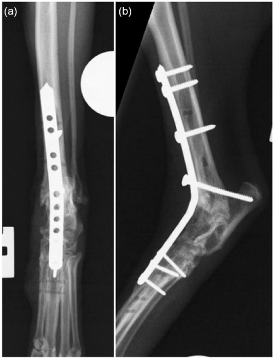
Tibiocalcaneal screws were placed engaging plate holes in five tarsi. A single calcaneotibial screw was placed through a plantar stab incision following breakage of the drill bit in the tibia during drilling in the cranial to caudal direction (cat 1; Figure 1). A calcaneotibial screw was not placed in cat number 6. The clinical notes do not record the reason for the omission of this component of the procedure. However, in this case a total of four screws were placed directly into the tarsal bones via the plate holes and the surgeon may have considered this to give adequate stability at this level. Information regarding plate type, PTA angle, tibiocalcaneal screw placement, number of screws placed in tibia, tarsus and metatarsus, and the percentage of the tibial and metatarsal length engaged with the fixation (span) were recorded and are given in Table 2. Mean PTA angle was 136° (range 116–166°). The mean number of the screws placed through the medial plate into each section of the limb were as follows: tibia 2.9 (range 2–3), tarsus 0.9 (range 0–2) and metatarsus 2.4 (range 1–4). The mean number of screws placed through the dorsal plate were as follows: tibia 3.6 (range 3–4), tarsus 0.9 (range 0–2) and metatarsus 2.1 (range 1–3). Mean percentage of bone length spanned by the medial implant was as follows: tibia 26% (range 17–32%) and metatarsus 36% (range 15–51%). Mean percentage of bone length spanned by the dorsal implant was as follows: tibia 36% (range 23–43%) and metatarsus 33% (range 13–60%). Intraoperative complications were recorded in two cases (cats 1 and 4). Both involved breakage of the drill bit during drilling for the calcaneotibial screws.

Caudocranial (a) and mediolateral (b) immediate postoperative radiographic views of the right tarsus of cat 1 showing orthogonal veterinary cuttable plates applied to stabilise pantarsal arthrodesis. Note broken drill bit in distal tibia and caudal-to-cranial orientation of calcaneotibial screw and radio-opacity dorsal to the joint consistent with demineralised bone matrix allograft in the caudocranial view
Data taken from postoperative images

Tarsal angle is the angle measured in immediate postoperative mediolateral radiographs between long axis of tibia and long axis of metatarsal. Tibial/metatarsal span refers to the length of plate overlying bone/length of bone measured along the long axis × 100
VCP = veterinary cuttable plate
Closure of the approach was performed with little concern regarding tension overlying the tibia. However, skin tension was appreciable over the tarsus and proximal metatarsus such that fascial closure was not possible in some cases, in which skin apposition alone was performed. Closure at this level was considered similar to other situations in which plates are applied to the lateral or medial metatarsus (eg, partial tarsal arthrodesis).
Re-check evaluations were performed in cats 1 and 3, at 9 and 29 weeks after surgery, respectively. The other four owners declined recheck evaluation, although the owners reported unremarkable recoveries by telephone during the weeks following surgery. In cats 1 and 3 radiographs revealed no implant-related complications and good progression of arthrodesis rates. Cat 1 showed new cancellous bone bridging in the tarsocrural and tarsometatarsal joint spaces, but joint spaces were still visible; cat 3 showed solid fusion of the tarsocrural and tarsometatarsal joints with modelling of bone and loss of subchondral bone plate. PTA angles were assessed on the follow-up radiographs: PTA angle for cat 1 was 118°; for cat 3 the left PTA angle was 131° and the right PTA angle was 123°. Orthopaedic examination of cat 1 9 weeks postoperatively revealed no hindlimb lameness with no discomfort on palpation and no instability on manipulation of the PTA. Cat 3 had a reportedly uneventful recovery and returned to normal outdoor activity but was presented 28 weeks following surgery for worsening of hindlimb function from 16 weeks postoperatively. Examination of this cat revealed moderate right hindlimb lameness with no discomfort or instability on palpation and manipulation of either PTA surgical site. A firm linear mass was palpable in the region of the right common calcaneal tendon proximally. Ultrasound examination (5–13 MHz linear array transducer [Logiq 9; GE Healthcare]) of the soft tissues caudal to the right crus revealed heterogeneous echogenicity and a focal loss of the normal myofibre pattern at the level of the myotendinous junction, alongside ill-defined hyperechoic areas, which suggested fibrosis; this was considered to be consistent with myotendinopathy of the right gastrocnemius musculotendinous unit. Involvement of the other components of the common calcaneal tendon was uncertain. Further investigation or treatment was declined at that stage and management continued with intermittent oral meloxicam. Exercise management recommended for both cases was a further 4 weeks of confinement to the house prior to a reintroduction of outdoor activity.
Cat 3 experienced a further complication approximately 13 months following surgery when the owners noticed the exposure of a screw head on the medial aspect of the left tarsus (Figure 2). Skin around the screw head appeared healthy. The protruding screw was a 2.0 mm screw in the medial plate and through the metatarsal bases. This complication was treated by removal of the medial plate and screws; the protruding screw was found not to have loosened (Figure 3). The dorsal plate was left in situ. During anaesthesia for removal of the medial plate from the left PTA, the owners gave permission for surgical exploration of the right gastrocnemius muscles and tendon of insertion. Grossly there was significant replacement of normal muscle with tough pale tissue considered to represent fibrous degeneration. The abnormal gastrocnemius tendon of insertion was resected from the level of the distal third of the tibia up to and including the majority of the fibrous gastrocnemius muscles, preserving the combined (biceps femoris, gracilis, semitendinosus) and superficial digital flexor tendons within the common calcaneal tendon. Histopathology was not performed. The owners were contacted by telephone 2 weeks following the second surgery, at which stage they reported a rapid restoration of symmetrical hindlimb function despite discontinuation of non-steroidal anti-inflammatory drugs. The owners were contacted for completion of the FMPI questionnaire 10 weeks following the second surgery, which was 16 months following the initial bilateral single-session PTA.

Caudocranial (a) and mediolateral (b) radiographic views of the left tarsus of cat 3 14 months following surgery. The head of the screw in the medial plate engaging the metatarsal bases was protruding from the skin. This screw is larger than the other screws in the metatarsals (2.0 mm vs 1.5 mm), which may have contributed to the development of this complication
Caudocranial (a) and mediolateral (b) radiographic views of the left tarsus of cat 3 14 months following surgery immediately following removal of the medial plate. The mediolateral image, in particular, allows assessment of progression of arthrodesis, which appears to be complete at all joint levels. Removal of the dorsal plate was not considered necessary and therefore was not performed
FMPI assessment was performed for all six cats a mean 8.8 months (range 6.0–16.0 months) following surgery. Results of the FMPI assessments are given in Table 3. Mean score for part 1 was 92.2% (range 80.9–97.1%). Mean score for part 2 was 95.8% (range 87.5–100%). Mean score for the overall score was 92.3% (range 81.6–97.4%). For all cats scores in part 1 were lowest for activities related to jumping and lying/sitting down. The cat with the lowest overall FMPI score was cat 1.
Results of telephone questionnaires (Feline Musculoskeletal Pain Index [FMPI]) performed for six cats following pantarsal arthrodesis by orthogonal veterinary cuttable plating

Discussion
This case series documents orthogonal plating using VCPs as an alternative for PTA in cats with talocrural injuries. The technique showed good long-term outcomes in terms of limb function and comfort. Outcomes assessed by FMPI questionnaires appeared consistent with subjective outcomes reported for a percutaneous technique of dorsal plating for PTA in cats, 3 and superior to the outcome in a previous report of canine PTA in which fewer than 50% of cases returned to pain-free limb function according to telephone contact and use of subjective owner questionnaires. 16
The optimal angle of PTA in cats has been suggested to be 100–125º.1,32 This may have been extrapolated from the tarsal standing angle for this species. However, we are unaware of studies demonstrating the importance of PTA within this range. We used the quoted range for PTA planning, rather than measuring the standing angle of the contralateral limb, although the latter method might be superior to achieve a bespoke angle for each individual. Given the potential to contour implants for each individual cat, matching the PTA angle to the standing angle of the contralateral tarsus is an option using the described orthogonal plating technique. It is difficult to be certain why a large range of postoperative tarsal angles was achieved, given the target of 100–125º. Given the relatively low stiffness of VCPs, loss of the angled contouring of the dorsal plate may have been associated with screw application. In the current study, FMPI results appeared to be unrelated to tarsal angle, with the highest FMPI outcome scores in case 5 in which tarsal angle was 166º, well outside the quoted optimal range. While achieving a natural standing angle still remains an appropriate aim, good hindlimb function in cats may be achieved through a larger range of tarsal angles than previously thought.
From these results no recommendations can be made for optimal metatarsal and tibial span and number of screws that should be placed in each bone; however, we recommend a span to encompass at least a third of the tibia and a third of the metatarsus to reduce implant strain and the potential for bone failure (particularly in the situation of the metatarsus distal to the final screw).
Intraoperative complications were observed twice, both involving breakage of drill bits during placement of tibiocalcaneal screws. Placing a positional screw between the distal tibia and calcaneus has been recommended to provide more mechanical stability and protection against plate failure in PTA in dogs, as in the absence of such a screw there is little opportunity for fixation of the calcaneus. 11 The calcaneus increases the working distance of the tarsal extensor muscles, and likely undergoes significant force during activity, which may contribute to ongoing instability of the talocalcaneal and calcaneoquartal joints. There is no specific requirement for the screw to be placed tibiocalcaneal vs calcaneotibial. Technically, it is simpler to place the screw from the plantar aspect of the calcaneus. However, calcaneotibial screw placement requires an additional surgical approach, and carries the risk of interference between the screw tip and dorsal plate. It is likely that the intraoperative complication of drill bit failure results from attempts to direct a 1.5 mm drill bit into the optimal location in the dense bone of the calcaneus having purchased the centre of the distal tibia. Another factor implicated in drill bit failure is repeat use of drill bits. There are no studies that describe the incidence of drill bit failure in veterinary orthopaedics, while in human orthopaedic surgery one study revealed that drill bits were the instrument with largest proportion of breakage. 33
The low profile of the 2.0 mm VCP may help to minimise soft tissue tension during closure of the surgical site and therefore reduce the risk of wound dehiscence and secondary postoperative infection, which has been previously reported with the use of DCPs for arthrodesis. 34 Nonetheless, the presence of two plates undoubtedly increases risk of skin tension in this site; it may be assumed that tension in the skin overlying the screw head on the left tarsus in cat 3 contributed to its emergence through the skin several months following surgery. Wound dehiscence was observed in one case in a previous series of 11 PTAs in cats, 3 despite a percutaneous soft-tissue sparing approach, but was not seen in the present cases. The larger head of the 2.0 mm screw at the level of the metatarsal bases was more prominent than the adjacent 1.5 mm screws. This may have contributed to a focal increase in skin tension, resulting in emergence of the screw head over time. It is reasonable to place a 2.0 mm screw at this level as opposed to a 1.5 mm screw, given the significantly greater dorsoplantar dimensions of the metatarsal bases in comparison with the metatarsal bodies. However, the increased screw size may not significantly increase the overall construct strength in this situation, as sufficient metatarsal screws may be placed in parallel (in the same plate) or orthogonally (in the orthogonal plate).
Another reported soft tissue complication of PTA in dogs is plantar necrosis. 13 This complication involves devitalisation of digital skin particularly medially. The arterial supply to the plantar aspect of the hindpaw is similar in the dog and cat, with the primary supply being via two arteries: the perforating metatarsal artery (originating from the dorsal pedal artery and passing between the bases of the second and third metatarsal bones) and the caudal branch of the saphenous artery. Plantar necrosis was hypothesised to occur as a result of trauma to the perforating metatarsal artery, which is vulnerable dorsomedially at the level of the metatarsal bases. 13 This complication has not been reported in cats, to our knowledge. Despite a relatively extensive approach to this region, and application of dorsal and medial plates, there were no instances of plantar necrosis in the cases reported in this series. It is possible that the arterial supply via the caudal branch of the saphenous artery is more robust in the cat and that trauma to the perforating metatarsal artery is of less concern in this species; however, this is merely conjecture.
Delayed union was suggested to be the major factor in implant failure in one study of canine PTA. 16 Insufficient stability may result in an increased time to arthrodesis and subsequent cyclical failure of the construct. 16 Orthogonal plating has been shown to be biomechanically beneficial for fracture stabilisation, but orthogonal plating for PTA has not been biomechanically tested. 22 Theoretically, orthogonal plating reduces the plate strain and increases the implant stiffness, and the medial plate is edge-loaded. This should increase the area moment of inertia and the longevity of the implants, helping to prevent the implant failure of the dorsal plate, which is subjected to significant compressive bending forces when weight is placed on the limb. No instances of failure of union, and no instances of implant failure were recorded in the three tarsi with radiographic follow-up in the present series of feline PTA. It is thought that the mechanical construct described is therefore appropriate for this procedure in cats. A further advantage to medial plating is that it allows placement of screws in the bases of all metatarsals, rather than just the third metatarsal, as with dorsal fixation alone. The third metatarsal was suggested to undergo maximal stress in cases with a dorsal plate. 14
External coaptation has been recommended following PTA to provide adjunctive stability and to relieve stress on the primary fixation. 16 However, it has been reported that external coaptation with a polyurethane cast has a high incidence of soft-tissue injuries. 29 External coaptation was not performed in any cases of PTA in cats in a previous study, 3 or in the current study. We consider that the orthogonal plating technique using VCPs for PTA in cats, as performed in this study, provides sufficient stability to achieve arthrodesis in the absence of the requirement for postoperative external coaptation, therefore avoiding the risk of associated complications.
Long-term postoperative complications were recognised only in cat 3, the case of single-session bilateral PTA. In one report of chronic gastrocnemius tendinopathy in the dog following PTA, the aetiopathogenesis was suggested to be altered tension resulting in fibroid metaplasia in the musculotendinous unit of the gastrocnemius. 13 There was no report of its management in the single reported case in that study. 13 Pathology of the gastrocnemius myotendinous unit was also reported in 3/13 dogs undergoing PTA, one of which underwent transection of the common calcaneal tendon with a resulting improvement in limb function. 11 In the current series, diagnosis of gastrocnemius myotendinopathy was performed ultrasonographically, and treatment by excision of the affected tissue appeared to result in a significant improvement in function. In the cat the soleus muscle (originating on the fibular head and merging with the lateral part of the gastrocnemius muscle or tendon, and absent in the dog) is an important extensor of the hock. We presume that excision of the fibrous tissue within the common calcaneal tendon was likely distal to the merging of this muscle with the lateral part of gastrocnemius as no division was evident. Predisposing factors for gastrocnemius tendinopathy following PTA are yet to be determined.
The complication of protrusion of a screw head from the skin in cat 3 resulted in concerns regarding the potential for introduction of infection, implant-associated biofilm/glycocalyx formation and osteomyelitis. However, the skin in the region of the screw head appeared healthy, with no evidence of infection. Implant removal resulted in uneventful healing of the skin. We are aware of no other cases in the literature of single-session bilateral PTA in a cat. Given that both major complications in this series were seen in the only cat with single-session bilateral surgery it may be prudent to suggest that this be avoided where possible.
Functional scores in all cases were favourable, indicating very good outcomes in terms of ability of the cat to perform normal feline activities and absence of pain in the affected limb. Jumping and sitting/lying down were recognised as the most commonly affected functions obtaining lower scores than other functions following feline PTA in this case series, although this is likely to be a feature of PTA, regardless of the mechanism by which it is achieved, owing to the loss of motion of the hock. It is unclear why cat 1 had a significantly lower overall FMPI outcome score.
Limitations of this study include its retrospective nature, resulting in incomplete clinical and radiographic follow-up, and the small number of cases reported with no comparator. Further studies should aim to compare the various forms of fixation described for feline PTA to inform decision-making when performing this technique and further study on the optimal PTA angle for cats.
Conclusions
PTA was performed using orthogonal plating (VCPs), resulting in good outcomes in seven cases (six cats). Excision of the gastrocnemius tendon resulted in resolution of lameness in a single case with gastrocnemius tendinopathy following feline PTA.
Footnotes
Conflict of interest
The authors declared no potential conflicts of interest with respect to the research, authorship, and/or publication of this article.
Funding
The authors received no financial support for the research, authorship, and/or publication of this article.