
Abstract
A 6-year-old male castrated Chartreux cat was referred for recurrence of an injection site sarcoma at the base of the tail 7 months after the initial surgery. Upon presentation, the physical examination was unremarkable except for a non-painful, subcutaneous mass, 2 cm in diameter, firmly attached to the underlying tissue on the left lateral side of the tail base. Complete blood count, biochemistry and urinalysis were within normal limits; thoracic radiographs and abdominal ultrasound showed no evidence of metastatic disease. After removing the mass with 3 cm margins laterally and two deep fascial planes, the defect was reconstructed after tail amputation using a coccygeal axial pattern flap based on the lateral coccygeal arteries and veins. There were no complications with wound healing and the only visible change was a difference in hair coat direction at the 1 month re-check. This is the first report to describe the utility and feasibility of the coccygeal axial pattern flap to reconstruct a large cutaneous defect over the caudodorsal pelvic region in a cat.
Case Report
Surgical reconstruction of large cutaneous defects in the caudodorsal pelvic region following trauma or tumour removal can be challenging. Reconstruction options include random subdermal plexus flaps, full-thickness skin grafts, axial pattern flaps or a combination of techniques. 1
Axial pattern skin flaps are local skin flaps incorporating a direct cutaneous artery and vein, 2 allowing for the transfer of an extensive segment of skin in a single stage. 3 Owing to the incorporation of direct cutaneous vessels, immobilisation of the area is not required for successful healing. 1
Axial flaps are easy to develop and to rotate, with survival rate ranging between 87% and 100%.3–6 However, the cosmetic appearance may be unacceptable to owners owing to the direction of hair growth and coat colour of the flap. 2
In dogs and cats, different axial pattern flaps have been described, based on the following direct cutaneous arteries: angularis oris artery, superficial temporal artery, caudal auricular artery, omocervical artery, thoracodorsal artery, superficial brachial artery, cranial and caudal superficial epigastric arteries, ventral and dorsal deep circumflex iliac artery, and genicular artery. 2
In an experimental study in dogs, Saifzadeh et al 1 demonstrated the utility of the coccygeal axial pattern flap, and reported an average flap survival of 78% of the length of the tail. Use of the coccygeal axial pattern flap for reconstruction of a cutaneous defect in the cat has not been previously reported.
A 6-year-old male castrated Chartreux cat was referred for the recurrence of an injection site sarcoma (ISS) at the base of its tail. A 5 mm mass had been noticed by the owner 1 year before the first surgery. After doubling in size, the mass was removed by the referring veterinarian with a marginal excision. The histopathology results were compatible with ISS. Seven months after the first surgery a rapidly growing mass recurred at the surgical site, and the patient was referred. On presentation, the physical examination was unremarkable except for a 2 cm, non-painful, subcutaneous mass firmly attached to the underlying tissue on the left lateral side of the tail base. The cat was admitted for further investigation; complete blood count, biochemistry and urinalysis were within normal limits, and thoracic radiographs and abdominal ultrasound showed no evidence of metastatic disease. Digital rectal examination was unremarkable. Cytology of the mass was compatible with malignant mesenchymal neoplasia. A computed tomographic scan of the pelvis for surgical planning was requested but the owner declined. The cat was anaesthetised and a dorso-ventral radiograph of the pelvic area, marking the surgical margins with skin staples, was taken to aid surgical planning. In addition, the skin of the tail was marked with staples to measure the length needed to cover the defect considering the cone area measurement rule (Figure 1a).

Pre- and intraoperative photographs. (a) Dorsoventral radiographs with skin staples around the mass and tail to aid in surgical planning; (b) defect created after excision of the mass; (c) view following dorsal tail incision; (d) suction drain placement after tail amputation; (e) dorsal view after reconstruction and closure of the subcutaneous tissue and skin
In the operating theatre the cat was placed in sternal recumbency with its tail hanging. The mass was removed en bloc with 3 cm margins laterally and with two deep fascial planes (Figure 1b).
Gloves and surgical instruments were changed before proceeding with the reconstruction.
A dorsal tail skin incision was made, and care was taken to preserve the lateral coccygeal arteries and veins (Figure 1c). Soft tissue was gently and thoroughly dissected as close as possible to the coccygeal bones to avoid damaging the vessels, and the tail was amputated between the second and third coccygeal vertebrae (Figure 1d). The flap was then rotated 180º dorsally, positioned over the skin defect and trimmed to fit the defect (Figure 1e). The total length of the flap used was approximately 55% of the entire tail length. An active Redon suction drain was placed, and the flap was sutured using a simple continuous suture with 4/0 monofilament absorbable material (poliglecaprone 25) in the subcutis and skin staples. The active suction drain was sutured to the skin with a Chinese finger trap suture. Surgical time was 40 mins and anaesthesia time was 60 mins.
The cat recovered uneventfully and was kept hospitalised for pain management (buprenorphine 10 μg/kg q12h) and maintenance of the active suction drain. Two days after surgery and after active suction drain removal, the cat was discharged with instructions to wear an Elizabethan collar for 10 days, and prescriptions for clavulanic acid potentiated amoxicillin for 7 days (12.5 mg/kg q12h) and non-steroidal anti-inflammatory drugs for 5 days (meloxicam, 0.05 mg/kg q24h). The flap was clinically evaluated at 2 days, when the drain was removed, and before discharge, and then at the following intervals post-surgery: 10 days (at suture removal), 30 days, 180 days and 365 days. Thirty days after surgery and at subsequent check-ups, the patient was in good condition and the only visible change was the different direction of hair growth in the flap compared with the surrounding coat (Figure 2). Histopathological examination confirmed the previous diagnosis of ISS with complete surgical margins. The owners were happy with the cosmetic appearance, and no clinical relapse or distant metastasis were recorded.
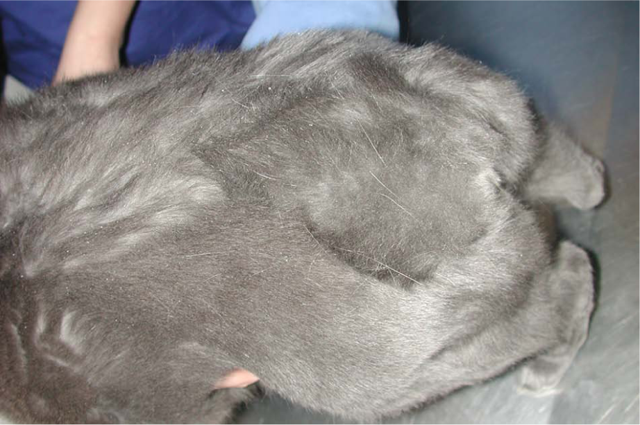
Patient at the 30-day re-check; note the different direction of hair growth between the reconstructed area and the surrounding area
In this case, a coccygeal axial pattern flap based on the lateral coccygeal artery was effective in closing a large defect at the base of the tail after tumour excision.
Regardless of the cause of injury, most skin defects can be reconstructed using advancement flaps, transposition flaps, rotation flaps, axial pattern flaps or a combination of these.
Axial pattern flaps suitable for closure of caudo-dorsal skin defects include those based on the ventral branch of the deep circumflex iliac artery and vein, and caudal superficial epigastric artery and vein. 7 However, some dorsal skin defects are not easily reconstructed with these type of flaps. The length of the caudal superficial epigastric axial pattern flap necessary to reach these defects may exceed the limit of flap length and result in distal flap necrosis. 1
The paired lateral coccygeal arteries originate from the caudal gluteal arteries and, combined with the median caudal artery, provide the majority of blood supply to the tail. 8
In an experimental study in dogs, the coccygeal axial pattern flap based on the lateral coccygeal arteries was used for reconstruction of defects on the caudodorsal pelvic region. This study revealed a mean failure of 22% of total flap area and length. The authors suggested, for clinical purposes, that the maximum flap length should not exceed 78% of the tail length in order to minimise the risk of distal flap necrosis; 1 in fact, for clinical use in this case 55% of the tail length was used.
After presurgical planning in this case, it was decided that the coccygeal axial pattern flap was most appropriate (Figure 1a) owing to the location of the tumour and to the defect created in the dorsal pelvic region (Figure 1b).
Alternate techniques were considered for reconstruction in this case but were considered to be less feasible.
Axial pattern flap based on the ventral branch of the deep circumflex iliac artery and vein, and the caudal epigastric artery and vein was considered too short to completely cover the defect; moreover, it was questionable whether the circumflex artery would still be present after the first surgery. The possibility of using a rotation skin flap was dismissed because the defect created reached the sacrococcygeal joint and the tail movement may lead to wound dehiscence. A full-thickness skin graft was not considered owing to the inability to immobilise the area.
The coccygeal axial pattern flap, in contrast with other skin flaps, is well characterised by the anatomical conformation of the tail as the cone shape of the structure confirms a larger base of the flap with a thinner apex. The cone area measurement (π × length of the tail × tail base radius [easily calculated by half tail base diameter]) was really useful in the decision-making process during surgical planning. The use of this kind of flap was also suitable in reducing associated anaesthesia time and risks involved with longer surgery, as well as cost. As with any plastic reconstructive surgery, the procedure proposed must be preceded by precise measurement of the defect and the involved tissue in order to gain the correct amount of donor skin.
Conclusions
The use of a coccygeal axial pattern flap was useful inthe decision-making process during surgical planning, and was an easy technique for the closure of a left dorso-lateral defect at the base of the tail without any short- or long-term complications. The total length of the flap used was approximately 55% of the entire tail length; therefore, according to Saifzadeh et al, 1 the risk of distal necrosis was minimal. According to the owner, the overall cosmetic aspect of this flap in this cat was excellent, even given the change in the direction of the hair coat, which was noticeably different in the reconstruction area compared with the surrounding area (Figure 2). The coccygeal axial pattern flap based on the lateral coccygeal arteries was a useful and easy technique for the coverage of the gluteal, ischial and dorsal areas after tumour excision in a cat.
Footnotes
Acknowledgements
We gratefully acknowledge Professor Paolo Buracco and Samuel Sharp for their assistance with the preparation of the manuscript.
Conflict of interest
The authors do not have any potential conflicts of interest to declare.
Funding
The authors received no specific grant from any funding agency in the public, commercial or not-for-profit sectors for the preparation of this case report.