
Abstract
Interpretation of ultrasonographical measurements requires an understanding of the source and the magnitude of variation. A substantial part of the variation can be attributed to the observer, the equipment or the animal. The aim of this study was to evaluate which adrenal gland measurement is the least variable within and between observers. Three experienced ultrasonographers examined six cats at three different times on the same day, more than 1 h apart, according to a strict scanning protocol. Seven ultrasonographical measurements were performed on each adrenal gland (maximal length on sagittal images, maximal height at the cranial and caudal poles on sagittal and transverse images, and maximal width of the cranial and caudal poles on transverse images). Height measurements in both planes showed the lowest variability within and between observers compared with length and width measurements. Descriptive ultrasonographical features, such as echogenicity of the gland, presence of hyperechoic spots or layering assessment, demonstrated satisfactory-to-good intra- and inter-observer agreement, whereas the shape assessment showed very poor inter-observer agreement. The results of this study describe a reliable scanning protocol that can be the basis for future adrenal ultrasonographical examinations for cats suspected of adrenal disease (eg, hyperaldosteronism, hyperadrenocorticism, sex hormone-producing tumours).
Introduction
Ultrasonography of the adrenal glands is the most commonly used imaging modality in the diagnosis of adrenal diseases in cats. 1 Only two studies have examined the reliability of the ultrasonographical measurements of the feline adrenal glands in healthy cats.2,3 The first study concluded that ultrasonographical measurements underestimate the anatomic measurements (2 mm smaller). In the same study, the length and height were more reliable than the width. 2 In the second study, which concerned the reliability of ultrasonographical measurements, the coefficient of variation of ultrasonographical measurement of the adrenal glands was lowest for the short-axis measurement (4.4–7.2%). The maximal measurement variation in a day-to-day serial examination was 1.2 mm in 10 cats. 3
The aim of this study was to evaluate which adrenal gland measurement is the least variable within and between observers. This may help to determine if we can assess a more reliable normal and pathological threshold for the size of the feline adrenal glands.
Materials and methods
Six privately owned cats were involved (two castrated males and four neutered females), ranging in age from 3.0 to 9.0 years (mean 5.7) and weighing 3.0–5.0 kg (mean 4.2). The cats were clinically healthy, exempt of any past disease and until at least 6 months after the study.
The ultrasonographical examination was performed in dorsal recumbency without sedation using a multifrequency microconvex probe set at 8 MHz (IU22; Philips Medical). Three ultrasonographers were involved in the study, two European College of Veterinary Diagnostic Imaging diplomates (JHS, EV) and one final-year resident (AC), operating with the same machine settings. At three different times on the same day (>1 h apart), they scanned both adrenal glands according to a previously described technique.3,4 The shape (bi-lobed, fusiform or rounded), the echogenicity (hypo-, iso- or hyperechoic compared with the surrounding fat) and the presence of layers or hyperechoic spots, with or without distal acoustic shadowing, were recorded. According to a previously described protocol with strict scanning planes, 5 seven distances were measured (Figures 1–4): the maximal length (craniocaudal) in sagittal plane, the maximal width (mediolateral) of the cranial and the caudal poles in transverse plane, and the maximal height (ventrodorsal) of the cranial and caudal poles in sagittal and transverse planes. The difficulty in finding the adrenal gland and the behaviour of the cat were recorded. An additional external person (ES) assured operators were blind in regard to the cats being scanned.

Length (+) and height (x,⊙) ultrasonographical measurements of the left adrenal gland (LAG) in a sagittal plane
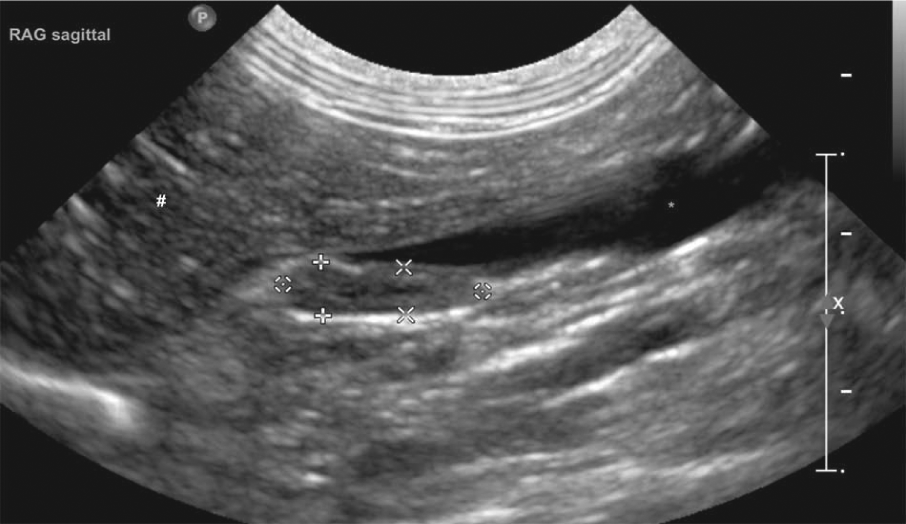
Length (+) and height (x,⊙) ultrasonographical measurements of the right adrenal gland (RAG) in a sagittal plane. # = liver; * = caudal vena cava

Height (+) and width (x) ultrasonographical measurements of the left adrenal gland (LAG) in a transverse plane. a = aorta

Height (+) and width (x) ultrasonographical measurements of the right adrenal gland (RAG) in a transverse plane. # = liver; * = caudal vena cava; § = duodenum
A random-effects model was used with cat, observer and measurement within observer as random effects. 6 Within the framework of the random-effect model, three variance components are estimated, that is, the variation within observers, σ2a, the variation due to observers, σ2i, and the variation due to cats, σ2c, using restricted maximum likelihood techniques. 7 Based on these variance component estimates, limits of agreement (LA) were derived that contain 95% of the measurements of the same cat by the same observer (within-observer LA); 95% of the measurements of the same cat, but assessed by different observers (between-observer LA); and 95% of the measurements of different cats assessed by different observers (between-cat LA). Assuming normally distributed measurements, the limits of agreement are obtained from the observed average value for the particular measurement, together with the appropriate sources of variation, that is, the variance components.
The cats’ behaviour and the difficulty in finding the adrenal gland were considered as covariates, and the effect of these covariates on the within-observer variance of the adrenal measurements was evaluated with a paired Student’s t-test, using cat and observer as a blocking factor.
For the descriptive characteristics (shape, echogenicity, hyperechoic spots, layering), the percentage of agreement both within and between observers was calculated and compared with the expected value under the hypothesis that the assessment was haphazard. Considering the shape or the echogenicity or the layering of the gland with three possible values, the expected value for pairwise agreement equalled 33% both within and between observers. Considering the presence or absence of hyperechoic spots, the expected value for pairwise agreement equalled 50% both within and between observers.
Results
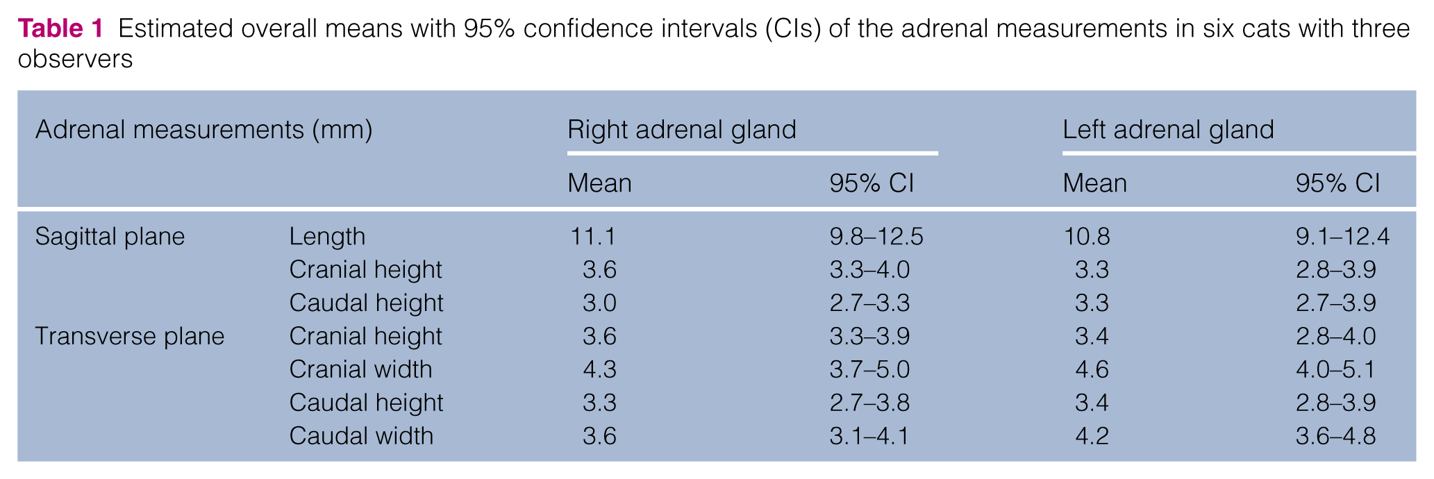
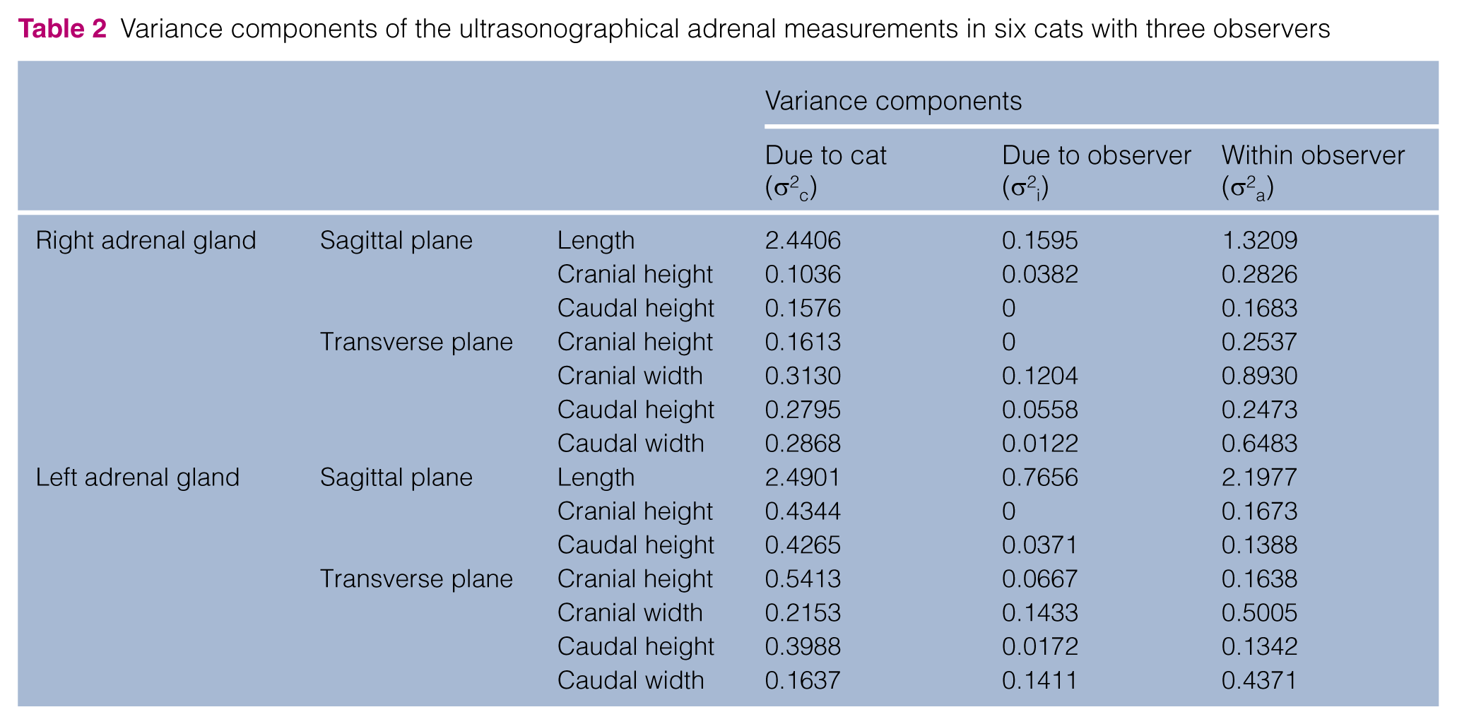
The estimated overall means (with 95% confidence intervals) of the adrenal gland measurements are presented in Table 1. The variance components are shown in Table 2 and the LA, as defined earlier, in Figure 5. For all measurements, the extra variation due to the observer is moderate compared with the variation within observers and the extra variation due to cat.
Estimated overall means with 95% confidence intervals (CIs) of the adrenal measurements in six cats with three observers
Variance components of the ultrasonographical adrenal measurements in six cats with three observers

Limits of agreement for measurements within observers (intra-observer), measurements between observers (inter-observer) both on the same cat, and measurements between observers on different cats. RAG = right adrenal gland; LAG = left adrenal gland; CrH = cranial height; CdH = caudal height; CrW = cranial width; CdW = caudal width; sag = sagittal plane; tr = transerve plane
As expected, length had the highest variability for both glands, with larger variability on the left. The within-observer LA equalled 9.3 (13.0) mm for the right gland and 8.3 (13.1) mm for the left one. The between-observer LA equalled 9.1 (13.1) mm for the right gland and 7.9 (13.6) mm for the left one, only slightly broader than the within-observer LA. The between-cat LA, however, was substantially broader – equal to 7.9 (14.4) mm for the right gland and 6.9 (14.6) mm for the left one.
The variance components of most of the short axis measurements were moderately larger in the right gland compared with the left one (Table 2).
The within- and between-observer variance components of the cranial height measurements were comparable if taken in a sagittal or a transverse plane. In the sagittal plane, the within-observer LA equalled 2.8 (4.5) mm for the right adrenal gland and 2.7 (4.0) mm for the left one, whereas the between-observer LA equalled 2.7 (4.6) mm for the right and 2.6 (4.0) mm for the left adrenal gland, only slightly broader than the within-observer LA. The between-cat LA was only slightly broader and equalled 2.6 (4.7) mm for the right adrenal gland and 2.0 (4.6) mm for the left one. With regard to the right adrenal gland, the within-observer LA was substantially broader for the cranial height measurements compared with the caudal height measurements in both scanning planes.
The within-and between-observer LA of the caudal height measurements are comparable if taken in a sagittal or a transverse plane. In the sagittal plane, the within-observer LA equalled 2.3 (3.7) mm for the right adrenal gland and 2.7 (3.9) mm for the left one, whereas the between-observer LA was the same, that is, 2.7 (3.7) mm for the right and only slightly broader, that is, 2.6 (4.0) mm for the left adrenal gland. A moderate increase was observed for the between-cat LA, with values equal to 2.1 (4.0) mm for the right adrenal gland and 2.0 (4.6) mm for the left one.
The within and between variance components were, in most cases, smaller for the height measurements than for the width measurements, except for the caudal width of the right adrenal gland in a transverse plane. On the contrary, the between-cat variance component was similar between height and width measurements.
The within-observer LA for cranial/caudal width measurements corresponded to 2.8 (5.9) mm/2.3 (4.9) mm for the right adrenal gland and to 3.4 (5.7) mm/3.1 (5.3) mm for the left one, whereas the between-observer LA for cranial/caudal width measurements corresponded to 2.7 (6.0) mm/2.3 (5.0) mm for the right and 3.2 (5.9) mm)/3.0 (5.5) mm for the left adrenal gland. The between-cat LA was 2.4 (6.2) mm)/2.0 (5.2) mm for the right and 3.0 (6.1) mm/2.8 (5.6) mm for the left adrenal gland.
The percentage of agreement both within and between observers about the descriptive ultrasonographical features of the adrenal glands are shown in Table 3. The echogenicity and layering description demonstrated very good within-observer agreement (93%). The shape description and the presence of focal spots demonstrated a moderate within-observer agreement (80% and 81%, respectively). The between-observer agreement on the shape of the adrenal gland was very poor (31%), even below the expected 33% if choices were made haphazardly, whereas the between-observer agreement for the presence of focal spots, the layering and the echogenicity was satisfactory (78%, 89% and 74%, respectively).
Percentage of agreement within and between observers for descriptive ultrasonographical features of the adrenal glands in cats. The expected value under the hypothesis of random choice equals 33% for the three category variables shape, echogenicity and layering pattern and 50% for the binary variable hyperechoic spots

Discussion
The agreement for the ultrasonographical adrenal measurements in healthy cats was high overall, both within and between observers. This result confirms the ease of this procedure. Although we only dealt with healthy cats in this study, the good agreement between observers in the present study would be interesting in a clinical situation, as different observers could reproduce the procedure without major variation.
A previous study described the repeatability of the ultrasonographical adrenal measurements in cats. It showed a small coefficient of variation on 10 consecutive measurements (2.5–13.5% for the length and 4.4–7.2% for the short-axis measurement) and the difference in adrenal measurements between two consecutive days ranged from 0.3 to 1.2 mm. However, the latter study did not report the effect of multiple observers. 3
The lack of standardised scanning protocol in previous studies of ultrasonographical adrenal measurements makes comparison between them and with our study difficult.2,3 Although the length is clearly anatomically defined, the short-axis measurements are more variable when called thickness, height or width. We constrained ourselves to strict scanning planes to define the height of the adrenal gland in a dorsoventral plane and the width in a mediolateral plane. In the present study, the width measurements had much higher within- and between-observer variability than the height measurements. A previous study with a very similar scanning protocol reports that the only significant differences between the post-mortem physical measurements and the ultrasonographical measurements are in width. 2 Comparing the maximum short-axis measurements (reported as 5.3 mm in a previous study 3 ), the present height measurements never reached 5 mm, whereas the width measurements reached 6 mm. The ultrasonographical adrenal width appears to be a less reliable measurement in cats. This result is likely owing to the ill-defined medial and lateral borders of the glands compared with the dorsal and ventral outline associated with the close contact with vascular landmarks (aorta and caudal vena cava for left and right adrenal glands, respectively).
Another hypothesis to explain the increased variability of the width measurement may be the use of a transverse scanning plane to measure the width. If the scanning plane is oblique, the true width of the gland could be overestimated. This finding is reported in ultrasonographic studies of the canine adrenal and thyroid glands.5,8 It appears difficult to obtain a true transverse plane of the gland, as the adrenal gland in cats is not exactly in the sagittal plane of the body, but slightly angled with a more laterally deviated cranial pole. 4 On the contrary, it is easier to obtain a true sagittal plane by aiming the maximum length of the gland. Considering the height measurements, the within- and between- observer variability is similar if measured in sagittal or transverse planes. Indeed, if the adrenal gland is parallel to the surface of the transducer, when the scanning orientation changes from a sagittal to a transverse plane, the height of the gland remains perpendicular to the transducer, so quite consistent, whereas the width depends on the degree of rotation of the transducer and is most likely to be more variable.
In addition, in the right adrenal gland, the cranial height measurement had broader within-observer limits of agreement than the caudal height measurement in any plane. These findings are similar to what is described in dogs and most likely due to the similar echogenicity and the close contact of the right adrenal gland, particularly the cranial pole, with the caudate lobe of the liver and the caudal vena cava. 5
The agreement on the descriptive ultrasonographical features of the feline adrenal gland is satisfactory to very good, except for the assessment of the shape between observers. Despite a consensual scanning protocol, the descriptive features are very subjective. To reduce variability, the internal echogenicity and echostructure of the gland may be compared with adjacent landmarks or organs. However, the changes in shape of the gland are minor and too subjective to be evaluated reliably with ultrasound.
Conclusions
The within and between limits of agreement of the ultrasonographical adrenal measurements in healthy cats are narrow overall, confirming the reliability of ultrasonography. The main precautions to reduce the variation are the use of a strict consensual scanning protocol and the same updated and calibrated ultrasound device. Considering the scanning protocol, height measurements in sagittal or transverse planes are more reliable than length or width measurements in the transverse plane.
Footnotes
Acknowledgements
We would like to thank Laure Gatel, Eva Vandermeulen and Miguel Campos for their contribution to the experiment.
Conflict of interest
The authors do not have any potential conflicts of interest to declare.
Funding
This research received no specific grant from any funding agency in the public, commercial or not-for-profit sectors.