
Abstract
There have been major concerns that the nephrotoxicity of commercial formulations of Roundup herbicide is due to the active ingredient glyphosate. We therefore investigated and compared the mechanisms underlining the nephrotoxicity of Roundup herbicide and glyphosate alone in rat. Fifty-six adult male rats randomized into 7 groups of 8 rats per group were exposed to Roundup formulation and glyphosate alone daily by gavage at 3.6, 50.4, and 248.4 mg/kg body weight (bw) of glyphosate concentrations for 12 weeks with distilled water administered to the control group. Kidney biomarker (serum urea and creatinine, plasma cystatin-C, and neutrophil gelatinase-associated lipocalin), oxidative stress indices in the kidney tissue, activities of kidney membrane-bound enzymes (Mg-adenosine triphosphatase [ATPase], Ca-ATPase, Na/K-ATPase, and total ATPase), and histopathological changes in the kidney were monitored. Glyphosate concentration in the kidney was quantified by high-performance liquid chromatography with ultraviolet detection. Significant (P < 0.05) alterations in the levels of the kidney biomarker, oxidative stress markers, and membrane-bound enzymes were observed in the rats exposed to Roundup compared to the rats exposed to glyphosate alone. Rats exposed to Roundup accumulated more glyphosate residue in their kidney tissue. Severe histopathological lesions were only seen in the kidneys of rats exposed to Roundup. The nephrotoxicity observed cannot be due to the active ingredient in the Roundup formulation, as glyphosate alone has virtually no effect on the renal function of the exposed animals. Therefore, the general claim attributing nephrotoxicity of a glyphosate-based herbicide to its active ingredient should be discouraged.
Introduction
The need to feed the world’s increasing human population has prompted the use of agrochemicals meant to increase food production and ensure the continuation of the human race. 1 Such agrochemicals include pesticides, several formulations of inorganic fertilizers, and herbicides marketed as harmless, with little or no side effects by the manufacturers. However, these agrochemicals have been viewed by some researchers as chemicals whose environmental fate is poorly understood, despite the rapidly increasing use. 2 An example of these agrochemicals is glyphosate-based Roundup herbicide manufactured by Monsanto. Roundup is a broad-spectrum herbicide and reportedly used in agriculture, horticulture, and silviculture on large farms, around homes, and gardens for the control of annual, biennial, and perennial grasses, sedges, and weeds. 3 It is also used as a desiccant on cereals, oilseed rape, maize, and sunflowers. 4 Commercial formulation of Roundup contains glyphosate (N-phosphonomethyl glycine) as its active ingredient 5 and a mixture of polyoxyethylene amine (POEA) with other unspecified surfactants 6 required for rapid penetration and absorption of the herbicide into the plant.
Glyphosate, which today has become worldwide the most widely used active substance for weed control, was reported to have been introduced into agricultural practice in the 1970s. 7,8 It has been reported to account for 35% of all pesticides used in agricultural production in Denmark. 9 In addition, Steinmann et al 10 reported that glyphosate is used on 4.3 (39%) million hectares of agricultural land each year in Germany. Also, 50% to 60% of sunflower crops in France, Romania, and Hungary are known to be treated before harvest with glyphosate, and it is the most commonly used herbicide in commercial fruit orchards in the United Kingdom. 11 It is the most commonly used herbicide in Africa, 12 and Chikoye et al 13 reported that Nigeria has achieved 55% increase in the yield of agricultural production due to the increasing use of the herbicide.
As a result of its use in desiccation, its residues have been reportedly detected in foodstuffs as well as drinking water contaminated via rain, surface runoff, and leaching into groundwater, thereby increasing the likely routes of animal exposure. 14,15
Studies have reported various toxicities of glyphosate-based herbicide in the exposed animals. For instance, hematological, biochemical, and oxidative alterations in mice exposed to 50 and 500 mg/kg body weight (bw)/day of glyphosate have been documented. 16 Irreversible liver damage in rats consuming 4.87 mg/kg bw, of glyphosate every 2 days over 75 days has been reported by Benedetti et al. 17 Romano et al 18 revealed an endocrine disruptive effect of glyphosate on rats exposed to 5, 50, and 250 mg/kg bw. Glyphosate has also been reported to induce oxidative stress due to the production of reactive oxygen species in animals. 19
Although the mechanisms of Roundup toxicity in mammals are still questioned, and there are mounting concerns that the reported toxicities could either be due to the active ingredient (glyphosate) in the herbicide or other surfactants that make up the herbicide formulation. However, Mesnage et al 20 stated that there is an unexpressed widely believed assumption that the active ingredient against plant metabolism is the most toxic compound of glyphosate-based Roundup herbicide formulation. Meanwhile, Jasper et al 16 reported that the mechanisms of toxicity of glyphosate formulations are complicated. Although a report has shown that several regulatory agencies and scientific institutions worldwide have concluded that both glyphosate alone and commercial formulations of Roundup pose no threat to human health, 21 the report was later challenged by Brausch and Smith 22 who noted that glyphosate alone is very slightly to moderately toxic to aquatic animals, but the commercial formulation of Roundup is considered more toxic due to the presence of the surfactant POEA.
While some scientific bodies have reportedly declared that glyphosate alone isn’t toxic and rather the adjuvants that are added to aid absorption into plant tissues are toxic, 23,24 a report by Williams et al 21 has classified these adjuvants as inert. This leaves an argument in the favor of selectively studying the toxicity of various constituents of glyphosate-based herbicides.
In an effort to understand what could be considered as responsible for Roundup toxicity and avoid ambiguity, Mesnage et al 5 who studied the damaging effect on liver and kidney in rats exposed to ultra-low dose (0.1 ppb of Roundup corresponding to 4 ng/kg bw/day of glyphosate consumed for 2 years) of Roundup herbicide and our recent study which indicated reproductive toxicity of Roundup herbicide at 3.6, 50.4, and 248.4 mg/kg bw of glyphosate doses in male albino rats 25 concluded that it is difficult to attribute the toxicity of the commercial formulation of Roundup to a given component and suggested more research testing for glyphosate alone.
Meanwhile, a study has reported the level of glyphosate in the kidney to be over 100-fold greater than the level found in fat muscles and most other tissues, 26 and most research showed that urine is the main route of glyphosate’s elimination from the body. 27 -29 The process of urine formation in the kidney has been well documented. Miller and Harley 30 summarized that blood pressure from the closed circulatory system forces blood through a membrane filter in a kidney, where it goes through the key processes of filtration, reabsorption, secretion, and excretion in which urine is voided from the body. Despite the huge attention given to the elimination of glyphosate from the body through urine, very little attention has been given to the possible effects of this glyphosate on the kidney which is the chief organ in urine formation.
Few studies have examined the effects of glyphosate-based herbicide on kidney function in animals. Increased incidence of anatomical sign of pathologies as well as alterations in urine biochemical parameters, indicative of renal dysfunction by glyphosate, have been reported 31 ; increased sign of kidney anatomorphological pathology by glyphosate has also been documented 32 ; human epidemiology studies indicate that unexplained chronic kidney disease due to glyphosate-based herbicide exposure killed thousands of rice farm workers in Sri Lanka 33 and sugarcane workers in Central America. 34 All the studies however attributed its findings on the kidney toxicity to glyphosate, the active ingredient, but each of the studies only tested the commercial formulations of glyphosate-based Roundup, advocating the need to consider more research testing for glyphosate alone. In an effort to better understand the renal toxicity of glyphosate-based herbicide, we therefore designed this study to assess and compare the effects and mechanisms of action of glyphosate alone and commercial formulation of Roundup on renal function in male albino rats.
Materials and Methods
Chemical
The herbicide product tested was the one commonly used in Nigeria: Roundup (Monsanto Europe S.A./N.V., Antwerp, Belgium) in a commercial formulation (360 g/L of glyphosate in the form of 441 g/L potassium salt) and glyphosate (Sigma-Aldrich subsidiary of Merck, St. Louis Missouri, USA). They were stored in their original container and kept in a cool, dry, and well-ventilated area at room temperature throughout the experiment.
Experimental Animals
A total number of 56 apparently healthy adult male albino rats weighing 200 to 210 g were used in this study. The rats were obtained from the breeding section of the animal house of the Department of Zoology and Environmental Biology, Olabisi Onabanjo University, Ago-Iwoye, Nigeria, and were initially acclimatized under laboratory conditions of 25°C ± 5°C and 65% ± 5% relative humidity in a well-ventilated experimental animal house for 1 week before the initiation of the study. The rats were allotted into individual wooden cages (65 × 35 × 50 cm). Standard laboratory rat chow and clean drinking water were supplied to ad libitum. The experimental protocol was conducted in accordance with the regulations of the local ethics committee in animal care unit of Olabisi Onabanjo University, Ago-Iwoye, Ogun State, Nigeria. We performed the animal experiment according to ethical guidelines of animal experimentation (regulation CEE 86/609).
Experimental Design
This study was designed according to our previous study 25 with little modifications. The rats were randomized into 7 experimental groups consisting of 8 rats per group. Three groups were administered by oral gavage with 0.01, 0.14, and 0.69 mL/kg bw/d of Monsanto Roundup herbicide corresponding to 3.6, 50.4, and 248.4 mg/kg bw/d of glyphosate, respectively. Another 3 groups were also treated by oral gavage with glyphosate alone at 3.6, 50.4, and 248.4 mg/kg bw/d, whereas the last (control) group was administered with distilled water for a period of 12 weeks. Roundup and glyphosate were diluted in an aqueous suspension and administered to the rats once per day in a volume of 0.25 mL/100 g of body weight as described earlier. 18 The dose regime was based on no-observed-adverse-effect level (NOAEL) for developmental toxicity in rats as reported. 17,21,35 The values are within the limits of NOAEL and equivalent to 1/1,000, 1/100, and 1/20 of lethal dose (LD50) in rats as described. 36 At the end of the exposure period, blood sample and kidney tissue were collected from the rats in each group for laboratory analysis.
Sample Collection and Laboratory Analysis
The blood samples were collected from the retro-orbital sinus into microhematocrit tubes. Serum samples were separated within 1 hour after collection. The kidney tissue was dissected and washed with saline solution. One of the excised kidneys of each rat was minced and homogenized in Tris-HCl buffer (pH 7.4) and centrifuged (3,000×g for 10 minutes), and the resulting clear supernatant was used for various enzymatic and nonenzymatic biochemical assays. The second portion of the kidney tissue was fixed in 10% neutral-buffered formalin and processed for histopathological examinations while the third portion was used for glyphosate estimation.
Analysis of Kidney Biomarker
Serum samples were analyzed for urea and creatinine using Chemistry Hitachi Model 917 multichannel analyzer (Roche Diagnostics, Indianapolis, Indiana). We checked for accuracy and precision with Bio-Rad quality controls (Bio-Rad Laboratories Inc. Hercules, Califonia). Meanwhile, cystatin-C (Cys-C) and neutrophil gelatinase-associated lipocalin (NGAL) in plasma were quantified using a commercial enzyme-linked immunosorbent assay kit (Bioporto Diagnostics Gentofte, Denmark) according to the manufacturer’s instruction.
Analysis of Antioxidant System Parameters in Kidney Tissue
Thiobarbituric acid reactive substance assay was carried out to measure malondialdehyde (MDA), a lipid peroxidation product, at 534 nm in the kidney tissue of the rats. Measurement was obtained at a temperature of 95°C for 30 minutes as described previously. 37 The level of reduced glutathione (GSH) was estimated in the kidney tissue of the rats based on the reduction of 5,5-dithiols-2 nitrobenzoic acid by sulfhydryl (SH) groups of glutathione to form 2-nitro-mercaptobenzoic per mole of glutathione. 38 For the analysis of catalase (CAT) activity, we used the method of Johansson and Borg. 39 However, the activities of glutathione peroxidase (GPx) and superoxide dismutase (SOD) were determined using the Cayman assay kit as described by the manufacturer.
Estimation of Membrane-Bound Adenosine Triphosphatase
The membrane-bound adenosine triphosphatase (ATPase) enzyme (Na+/K+-ATPase, Ca2+-ATPase, and Mg2+-ATPase) analysis was carried out in the kidney tissue of the rats using the method of Abramsa and Smith, 40 Hjerten and Pan, 41 and Ohnishi et al 42 with little modifications. Briefly, 0.1 mL of kidney homogenate was added to a mixture containing ice-cold Tris-HCl buffer (0.1 mol/L pH 7.4), 5 mmol/L of ATP, 2 mmol/L of CaCl2, 2 mmol/L of MgCl2, 60 mmol/L of NaCl, 20 mmol/L of KCl, and protein enzyme making 2 mL of assay mixture. The assays were carried out by estimating the amount of phosphorus liberated from the incubation mixture. Incubation of the tubes was at 37°C, and enzymes were inactivated by the addition of 1 mL cold 10% (w/v) trichloroacetic acid (TCA) after 25 minutes. The tubes were kept in ice for 20 minutes. Precipitated proteins were removed by centrifugation. A control was simultaneously setup by adding enzyme after TCA at the end of the incubation period. Protein concentration was estimated by the method of Lowry et al 43 using bovine serum albumin as the standard
Glyphosate Analysis
The second excised kidney samples of the rats were analyzed for glyphosate with the use of Agilent Technologies 1200 series high-performance liquid chromatography with ultraviolet detection (Agilent Technologies, Santa Clara, Califonia, USA) as described by Qian et al. 44 Isophritic mode of running on a Zorbax Eclipse XDB-C18 column (250 × 4.6 mm) (Thomas Scientific) was used. The mobile phase consists of methanol and water in ratio 50:50 with an injection volume of 5 μL and retention wavelength of 220 nm run for 10 minutes. A blank sample was spiked with prepared calibration standard glyphosate solutions of 6.25, 12.5, 25, 50, and 100 mg/kg and equilibrated. A calibration curve was obtained from the matrix-matched multilevel calibration solutions, and linearity was determined from the coefficient of association (R2).
Histopathological Studies
Our previous method 45 was adopted with little modifications for the qualitative analysis of the kidney histology in this study. Briefly, the tissue samples were fixed for 48 hours in 10% formalin–saline and dehydrated by passing successfully in a different mixture of ethyl alcohol and water, cleaned in xylene, and embedded in paraffin. Sections of the tissues (5–6-μm thick) were prepared using a rotary microtome. The obtained tissue sections were collected on glass slides, deparaffinized, and stained with Hematoxylin–Eosin (H&E) before mounting in neutral Dibutyl Phthalate Xylene (DPX) medium. Prepared slides were examined at 400× magnifications.
Statistical Analysis
All the data were presented as the Mean ± Standard Deviation (SD). Statistical analyses for all measurements were performed using Statistical Package for Social Sciences (SPSS) version 20.0. The mean, standard error of the mean, and analysis of variance were conducted. Post hoc test was performed using the Student-Newman-Keuls. P < 0.05 was considered to be statistically significant.
Results
Level of Kidney Biomarker
Figure 1 presents the serum levels of urea and creatinine in the rats exposed to Roundup formulation and glyphosate alone at varying concentrations. Level of serum urea was significantly higher (P < 0.05) in the rats exposed to Roundup at 248.4 mg/kg bw of glyphosate concentration. This was followed by those exposed to the formulation at 50.4 mg/kg bw of glyphosate. Level of serum urea was lowest in the control and not statistically significant (P > .05) compared to those exposed to Roundup formulation at 3.6 mg/kg bw of glyphosate concentration and those exposed to all the varying concentrations of glyphosate alone. However, there was no significant difference in the creatinine level in the control group and those of the other experimental groups except in the rats exposed to Roundup formulation at 248.4 mg/kg bw of glyphosate concentration, where the level of creatinine was observed to be significantly (P < 0.05) higher.
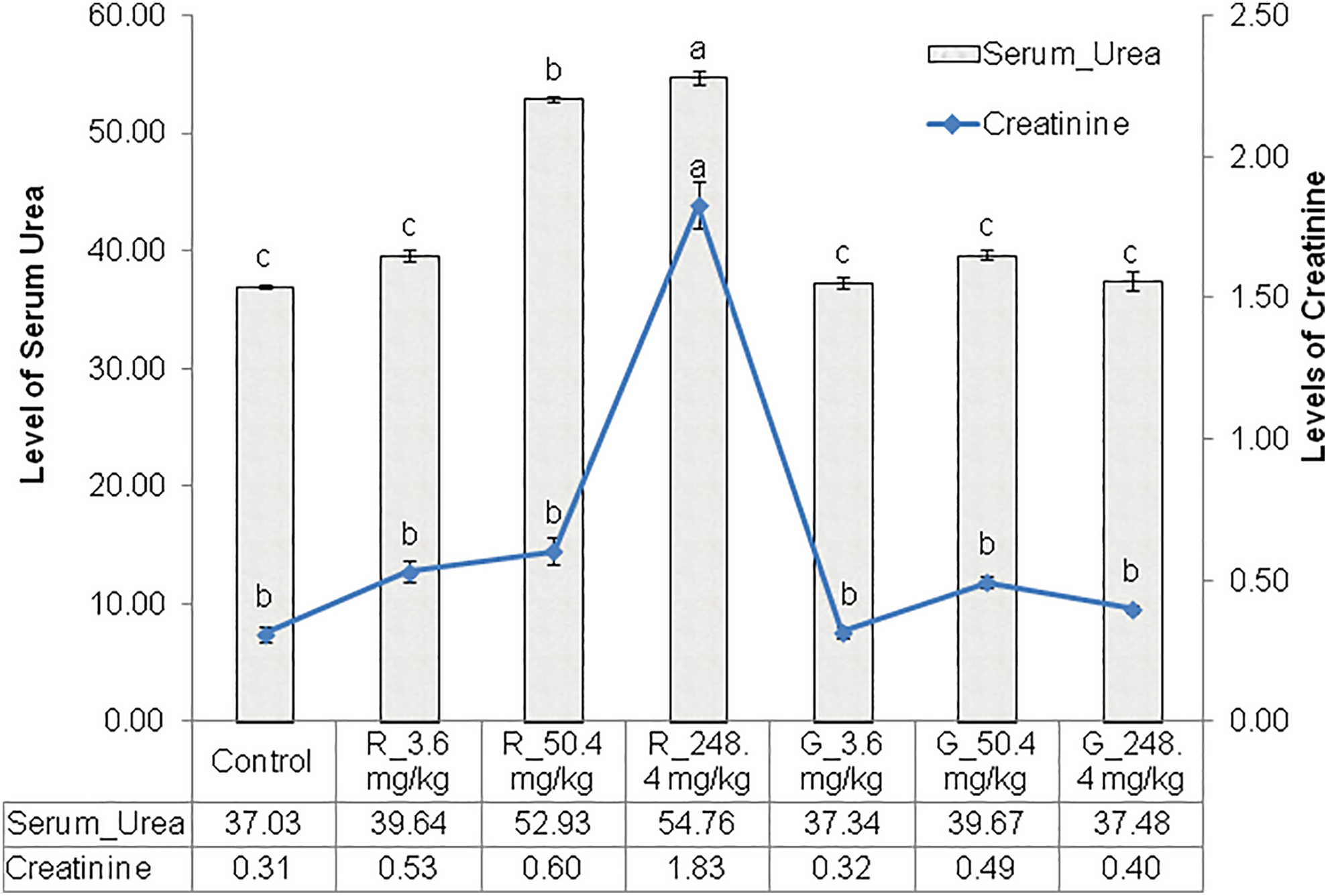
Levels of serum urea and creatinine (mg/dL) in the rats exposed to Roundup formulation and glyphosate alone at varying concentrations. G_ indicates pure glyphosate exposure; R_, Roundup herbicide exposure. Error bars represents standard deviation.
The levels of plasma NGAL and Cys-C in the rats exposed to Roundup formulation and glyphosate alone at varying concentrations are represented in Figure 2. Level of plasma NGAL was significantly higher (P < 0.05) in the rats exposed to Roundup at 284.4 mg/kg bw of glyphosate concentration and followed by those exposed at 50.4 mg/kg bw of glyphosate concentration. However, there was no significant difference (P > 0.05) in the level of plasma NGAL between the control, those exposed to Roundup at 3.6 mg/kg bw of glyphosate concentration and those exposed to all the varying concentrations of glyphosate alone. On the other hand, there was no significant difference in the level of plasma Cys-C in control and those of the other experimental groups except in the rats exposed to Roundup formulation at 248.4 mg/kg bw of glyphosate concentration, where plasma Cys-C level was observed to be significantly (P < 0.05) higher.
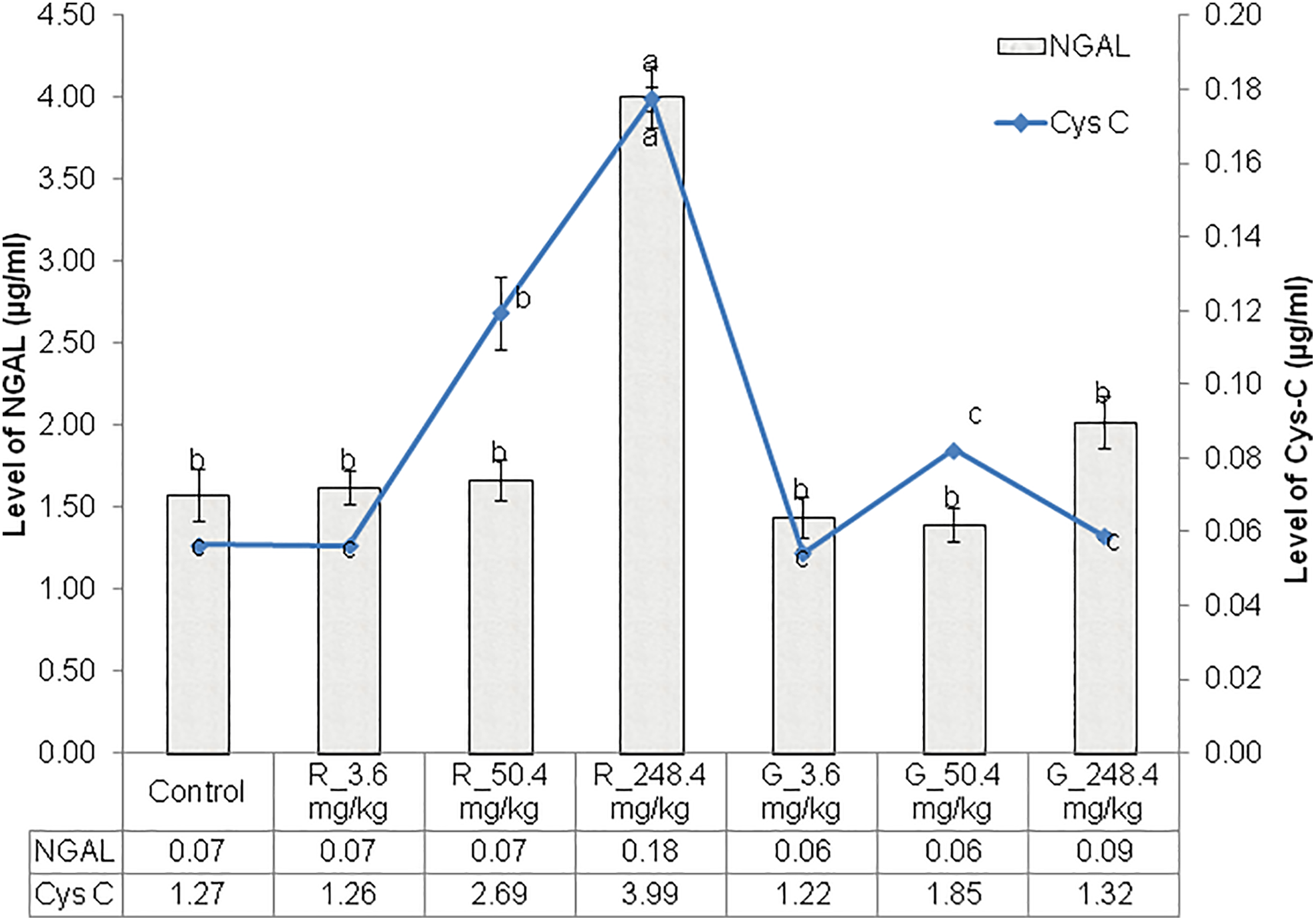
Levels of plasma NGAL and Cys-C (μg/mL) in the rats exposed to Roundup formulation and glyphosate alone at varying concentrations. Cys-C indicates cystatin-C; R_, Roundup herbicide exposure; G_, pure glyphosate exposure; NGAL, neutrophil gelatinase-associated lipocalin. Error bars represents standard deviation.
Level of Oxidative Stress Markers in the Kidney
Table 1 shows the level of oxidative stress markers in the kidney of rats exposed to Roundup formulation and glyphosate alone at varying concentrations. Levels of SOD and CAT in the kidney tissue of rats exposed to Roundup formulation were observed to be significantly (P < 0.05) reduced compared to the control and those exposed to glyphosate alone. These values were significantly higher (P < 0.05) in the rats exposed to glyphosate alone at 3.6 mg/kg bw. On the other hand, there was no significant difference (P > 0.05) in the levels of SOD and CAT between the control, those exposed to Roundup formulation at 3.6 mg/kg bw of glyphosate concentration, and those exposed to glyphosate alone at 50.4 and 248.4 mg/kg bw. Activities of SOD and CAT were significantly lowest in the rats exposed to Roundup formulation at 248.4 mg/kg bw of glyphosate concentration. In a similar trend, GPx activity and GSH concentration were significantly higher in the rats exposed to glyphosate alone at 3.6 mg/kg bw but lowest in the rats exposed to Roundup formulation at 248.4 mg/kg bw of glyphosate concentration. On the other hand, the level of MDA was significantly higher (P < 0.05) in the rats exposed to Roundup formulation at 248.4 mg/kg bw of glyphosate concentration. There was however no significant difference (P > 0.05) in the level of MDA between the rats exposed to Roundup formulation at 3.6 and 50.4 mg/kg bw of glyphosate concentration and those exposed to glyphosate alone at 50.4 and 284.4 mg/kg bw. The level of MDA was however significantly lowest in control and those exposed to glyphosate alone at 3.6 mg/kg bw.
Level of Oxidative Stress Markers in the Kidney Tissue of the Rats Exposed to Roundup Formulation and Glyphosate Alone at Varying Doses.a
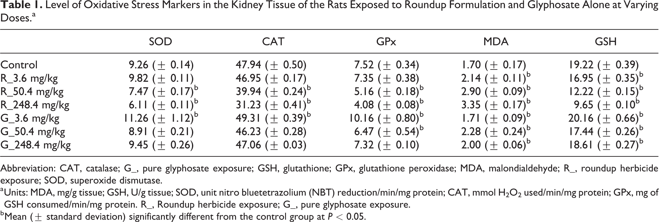
Abbreviation: CAT, catalase; G_, pure glyphosate exposure; GSH, glutathione; GPx, glutathione peroxidase; MDA, malondialdehyde; R_, roundup herbicide exposure; SOD, superoxide dismutase.
a Units: MDA, mg/g tissue; GSH, U/g tissue; SOD, unit nitro bluetetrazolium (NBT) reduction/min/mg protein; CAT, mmol H2O2 used/min/mg protein; GPx, mg of GSH consumed/min/mg protein. R_, Roundup herbicide exposure; G_, pure glyphosate exposure.
b Mean (± standard deviation) significantly different from the control group at P < 0.05.
Level of Membrane-Bound Enzymes in the Kidney
The activity of membrane-bound enzyme in the kidney of the rats exposed to Roundup formulation and glyphosate alone at varying concentrations is presented in Table 2. The activity of the membrane-bound enzymes evaluated (Mg2+-ATPase, Ca2+-ATPase, Na+/K+-ATPase, and total ATPase) followed a similar trend in the experimental groups. Activities of each membrane-bound enzymes recorded in the control group were not significantly different (P > 0.05) from those recorded in the rats exposed to Roundup at 3.6 mg/kg bw of glyphosate concentration and those exposed to all the concentrations of glyphosate alone. These were however significantly higher (P < 0.05) in rats exposed to glyphosate alone at 50.4 mg/kg and 248.4 mg/kg bw than those recorded in the rats exposed to Roundup formulation at similar concentrations of glyphosate.
Level of Membrane-Bound Enzymes (μg Inorganic Phosphate Liberated/min/mg Protein) in the Kidney of the Rats Exposed to Roundup and Glyphosate Alone at Varying Doses.
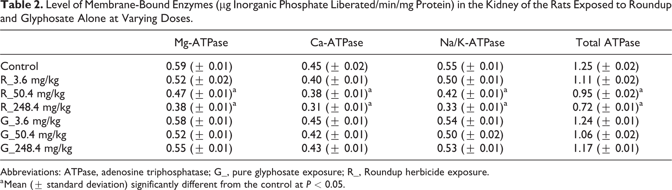
Abbreviations: ATPase, adenosine triphosphatase; G_, pure glyphosate exposure; R_, Roundup herbicide exposure.
a Mean (± standard deviation) significantly different from the control at P < 0.05.
Level of Glyphosate Residue and Percentage Accumulation in Kidney Tissue of the Rats
Figure 3 present the levels and percentage accumulation of glyphosate in the kidney tissue of the rats exposed to Roundup formulation and glyphosate alone at varying concentrations, respectively. Glyphosate was not detected in the kidney of the control group and those exposed to glyphosate alone at 3.6 mg/kg bw. Mean glyphosate accumulation in the kidney was highest in the rats exposed to Roundup at 248.4 mg/kg bw of glyphosate concentration and significant (P < 0.05). This was followed by those exposed to Roundup at 50.4 mg/kg bw of glyphosate concentration. There was no significant difference (P > 0.05) in the mean glyphosate residue between the rats exposed to glyphosate alone at 50.4 mg/kg and 248.4 mg/kg bw. Glyphosate accumulation was however lowest in the kidney of the rats exposed to Roundup at 3.6 mg/kg bw of glyphosate concentration. On the other hand, percentage of glyphosate accumulation in the kidney was significantly higher (P < 0.05) in the rats exposed to Roundup at 3.6 mg/kg bw of glyphosate concentration and followed by those exposed at 50.4 mg/kg bw of glyphosate concentration. Percentage of glyphosate accumulation was not significantly different in the kidney of the rats exposed to Roundup at 248.4 mg/kg bw of glyphosate concentration and those exposed to glyphosate alone at 50.4 mg/kg bw. Percentage glyphosate accumulation was however lowest in the kidney of rats exposed to glyphoste alone at 248.4 mg/kg bw.
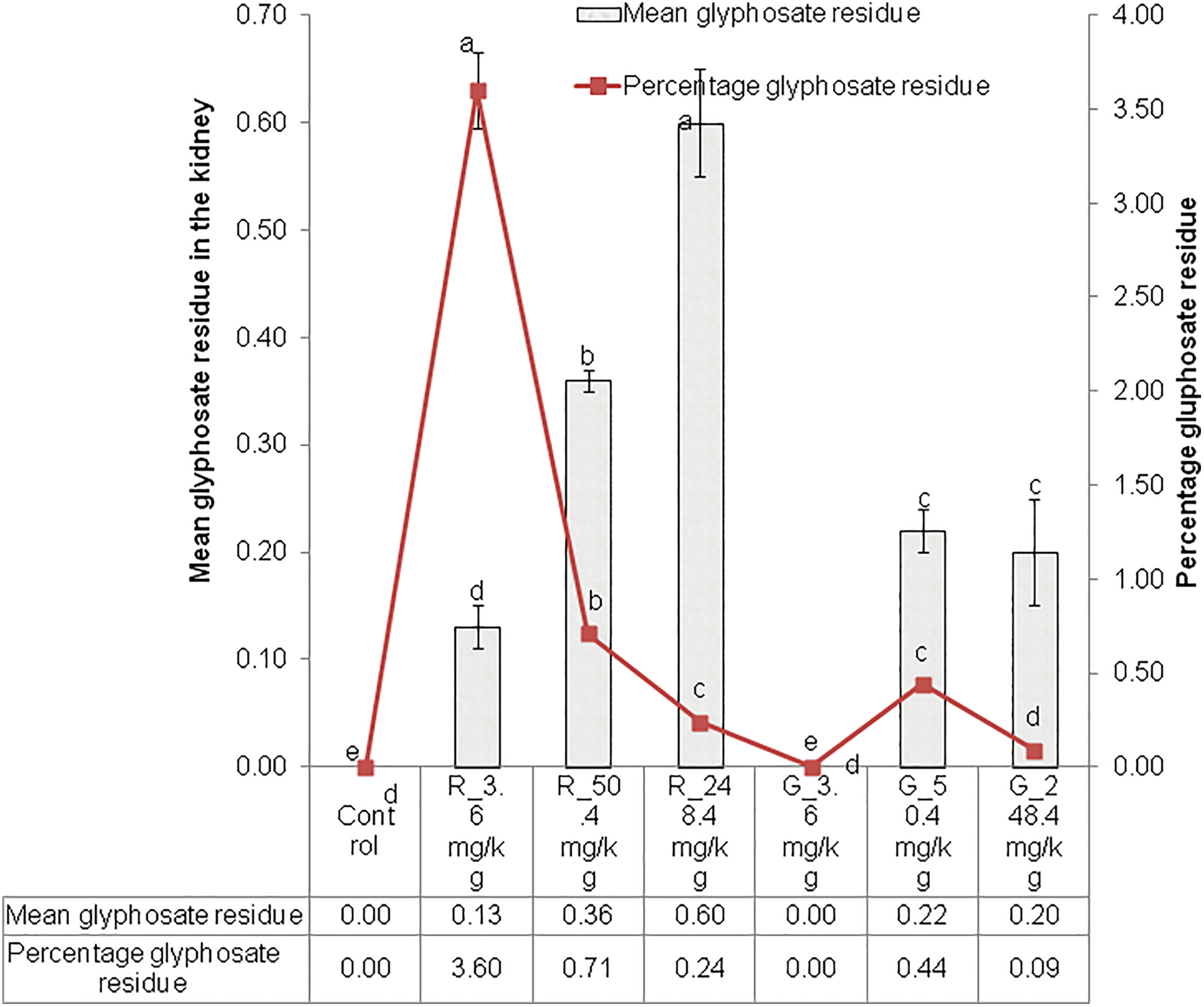
Glyphosate residues and percentage accumulation in the kidney tissue of rats exposed to Roundup formulation and glyphosate alone at varying concentrations.
Histopathological Assessment of Kidney
Photomicrographs of the rat kidney sections exposed to Roundup formulation and glyphosate alone at different concentration are shown in Figure 4. Normal histoarchitecture of renal cortex, renal corpuscles, proximal convoluted tubules, and distal convoluted tubules were observed in the control group. Kidney sections of rats exposed to Roundup at 3.6 mg/kg bw of glyphosate concentration showed distorted renal cortical histoarchitecture, expanded urinary space due to glomerulosclerosis, and tubular necrosis. These lesions were more severe in the rats exposed to Roundup formulation at 50.4 and 248.4 mg/kg bw of glyphosate concentrations, respectively. However, kidney sections of rats exposed to all the concentrations of glyphosate alone revealed normal histoarchitecture of renal cortex, renal corpuscles, proximal convoluted tubules, and distal convoluted tubules with no pathology.
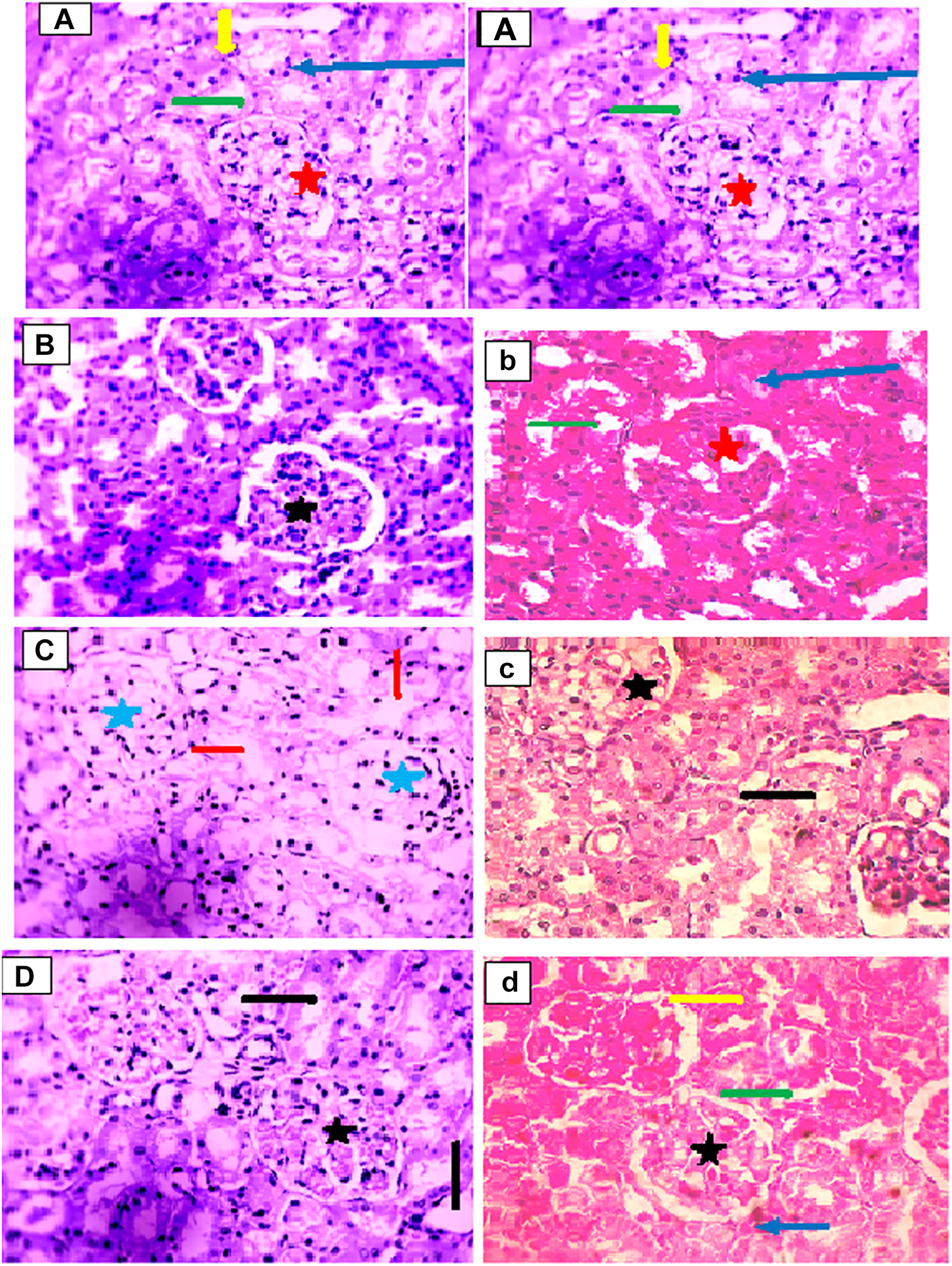
Photomicrographs of the rat’s kidney sections exposed to Roundup formulation and glyphosate alone. Hematoxylin-Eosin stain ×400. (A) Control showing normal histoarchitecture of the renal cortex (long blue arrow), renal corpuscles (red star), proximal convoluted tubules (yellow arrow), and distal convoluted tubules (green arrow). (B) Exposed to Roundup at 3.6 mg/kg body weight (bw) of glyphosate concentrations showing mild distorted renal cortical histoarchitecture characterized by expanded urinary space (black star). (C) Exposed to Roundup at 50.4 mg/kg bw of glyphosate revealing severe distorted renal cortical histoarchitecture due to glomerulosclerosis (blue star) and tubular necrosis (red arrow). (D) Exposed to Roundup at 248.4 mg/kg bw of glyphosate showing mild distorted renal cortical histoarchitecture (black star), and mild tubular necrosis (black arrow). b, Exposed to glyphosate alone at 3.6 mg/kg bw showing normal renal cortex (blue arrow), renal corpuscles (red star), and distal convoluted tubules (green arrow) c, Exposed to glyphosate alone at 50.4 mg/kg bw revealing mild distorted renal cortical histoarchitecture characterized by expanded urinary space (black star) and mild tubular necrosis (black arrow). d, Exposed to glyphosate alone at 248.4 mg/kg bw showing mild distorted renal cortical histoarchitecture (black star), normal histoarchitecture of the renal cortex (blue arrow), proximal convoluted tubules (yellow arrow), and distal convoluted tubules (green arrow).
Discussion
In an effort to clarify the nephrotoxicity status of the active ingredient in Monsanto Roundup herbicide, this study was therefore designed to assess and compare the effects and mechanisms of action of glyphosate alone and commercial formulation of Roundup herbicide on the renal function in male albino rats using standard markers.
Creatinine, a breakdown product of creatine phosphate, is excreted via the kidney, and the serum creatinine level have been reported to reflect the rate of renal (glomerular) filtration. 46,47 In addition, an elevation in the blood level of urea, described as an end product of metabolism, 48 was identified as a good indicator for kidney dysfunction. 49 Sugam et al 50 earlier reported that an increase in serum urea level is seen when there is damage to the kidney or the kidney is not functioning properly. Higher levels of serum urea and creatinine observed in the rats exposed to Roundup herbicide formulation at varying concentrations of glyphosate in this study could, therefore, be an indication of kidney malfunctioning or damage.
Although the use of serum creatinine and urea concentrations as markers of kidney function have been established over the years, but due to their late reflection of reduced glomerular filtration rate (GFR), the search for novel markers of kidney function has intensified. As a result, Cys-C and NGAL have been recently proven to be a better marker of both early acute and chronic kidney diseases. 51 Neutrophil gelatinase-associated lipocalin is a 25-KDa protein and a member of lipocalin family, 52 which is secreted by the epithelia cells of the kidney. Synthesis and levels of NGAL in plasma and urine have been reported to increase in response to any tubular injury after acute kidney disease, 52 and Mitsnefes et al 53 however suggested that NGAL could be a better indices of kidney disease and severity. Our study has confirmed that level of plasma NGAL could be an early and better biomarker of kidney impairment than the serum creatinine, as we observed a marked increase in the level of plasma NGAL of the rats exposed to Roundup formulation. This could be an indication of tubular injury and kidney disease inflicted by the compositions of the Roundup herbicide. Cystatin-C is a 13-KDa protein that is freely filtered by the kidney and then metabolized in the lysosomes of the proximal convoluted tubules. It has been extensively studied, and its use as marker of kidney function has been favorably compared with standard markers. 54 Although, Cys-C has been reported to be a reliable marker of kidney function and can detect impairment of GFR earlier than serum creatinine, 55 but our finding in this study has shown otherwise as we observed notable increase in the level of plasma Cys-C in rats exposed to Roundup formulation only at 248.4 mg/kg bw of glyphosate concentration, indicating that plasma Cys-C may not be a better marker of early kidney injury than serum urea and creatinine as widely acknowledged. Plasma Cys-C and serum creatinine were similarly expressed in the rats exposed to Roundup formulation at 248.4 mg/kg bw of glyphosate concentration
The roles of antioxidant defense system (CAT, GPx, SOD, and GSH) in the body have been documented. 56,57 In this study, Roundup herbicide-induced oxidative stress in the kidney of the exposed rats through the observed depletion in their cellular activities of antioxidant defense system.
The high level of MDA observed in the kidney tissues of the rats exposed to Roundup formulation in this study may indirectly suggest an increased production of oxygen-free radicals that are known to act on unsaturated fatty acids of phospholipid components of membranes to produce MDA, a lipid peroxidation product. The mechanism underlining this peroxidation is attributable to a direct interaction of the herbicide’s compositions with the cytoplasmic membrane of the kidney cells which disrupt the membrane structure. This has been reported as the principal molecular mechanism associated with the toxicities of several organophosphorus compounds. 58,59 In addition, the higher lipid peroxidation observed could also be attributed to the depletion of antioxidant defenses in the kidney of the rats exposed to Roundup herbicide at the increasing concentration of glyphosate. Reports have shown that inverse relationship exists between lipid peroxidation and GPx activity during stress. 60,61 We have also associated a high level of lipid peroxidation with decreased antioxidant enzyme activities. 62 McCord 63 reported that under acute oxidative stress, the toxic effects of pollutants may overwhelm the antioxidant defense system. Roundup herbicide formulation could, therefore, be strong enough to force an acute oxidative stress on the normal kidney physiology by overwhelming and depleting its antioxidant defense system.
Roundup herbicide has the potential to reduce the activities of membrane-bound enzymes (Mg-ATPase, Ca-ATPase, Na/K-ATPase, and total ATPase) in the kidney of the exposed animal as shown in this study. It has been reported by Langeswaran et al 64 that ATPases are important enzymes that provide metabolic energy to the living processes, regulating membrane permeability and ion (such Na+, K+, and Ca2+) transport across the cellular membrane at the expense of ATP by hydrolysis. On the other hand, report has shown that inhibition of these ion-dependent ATPases leads to disturbances in ion homeostasis resulting in impaired signal transduction, alteration in cellular metabolism, changes in cell membrane integrity and permeability, a rise in membrane fluidity, and disturbances in vital functions. 65 Therefore, the reduction in the activities of membrane-bound ATPases of the kidney of the rats exposed to Roundup herbicide as observed in this study could be an indication of disturbances in the cellular metabolism of the kidney and changes in the kidney cell membrane and integrity induced by the herbicide compositions. In addition, it has been established that peroxidation of membrane phospholipids affects the activities of membrane-bound enzymes through alterations in the lipid milieu as well as the structural and functional integrity of cell membranes. 66,67 Reduction in the activities of membrane-bound ATPases as documented by Parthasarathy and Joseph 68 was associated with the loss of protein-SH, due to an increased lipid peroxidative damage of cell membranes. It is, therefore, reasonable to suggest similar mechanism among the rats exposed to the Roundup herbicide in this study.
This study has shown that glyphosate could be bioaccumulated in the kidney tissue of an exposed animal. Since studies have shown that urine is the main route of glyphosate’s elimination from the body 27 - 29 and the main organ responsible for urine formation is the kidney, 30 it is therefore not unexpected that some quantities of this herbicide could be bioaccumulated in the kidney tissues. The level of glyphosate accumulation in the kidney tissue of rats exposed to Roundup formulation was observed to be higher than those exposed to glyphosate alone. The reduction in the activities of the enzyme antioxidant defense system is a possibility, because higher activities of these antioxidants defenses were observed in the kidney tissue of rats exposed to glyphosate alone, hence, accumulated less glyphosate in their tissue. The percentage accumulation of glyphosate in the kidney was highest at 3.6 mg/kgbw and reduced with increasing concentration of exposure. The mechanism behind this has not been well elucidated. However, it is possible that at higher serum glyphosate concentration, the kidney maintains the role of elimination, whereas more reabsorption is done at low concentration. More researches are needed on this.
The reduction in membrane-bound enzymes of the kidney, increased lipid peroxidation, reduced antioxidant defences, increased serum urea and creatinine, and alteration in the plasma levels of Cys-C and NGAL observed in rats exposed to Roundup formulation in this study could have resulted in the various histopathological damages to their kidney tissues. Kidney sections of rats exposed to Roundup showed distorted renal–cortical histoarchitecture, expanded urinary space due to glomerulosclerosis, and tubular necrosis with severity directly proportional to the increasing exposure concentrations. Nanayakkara et al 69 earlier reported histopathological changes characterized by tubular interstitial nephritis associated with mononuclear cell infiltration, glomerular sclerosis, and tubular atrophy as markers for kidney disease. In a similar trend, Wunnapuk et al 70 identified that the modes of renal cell death occurred in tubules and glomeruli during the acute stages of Roundup toxicity. Therefore, oral administration of Roundup herbicide does not only affect the kidney, but it also has the potential to cause chronic kidney disease and total kidney death.
It is important to draw attention to the fact that commercial formulation of Roundup is a mixture of glyphosate, which is the active ingredient, with varying and unspecified surfactants such as POEA required for rapid penetration and absorption of the herbicide into the plant. Previous studies have attributed the renal damage cause by commercial formulations of Roundup to its active ingredient glyphosate, 31,32 but our finding in this study suggests otherwise. We observed that glyphosate alone has no effect on the renal function of the exposed rats when compared to the severe effect of commercial formulation of Roundup on the renal function of the exposed rats. Surfactants have been reported to probably contribute to the toxicity of glyphosate formulations. 71 In addition, commercial formulation Roundup has been considered more toxic due to the presence of the surfactant POEA. 22 Therefore, the nephrotoxicity observed in this study might be due to the surfactant POEA in the Roundup formulation but not glyphosate as generally claimed and believed. Mesnage et al 29 earlier reported that the toxicology of mixtures cannot be fully understood without knowing the differential toxicity of the various compounds of the formulations and their combined effects. Hence, there is need for more studies testing the nephrotoxicity of POEA alone and compared with the commercial formulations and glyphosate alone in other to ascertain the mechanism that underlines the renal toxicity of glyphosate-based herbicides in animal.
Conclusion
Our findings in this study revealed that exposure to a glyphosate-based commercial formulation of Roundup herbicide at different concentrations of glyphosate induced severe renal damage, caused by increased oxidative stress resulting from the accumulation of glyphosate residue, reduction in membrane-bound enzymes of the kidney, increased serum urea and creatinine, and disturbances in plasma Cys-C and NGAL of the kidney. The renal toxicity observed cannot be due to the presence of its glyphosate active ingredient in the herbicide, as glyphosate alone has virtually no effect on the renal function of the exposed animal. Based on this observation, it remains unknown whether glyphosate, when present in the Roundup formulation, is producing a synergistic effect or whether another component in the Roundup formulation is primarily responsible for the observed nephrotoxicity. Therefore, the general claim attributing the renal dysfunction of a glyphosate-based herbicide to its active ingredient should be discouraged.
Footnotes
Acknowledgments
The authors sincerely appreciate the permission given to them by the Department of Zoology and Environmental Zoology, Faculty of Science, Olabisi Onabanjo University to use its facilities. The authors also thank the laboratory technologist in the central biotechnology laboratory of the Federal University of Agriculture Abeokuta, University of Lagos and Olabisi Onabanjo University Ago-Iwoye, Nigeria for their technical assistance.
Authors’ Contributions
G. A. Dedeke contributed to conception and design, contributed to acquisition and analysis, and critically revised manuscript; F. O. Owagboriaye contributed to conception and design, contributed to acquisition, analysis, and interpretation, drafted manuscript, and critically revised manuscript; K. O. Ademolu contributed to design and critically revised manuscript; O. O. Olujimi contributed to design and critically revised manuscript; A. A. Aladesida contributed to design, contributed to analysis and interpretation, and critically revised manuscript. All authors gave final approval and agree to be accountable for all aspects of work ensuring integrity and accuracy.
Declaration of Conflicting Interests
The author(s) declared no potential conflicts of interest with respect to the research, authorship, and/or publication of this article.
Funding
The author(s) disclosed receipt of the following financial support for the research, authorship, and/or publication of this article: This study was funded through the 2013 needs assessment special presidential intervention fund in public Universities.