
Abstract
With the increasing use of mycophenolic acid (MPA) in solid organ transplantation, some clinical studies indicate that it is also a human teratogen. However, it is unknown by which mechanism MPA acts as a teratogen. Mycophenolic acid was a selective blocker of de novo purine synthesis, and its immunosuppressive effect is mediated by the inhibition of inosine monophosphate dehydrogenase, which could be a target for MPA-induced toxicity as well. The aim of our study was to examine the direct influence of MPA exposure on zebrafish (Danio rerio) embryos. Morphological defects including tail curvature and severe pericardial edema in zebrafish embryos caused by MPA (3.7-11.1 µmol/L) were found in a dose-dependent manner. The teratogenic index (25% lethal concentration value (LC25)/no observed adverse effect level ratio) was 16, which indicated MPA as a teratogen. Quantitative polymerase chain reaction analysis revealed that the expression level of impdh1b and impdh2 was significantly reduced by MPA treatment at 8 µmol/L (equals to LC25 level). All the toxic effects could be partially reversed by the addition of 33.3 µmol/L guanosine. Our results indicated that MPA impairs the development of zebrafish embryos via inhibition of impdh activity, which subsequently caused a guanosine nucleotide depletion in vivo.
Introduction
Mycophenolic acid (MPA) is a metabolized product and an active element of mycophenolate mofetil (MMF), which has been approved for the prevention of acute graft rejection in kidney, heart, and liver transplantation in 1995. 1,2 Mycophenolic acid is a known potent uncompetitive inhibitor of inosine monophosphate dehydrogenase (IMPDH), the rate-limiting enzyme for the de novo synthesis of guanosinenucleotides, 3,4 which plays crucial roles in cell proliferation and other cellular functions including DNA replication, RNA and protein synthesis, and cellular signaling. 5 Consequently, MPA can block T- and B-lymphocyte proliferation and clone expansion and prevent the generation of cytotoxic T cells and other effector T cells. Through depletion of guanosine nucleotides, MPA can suppress glycosylation and the expression of several adhesion molecules, thereby decreasing the recruitment of lymphocytes and monocytes into the sites of inflammation and graft rejection. 4 With the increasing use of MPA in solid organ transplantation, the most common adverse effects of MPA had been reported, including gastrointestinal disturbances and myelosuppression. 6,7 However, the use of MPA during pregnancy poses significant risk for patients and their unborn offsprings, 8 –11 so the importance of drug-related developmental toxicity became evident.
Zebrafish (Danio rerio) have been widely used by molecular geneticists and developmental biologists for studying the mechanisms of vertebrate development. Zebrafish have its advantages compared to the traditional in vivo model in which multiple organs can be observed. It has been reported that targeting impdh by MPA or morpholinos in zebrafish resulted in suppressed embryo angiogenesis without significant impact on the gross morphology. 12 Furthermore, sequence alignment revealed that humans have 2 isoforms of IMPDH, 13,14 namely IMPDH1 and IMPDH2, whereas the zebrafish genome contains 3 isoforms (impdh1a, impdh1b, and impdh2), possibly because of additional gene duplications. 15 The amino acid sequences are highly conserved from human to zebrafish (more than 90% identity). Expression analysis by RNA whole-mount in situ hybridization of the 3 genes in zebrafish embryos showed that impdh2 is mainly expressed in the ventral regions of the developing trunk. The other 2 isoforms are expressed in the superficial epithelial cells. Functional analysis revealed that impdh2 contributes to the daily rhythm of S phase in the cell cycle, whereas impdh1a contributes to ocular development and pigment synthesis and impdh1b serves to delay embryonic development. 11
According to our previous research, MPA induced significant growth defects during the development of zebrafish embryo, including tail defects and severe pericardial edema. However, it was unknown by which mechanism MPA acted as a teratogen. In this study, we focused on the developmental toxicity caused by MPA. We identified the target impdh genes of MPA in zebrafish embryo, discovering the link between impdh suppression and developmental defects in the zebrafish embryo. We also conducted RESCUE study of reversing the morphological defects caused by MPA to validate the molecular mechanisms underlining the developmental toxicity of MPA.
Materials and Methods
Zebrafish Maintenance
Wild-type zebrafish (Tübingen line) were obtained from Model Animal Research Center of Nanjing University. They were kept at 28.5°C as described. 16 The light–dark cycle was 14:10 hours. Wild-type fish were mated, and spawning was stimulated by the onset of light. Embryos were collected and placed at 28.5°C in Petri dishes containing embryo medium (0.2 g/L of Instant Ocean Salt in distilled water with 0.01% methylene blue). The zebrafish studies were approved by the Institutional Animal Care and Use Committee at Nanjing Tech University. Embryos and larvae were staged according to Kimmel et al. 17 The age of the embryos and larvae is indicated as hours postfertilization (hpf).
Substances
Mycophenolic acid (CAS No. 50-35-1), dimethyl sulfoxide (DMSO; CAS No. 67-68-5), and guanosine (CAS No. 118-00-3) were purchased from Sigma-Aldrich (St Louis, Missouri). Stock solutions were prepared by dissolving the pure chemicals in DMSO and then diluted to the desired concentrations in embryo medium. Final DMSO concentrations were 0.1% in the treatment solution. Dimethyl sulfoxide 0.1% was used as vehicle control during the assays.
Drug Treatment and Larvae Observation
Zebrafish embryos were distributed into 24-well plates with 20 embryos per well and 1 mL embryo medium. Survival and morphological defects were assessed using a dissecting microscope (SMZ745T; Nikon, Japan). Zebrafish embryos with any morphological defect were representative for the teratogenicity of MPA. For assessing both the lethal toxicity and the teratogenic toxicity, zebrafish embryos at 2 hpf were treated with various concentrations (0.5-50 µmol/L)of MPA for 70 hours. The number of dead embryos and embryos with any of the developmental defects were calculated. Image-Pro Plus 6.0 (Media Cybernetics, Rockville, Maryland) was used to define the tail curvature angle and the area of pericardial edema. GraphPad Prism 5.0 (San Diego, California) was used to calculate the 25% lethal concentration value (LC25), 50% lethal concentration value (LC50), 50% teratogenic concentration (EC50), and the concentration with no observed adverse effect level (NOAEL), respectively. Each experiment was done in triplicate and was repeated 3 times.
For the RESCUE study, 20 zebrafish embryos per well were treated with 33.3 μmol/L of guanosine and 8.0 μmol/L of MPA at 2 hpf. The evaluation of rescue phenotype was determined at 72 hpf. Each assay was done in triplicate and was repeated 3 times.
Reverse Transcription and Quantitative Real-Time Polymerase Chain Reaction Analysis
Total RNA from 10 embryos per well of 8.0 μmol/L of MPA-treated or control groups was isolated with TRIzol (Invitrogen, Cergy-Pontoise, France) and reverse transcribed by Moloney murine leukemia virus reverse transcriptase (Invitrogen) using oligo (dT) primers (Invitrogen). Real-time quantitative polymerase chain reaction (qPCR) was performed in an ABI 7900HT FastReal-Time PCR system (Applied Biosystems, Foster City, California) using the standard EvaGreen assay detection protocol. Briefly, all reactions were performed in a total volume of 30 μL containing 10 μL template, 1× PCR buffer, 0.2 mmol/L each deoxynucleotide nucleoside triphosphate, 2.5 mmol/L MgCl2, 0.75 U Taq polymerase, 0.5× EvaGreen (Biotium, Hayward, California), and 20 nmol/L fluorescein (Bio-Rad, Hercules, California). The expressions of β-actin under various treatment conditions during different time points were checked first to confirm that β-actin expressions were not changed by chemical treatment. The qPCR results were analyzed using β-actin as an internal standard, and fold of change was calculated relative to the untreated control using the ΔΔC t method. 18 The qPCR analysis on each RNA sample was performed in triplicate, and the result out of at least 3 independent experiments was shown. The primers for zebrafish are shown in Table 1.
The Primers Used for Zebrafish Embryos.
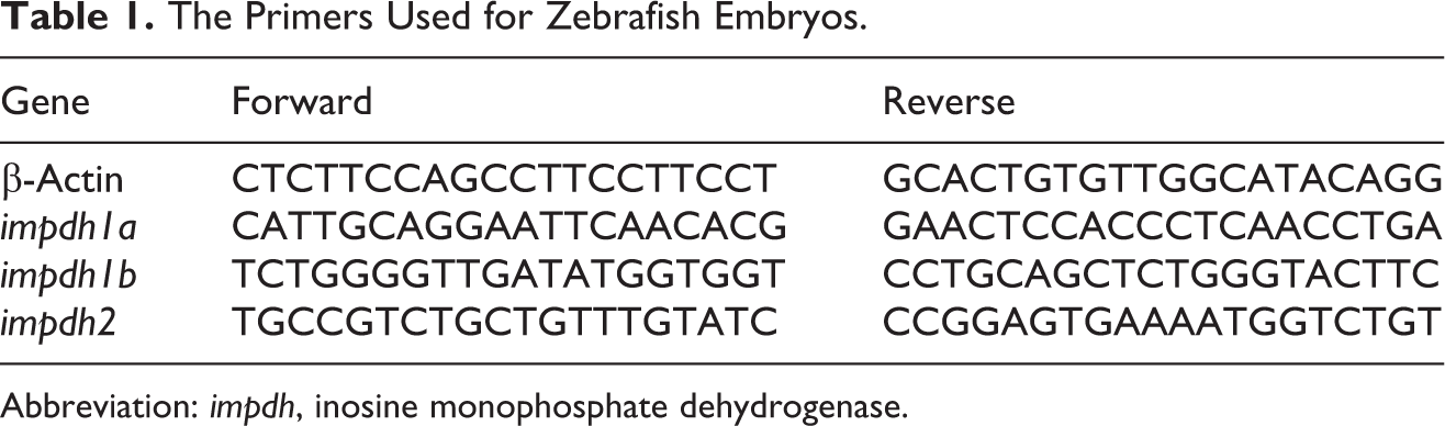
Abbreviation: impdh, inosine monophosphate dehydrogenase.
Statistical Analysis
All statistical analyses were expressed as mean ± standard error of the mean using GraphPad Prism 5.0. The decrease/increase in gene expression as determined by qPCR was analyzed using 2-way analysis of variance followed by Bonferroni posttests. Paired t test was used in all other experiments. Significance was considered when P values were lower than 0.05 (***indicates statistical significance P < 0.005, **P < 0.01, *P < 0.05).
Results
Lethality and Teratogenicity of MPA in Zebrafish Embryos
Mycophenolic acid treatment caused teratogenicity and lethal toxicity in zebrafish embryos in a dose-dependent manner (Figure 1). In the zebrafish model, compounds with a teratogenic index (LC25/NOAEL ratio) greater than or equal to 10 are classified as teratogens. 19 According to our assessment, the LC25, LC50, EC50, and NOAEL values were calculated as 8.0, 12, 6.9, and 0.5 μmol/L, respectively (Table 2). The LC25/NOAEL ratio was 16, which was greater than 10, so MPA could be classified as a teratogen.
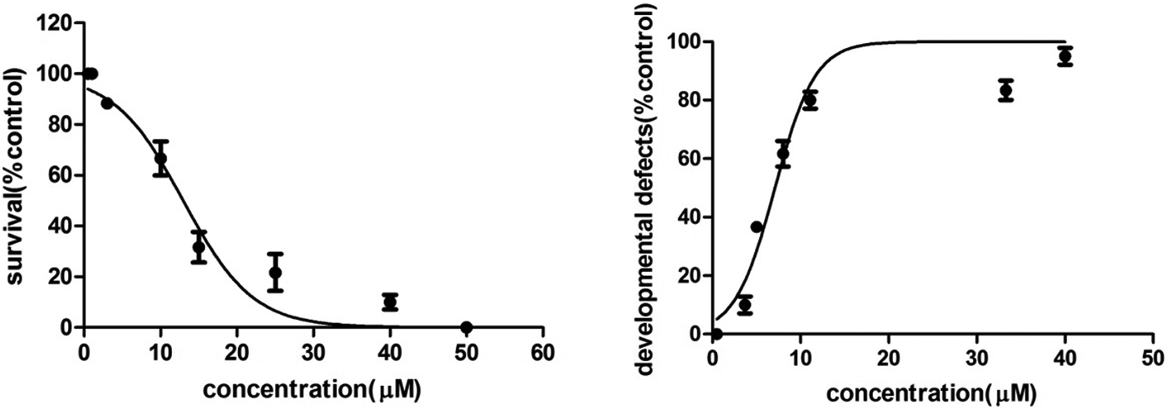
Mycophenolic acid (MPA) induced dose-dependent lethality and developmental defects at 72 hours postfertilization (hpf) in zebrafish.
LC25, LC50, EC50, and NOAEL Values of MPA.
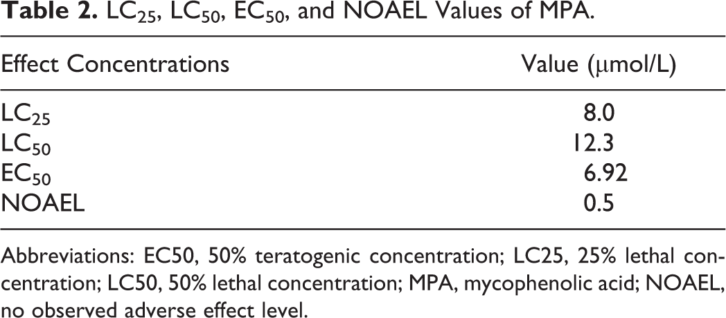
Abbreviations: EC50, 50% teratogenic concentration; LC25, 25% lethal concentration; LC50, 50% lethal concentration; MPA, mycophenolic acid; NOAEL, no observed adverse effect level.
The Developmental Defects Caused by MPA in Zebrafish Embryos
In the developmental toxicity assay, we found MPA induced significant morphological phenotypes, most prominently tail curvature and pericardial edema, in a dose-dependent manner (Figure 2A). Compared with the embryos in the control group, 8.0 µmol/L of MPA-treated embryos showed the severe ventral curvature with the average angle at almost 50° (Figure 2B), as well as the heart edema, which was 2-fold larger than that in the control embryos (Figure 2C).
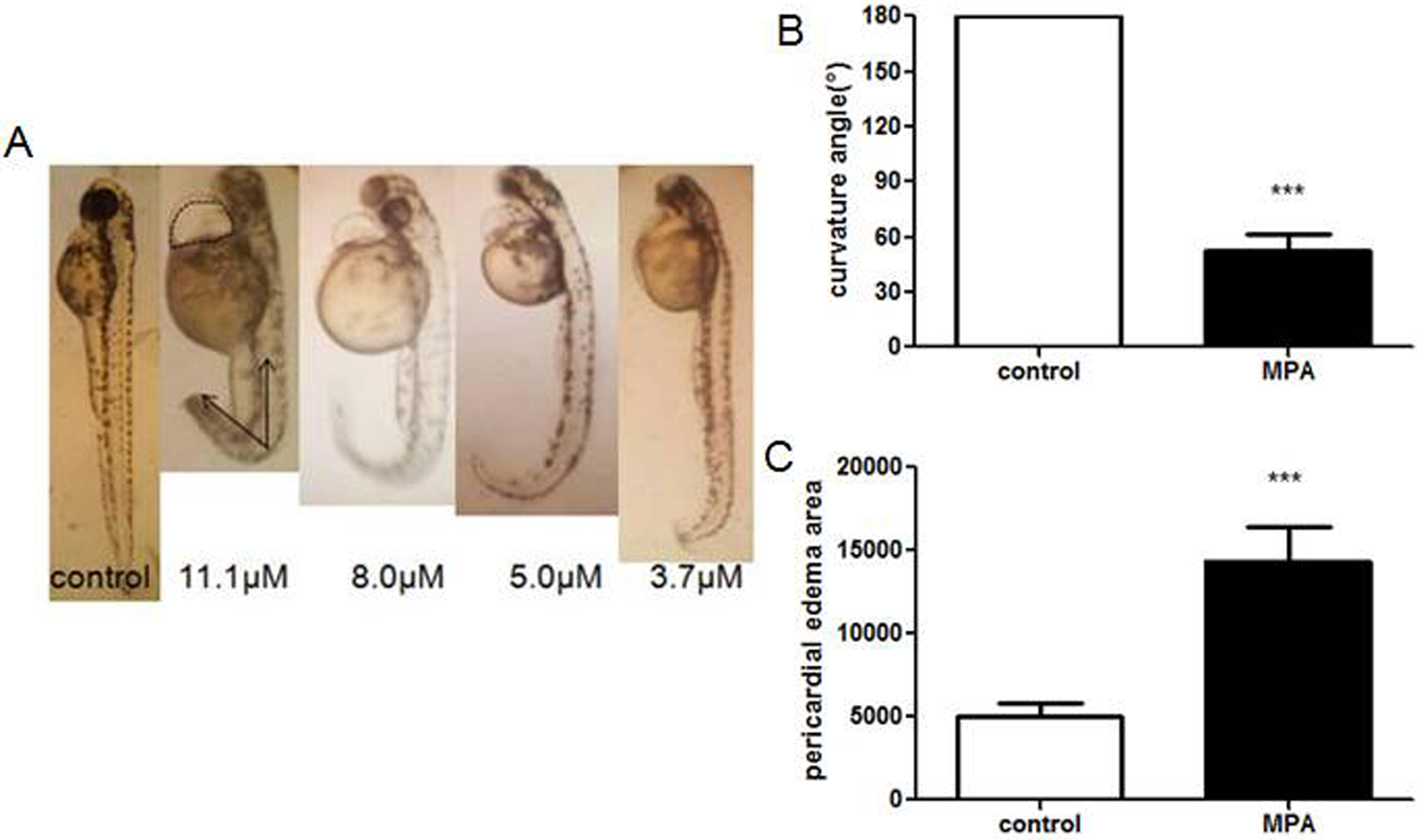
Zebrafish embryos were treated with mycophenolic acid (MPA) at 2 hours postfertilization (hpf) and observed at 72 hpf. Tail curvature angles are indicated by arrows and circled area was pericardial edema (A). Curvature angle (B) and the area of pericardial edema (C) in the 8.0 µmol/L of MPA-treated embryos were calculated with GraphPad Prism 5.0. ***Statistical significance P < 0.001.
The Expression of impdh Genes Altered by MPA
As MPA is a known inhibitor of IMPDH, we studied impdh genes in zebrafish to reveal the molecular mechanism of MPA toxicity. Zebrafish contains 3 isoforms of impdh—impdh1a, impdh1b, and impdh2. Gene expressions were measured at 2, 12, 24, 48, and 72 hpf for the control group and 8.0 µmol/L for the MPA treatment group. Mycophenolic acid treatment did not significantly alter the expression level of impdh1a compared with each control, respectively (Figure 3A). We detected high innate expression level of impdh1b at 12 hpf and significant downregulation by MAP at 24 and 48 hpf (Figure 3B). The innate expression of impdh2 was increasing gradually from 2 to 72 hpf. Significant downregulation by MPA was detected at 24 hpf (Figure 3C).
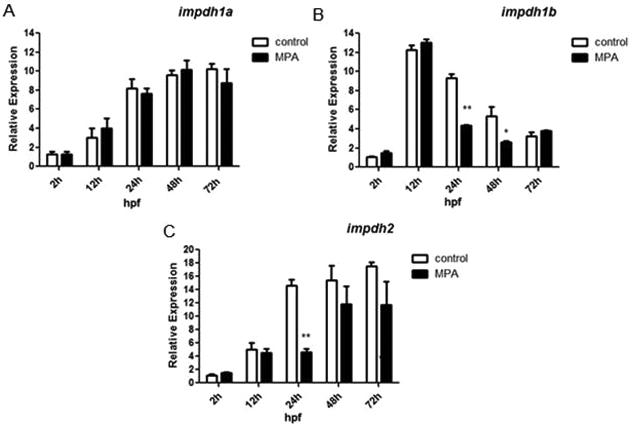
Real-time quantitative polymerase chain reaction (qPCR) analysis of impdh genes: (A) impdh1a, (B) impdh1b, and (C) impdh2 in the zebrafish embryos treated by mycophenolic acid (MPA) at 8.0 µmol/L. There is downregulation of impdh1b and impdh2 at different time points. Error bars represent means ± standard error of the mean (SEM) of 3 replicates. Significance at that particular time point was considered when P values were lower than 0.05. **Statistical significance P < 0.01. *P < 0.05.
RESCUE Study of External Guanine for MPA-Exposed Embryos
Our study suggested that the expression of impdh was inhibited by MPA treatment. Inosine monophosphate dehydrogenase inhibition could cause guanosine monophosphate (GMP) to deplete. Therefore, we hypothesized that external guanosine supply could rescue the morphological defects caused by MPA. 20 According to our tests, the sufficient dose of exogenous guanosine was 33.3 μmol/L. As expected, part of the embryos treated with both 8 μmol/L of MPA and 33.3 μmol/L of guanine recovered to normal phenotype (Figures 4A-C), and the normal rate of embryos was turned up from 20% to approximately 60% (Figure 4D). To examine the underlying mechanism of exogenous guanosine-induced phenotype rescue, we performed qPCR assays to measure impdh gene expression change. The results further revealed that guanosine alone at 33.3 μmol/L did not alter much of the expression levels of impdh1b and impdh2, except that it decreased impdh2 expression slightly at 72 hpf, which might be due to the prolonged exposure time. When coexposured to the embryos with MPA, guanosine significantly increased the expression levels of impdh1b and impdh2 almost to the normal level in the control group, especially at 24 hpf (Figures 5A and B).
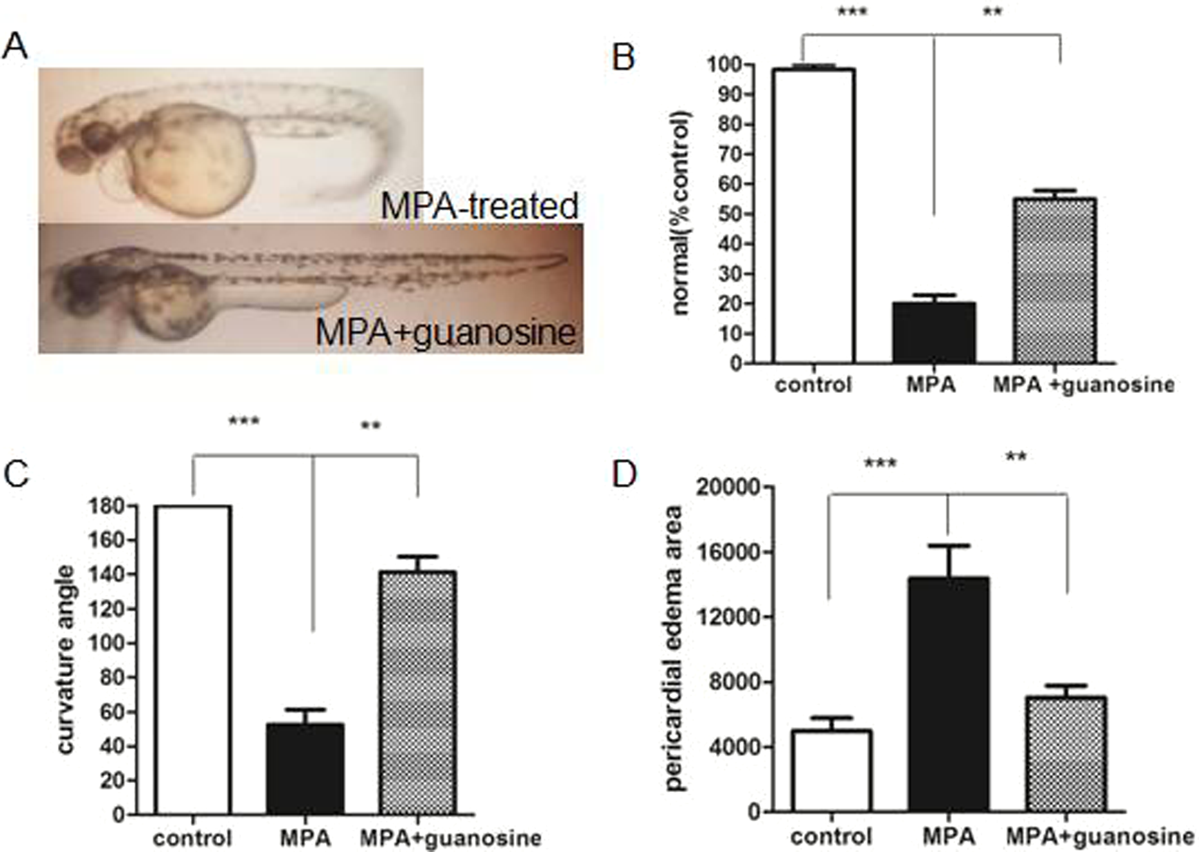
RESCUE study of guanosine supply on the development defects of mycophenolic acid (MPA)-treated embryos. (A) 8.0 µmol/L of MPA-treated embryos recovered at 72 hours postfertilization (hpf) after 33.3 µmol/L of guanosine supply. (B) Curvature angle and (C) the area of pericardial edema were calculated with GraphPad Prism 5.0 for different treatment groups. Data are presented as means ± standard error of the mean (SEM). Significance was considered when P values were lower than 0.05. ***Statistical significance P < 0.001. **Statistical significance P < 0.01.
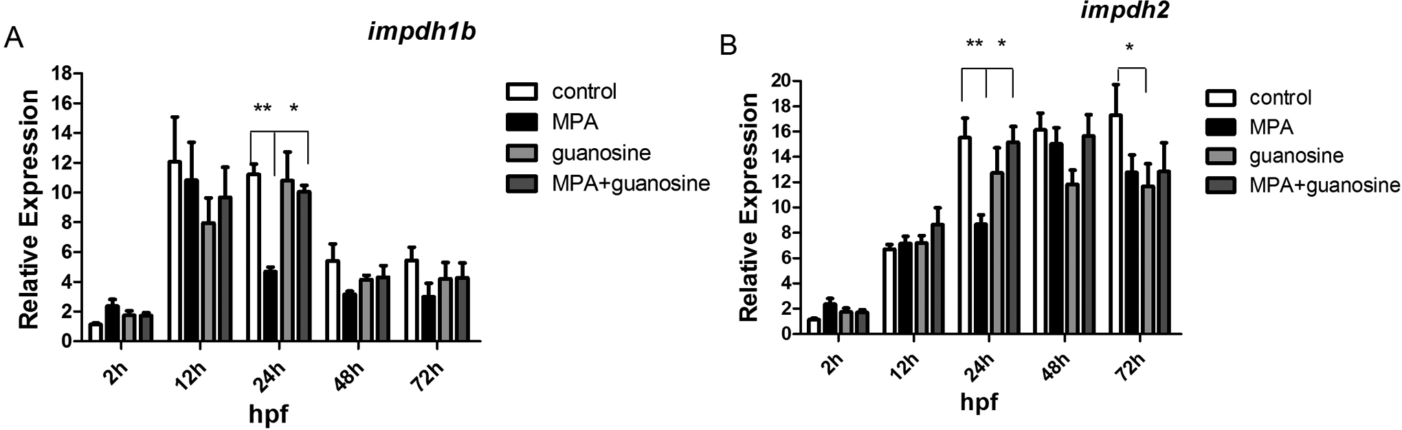
The relative expression levels of mycophenolic acid (MPA) target genes after external guanosine supply (33.3 μmol/L). (A) impdh1b and (B) impdh2. Data are presented as means ± standard error of the mean (SEM). Significance at that particular time point was considered when P values were lower than 0.05. **Statistical significance P < 0.01. *P < 0.05.
In summary, our results demonstrated at morphological level and molecular level that the additional guanosine supply can partially rescue zebrafish embryos from developmental defects caused by MPA treatment.
Discussion
Both animal and human studies revealed that MPA is a teratogen. Echardt group has reported the growth and development defects of rat embryos using whole embryo culture with MPA, and the effects were predominantly localized in the body regions. 21 Additionally, teratology studies of MPA in rabbits also showed fetal resorptions and malformations. 22 A prospective study of the European Network of Teratology Information Services has confirmed that mycophenolate is a human teratogen; there is a high incidence of major malformations (26%) after the first trimester exposure to mycophenolate. 23 The American National Transplantation Pregnancy Registry gathered pregnancy outcomes in female patients after organ transplantation and intake of MPA during gestation. Within this series of cases, 49% of all examined pregnancy outcomes were spontaneous abortions. Birth defects such as microtia and other deformities were also observed. 24 Using the zebrafish embryos, we also found MPA induced significant developmental defects, including tail defects and severe pericardial edema. The teratogenic index (LC25/NOAEL ratio) of MPA was greater than 10, a value representative of a teratogen in zebrafish model. Our results showed the same teratogenic toxicity of MPA as in other reports. Thus, it is concluded that teratogenic toxicity in the zebrafish model has a good correlation with other mammalian models.
Mycophenolic acid caused morphologic toxicity in zebrafish embryo in a dose-dependent manner. As IMPDH was the well-known target of MPA, we conducted qPCR assay to evaluate the expression change of impdh genes in zebrafish after MPA treatment. We indeed found MPA downregulated impdh1b and impdh2 at different time points, whereas the expression of impdh1a was untouched. According to literatures, 11,21 the 3 impdh gene homologs have different tissue-specific functions. Impdh1a is likely to have an eye-related function and impdh2 may function in metabolically active tissues. Impdh1b shows high expression in the brain, eye, heart, and muscle, whereas expression levels of impdh2 in the heart, muscle, and liver were much higher. Although the detailed functions of impdh1b are still unclear, it seems that it may have the function to inhibit early growth. Taken together, impdh1b and impdh2 are involved in the regulation of developmental process. Therefore, we speculated that the tail curvature was caused by the decrease in impdh1b and the pericardial edema was due to the decrease in impdh2. Further whole-mount in situ hybridization studies will be needed to investigate special and temporal impdh expression after MPA treatment.
Mycophenolic acid is a potent, selective, and reversible inhibitor of IMPDH, which catalyzes the oxidation of inosine-5′-monophosphate to xanthosine monophosphate (XMP), a precursor of GMP. Mycophenolic acid exposure to human IMPDH results in cessation of cellular division limited to those cells that do not have the salvage pathway for guanine synthesis. 25 Interestingly, there are few evidence describing developmental toxicity dependent on this pathway. Here, we demonstrated that the impact of MPA on zebrafish was reversible by the addition of guanosine, the substrate of the salvage pathway of guanosine triphosphate (GTP) synthesis. 20 This result may confirm that MPA affects zebrafish development by guanosine nucleotide depletion due to the direct inhibition of impdh. The reason why zebrafish embryos are sensitive to GTP withdrawal needs further investigation.
However, external guanosine addition could not totally rescue the development defect phenotype caused by MPA. We speculated there might be other events related to MPA effects on zebrafish. Our further goal will be discovering the remaining molecular targets of MPA in order to provide more mechanistic information useful for the risk assessment of its clinical use.
Although zebrafish has been recognized as a good animal model for the study of “predictive toxicology” (http://www.fda.gov/forconsumers/consumerupdates/ucm343940.htm), the equivalent concentration conversion between zebrafish and human is still lacking. Mycophenolic acid is available in 2 branded products: MMF (CellCept; Genentech, San Francisco, California) and enteric-coated MPA (Myfortic; Novartis, East Hanover, New Jersey). The clinic dose of MMF and enteric-coated MPA for humans is 1 to 2 g/d and 1.44 g/d, respectively. 26 After oral exposure of 1 g MMF, the mean maximum plasma MPA concentration (C max) in healthy individuals is about 25 mg/L, and concentrations of 1.3 to 3.5 mg/L are recommended in patients treated. In the study of humans, any exposure to MPA during the first trimester may be harmful, and the degree of toxicity may depend on the degree of MPA exposure. 27 Prenatal multiple fetal malformations were reported in the child of a renal transplant recipient who was exposed to a total daily dose of only 500 mg of MMF throughout the first trimester. 28 Although the concentration of MPA used in our study, 8 μmol/L (equivalent to 2.56 mg/L), falls into the plasma MPA concentration range in the clinic, in order to estimate how the treatment concentrations used in this study relate to human exposure populations, the bioconcentration of MPA in zebrafish embryos should be determined. We previously reported the bioconcentration of 2 small molecules, namely triptolide and gambogic acid; the bioconcentration of zebrafish embryos depended largely on the chemical property of the tested molecule and the exposure window. 29,30 So, our future goal is to determine the bioconcentration of MPA in zebrafish embryo during the current exposure window.
Footnotes
Author Contributions
Ling-Ling Jiang and Ming-Fang He contributed to conception and design; contributed to acquisition, analysis, and interpretation; drafted the manuscript; and agrees to be accountable for all aspects of work ensuring integrity and accuracy. Mei-Hui Liu and Jian-Ying Li contributed to acquisition and analysis. Zhi-Heng He critically revised the manuscript. Huan Li contributed to the guidance of real-time PCR experiments. Ning Shen contributed to the zebrafish care, breeding, and embryo culture. Ping Wei contributed reagents.
Declaration of Conflicting Interests
The author(s) declared no potential conflicts of interest with respect to the research, authorship, and/or publication of this article.
Funding
The authors disclosed receipt of the following financial support for the research, authorship, and/or publication of this article: This work was supported by the Key R & D Special Funds of Jiangsu Province (BE2015696) and New Teacher Fund from Chinese Ministry Education (20123221120005).