
Abstract
Talc is a mineral that is widely used in cosmetic products, antiseptics, paints, and rubber manufacturing. Although the toxicological effects of talc have been studied extensively, until now no detailed inhalation study of talc focusing on oxidative stress has been done. This repeated 4 weeks whole-body inhalation toxicity study of talc involved Sprague-Dawley rats. Male and female groups of rats were exposed to inhaled talc at 0, 5, 50, and 100 mg/m3 for 6 hours daily, 5 days/week for 4 weeks. The objective was to identify the 4-week inhalation toxicity of talc and investigate antioxidant activity after exposure to talc. There were no treatment-related symptoms or mortality in rats treated with talc. Glucose (GLU) was decreased significantly in male rats exposed to 50 and 100 mg/m3 of talc. Histopathological examination revealed infiltration of macrophages on the alveolar walls and spaces near the terminal and respiratory bronchioles. In male and female rats exposed to 100 mg/m3 talc, expression of superoxide dismutase 2, a typical biological indicator of oxidative damage, was significantly increased. Thus, inhalation of talc induces macrophage aggregations and oxidative damage in the lung.
Keywords
Introduction
Talc, also called talcum powder, is a mineral widely used in cosmetic products, antiseptics, ceramic industry, paints, confectionery food products, heat-resistant products, and rubber manufacturing. 1 –3 The chemical formula of talc is Mg3Si4O10(OH)2 and it is a relatively pure substance although it can contain small quantities of aluminum, chromium, iron, manganese, and nickel. 4,5 Talc feels greasy because its hardness is low, and talc particles have usually thin plate-like shape, but the dimensions of each plate alter among diverse body of ore. 6
Inhalation is the major pathway of human exposure of powdered toxic materials, and the lung is the first target organ. Adverse effects of inhaled toxicants include pulmonary disorders, lipid peroxidation, and DNA breakage. 7 –10 Inhalation of a large quantity of talcum powder can cause short-term pulmonary talcosis. 1 One case report described that talc-induced pneumoconiosis was due to repeated abuse of talc-adulterated marijuana, and others reported that inhalation of talc caused lung diseases such as talc pneumoconiosis, bronchiolar obstruction and bronchiolitis, or even deaths. 1 –3,11 –19 Moreover, perineal talc use has been linked with ovarian cancer. 20 –23
Animal studies have shown that inhalation of talc can lead to lung injuries that include pulmonary inflammation, impaired phagocytosis, interstitial fibrosis, epithelial hyperplasia, and pulmonary edema. 24 –27 Focal areas of papillary change in the surface epithelium of the ovary were described following intrabursal injection in rats. 28 Hemorrhage, edema, inflammation, proliferation, and fibrosis in lungs of rats exposed intrapleurally to talc slurry have been described. 29
In contrast, no inhalation study has focused on oxidative stress. Oxidative stress induced by an inhaled toxicant could produce substantial quantities of superoxide anion, and the antioxidant enzyme superoxide dismutase (SOD) plays an important role in protection from the toxicity of the superoxide free radical. 30 –32 Another important antioxidant enzyme in biological systems is glutathione peroxidase (GPx), which scavenges hydrogen peroxide and lipid peroxides. 33 Glutathione peroxidase is a selenium-containing enzyme that uses the sulfhydryl groups of reduced glutathione (GSH) as a hydrogen donor, catalyzing the formation of oxidized glutathione or glutathione disulfide. 34
The present study was undertaken to identify the 4-week inhalation toxicity of talc and to investigate the expression of antioxidant enzymes in rat lung after inhalation exposure to talc. While many studies have shown the hazardous effects due to inhalation of talc in rats, no study has dealt with the oxidative effects in tissues of rats. In addition, hematological, biochemical, and histopathological examinations were done, and talc components were analyzed.
Material and Methods
Analysis of Talc Sample
Talc (nonasbestos form) was provided as ultra-fine white talcum powder from Rex Material (Korea) and was analyzed by inductively coupled plasma-atomic emission spectrometry (ICP-AES). Structure and morphology of the particles were measured by field emission scanning electron microscopy (Quanta, Oregon) at an accelerating voltage of 20 kV. Talc samples were gold coated before measurement.
Animals and Treatments
Male and female 7-week-old Sprague-Dawley rats purchased from Orient Bio (Korea) were housed under standard laboratory conditions (temperature 22°C ± 3°C, humidity 50% ± 10%, and 12-hour day/night cycles). Animals were acclimatized to the facility for 1 week and observed for irregular behavior. Diets were provided ad libitum to the rats, except during exposure to talc. All animal experiments complied with the guidelines of the Institutional Animal Care and Use Committee. The rats were exposed to talc in a stainless steel inhalation chamber (Sibata, Japan) with a capacity of 1 m3. Temperature and relative humidity were maintained at 22°C ± 3°C and 50% ± 20%, respectively. Talc aerosol was generated by a dust generator (Sibata) and was delivered into the chamber with air purified through a high-efficiency particulate air filter. The talc flow rate into the chamber was maintained by continuously controlling talc particle counts per minute. Talc concentrations in the chamber were measured by collecting samples with a SIP-32L sampler (Sibata). Groups of 6 male and 6 female rats were exposed to 0, 5, 50, and 100 mg/m3 talc for 6 hours daily, 5 days/week for 4 weeks. Each rat was examined daily during exposure and postexposure to observe any symptoms related to talc exposure. After the terminal exposure, the rats were fasted for about 16 hours before necropsy.
Hematological and Biochemical Analyses
Blood samples were collected from the ventral aorta under isoflurane anesthesia. The blood samples were collected into complete blood count (CBC) bottles CBC bottles containing EDTA-2K and were analyzed using a Hemavet automatic hematology analyzer (Drew Scientific, Florida). The following parameters were determined: white blood cell count, neutrophil count, lymphocyte count, monocyte count, eosinophil count, basophils count, red blood cell (RBC) count, hemoglobin concentration, hematocrit, mean corpuscular volume, mean corpuscular hemoglobin (MCH), MCH concentration, RBC distribution width, platelet (PLT) count, and mean plasma volume (MPV).
Blood samples for biochemical analyses were also collected from the ventral aorta in the plain tubes and were kept at room temperature so as to be clotted, and the sera were obtained by centrifugation of the blood samples at 1580 g for 10 minutes after clotting. Serum biochemistry parameters examined using a Drichem4000 automatic serum analyzer (Fuji Photo Film, Japan) were alkaline phosphatase, lactate dehydrogenase, GLU, total cholesterol, aspartate aminotransferase, triglyceride, alanine aminotransferase, urea nitrogen in blood, gamma-glutamyl transferase (GGT), albumin, total protein, creatinine, and total bilirubin.
Histopathological Examination
Lung tissues of 3 rats in each male and female group were fixed by inflation with 10% neutral buffered formalin. The tissues were embedded in paraffin, and sections 3 to 5 µm in thickness were placed on glass slides. After staining with hematoxylin and eosin (H&E), the tissue sections were examined using a light microscope (Olympus, Japan), and photomicrographs were taken with a built-in DP70 digital camera.
Western Blotting
Lungs of 3 rats in each male and female group were frozen with liquid nitrogen and ground to powder by mortar and pestle. The powder was sonicated in radioimmunoprecipitation assay tissue lysis buffer (Santa Cruz Biotechnology, Santa Cruz, California). Protein contents were determined by using the BCA protein assay kit (Thermo Scientific, Rockford, Illinois). Proteins separated using 12% sodium dodecyl sulfate–polyacrylamide gel electrophoresis were transferred to a polyvinylidene difluoride membrane (Millipore, Billerica, Massachusetts) for 2 hours at 400 mA. After transfer, the membrane was incubated in blocking solution (5% skim milk in phosphate buffered saline + 1% Tween, PBS-T), and primary antibody was applied for 2 hours. Primary antibodies used in this study were SOD2 (1:1000 dilution; Abcam, Cambridge, United Kingdom), GPx-1 (1:500 dilution; Abcam), and β-actin (1:1000 dilution; Cell Signaling Technology, Danvers, Massachusetts). The membrane was then incubated for 1 hour in a 1:2000 dilution of horseradish peroxidase-conjugated secondary antibody (Santa Cruz Biotechnology). The membrane was washed by PBS-T 3 times, and target proteins were visualized using chemiluminescence (Pierce, Rockford, Illinois).
Statistical Analysis
All data are expressed as mean ± standard deviation (SD) unless otherwise specified. Statistical analyses were performed using SPSS version 12 (SPSS, Illinois). Statistical significance between the groups was analyzed by one-way analysis of variance followed by Tukey test. The level of statistical significance was P < 0.05.
Results
Chemical Composition, Structure, and Actual Concentration of the Talc
Talc contained 64.1% SiO2, 32.6% MgO, 2.76% CaO, and 0.27% Na2O, also including trace amounts of Fe2O3, Al2O3. and MnO. The crystal structure of talc particles was monoclinic and prismatic after analyzing by scanning electron microscopy (Figure 1). In addition, we confirmed that there was no asbestos in the talc sample we used in this study. Mass median aerodynamic diameter of talc aerosol generated in the chamber was 3.88 µm with a geometric SD of 1.86. Target concentration of talc in our study was 5, 50, and 100 mg/m3 and we confirmed that the actual concentrations over the duration of the exposure period were 4.8 ± 0.7, 54.2 ± 7.5, and 101.5 ± 8.6 mg/m3 at each of the exposure group.
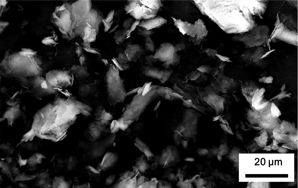
Scanning electron microscopy of the talc. Length of bar at right lower part in photo: 20 µm. Image was taken at 2000× magnification.
Body Weight and Organ Weight Relative to Body Weight
There were no treatment-related adverse symptoms or deaths associated with inhaled talc during the experimental period. We measured body weight of experimental animal every 3 days. Body weight was slightly decreased on third day after exposure in 50 and 100 mg/m3 exposure group of both male and female rats; however, there were no significance when compared with the control group (data not shown). There were no significant differences in the relative weight of the heart, thymus, kidney, liver, lung, and spleen compared to body weight (data not shown).
Hematological and Biochemical Analyses
Hematology evaluations for the male and female rats at the end of the study are shown in Tables 1 and 2, respectively. The MPV in males exposed to 5 mg/m3 of talc and PLT count in females exposed to 50 mg/m3 were increased significantly when compared with control rats. However, all hematological changes fell within the range of the historical control values. Glucose was decreased significantly in male rats exposed to 50 and 100 mg/m3 (Table 3). Triglyceride was decreased in all male rats exposed to talc, with a significant difference only in the 50 mg/m3 exposure group (Table 3). Aspartate aminotransferase and GGT were increased significantly in female rats exposed to 50 mg/m3 compared with the control group (P < 0.05; Table 4). No other hematological and biochemical significant differences were evident in talc-exposed male and female rats.
Hematological Values of Male Rats Exposed to Talc for 4 Weeks by Inhalation.a

Abbreviations: WBC, white blood cell; NE, neutrophil; LY, lymphocyte; MO, monocyte; EO, eosinophil; BA, basophils; RBC, red blood cell; HB, hemoglobin; HCT, hematocrit; MCV, mean corpuscular volume; MCH, mean corpuscular hemoglobin; MCHC, mean corpuscular hemoglobin concentration; RDW, red blood cell distribution width; PLT, platelet; MPV, mean plasma volume.
aSize of each group (6).
bSignificantly different with control (P < 0.05).
Hematological Values of Female Rats Exposed to Talc for 4 Weeks by Inhalation.a
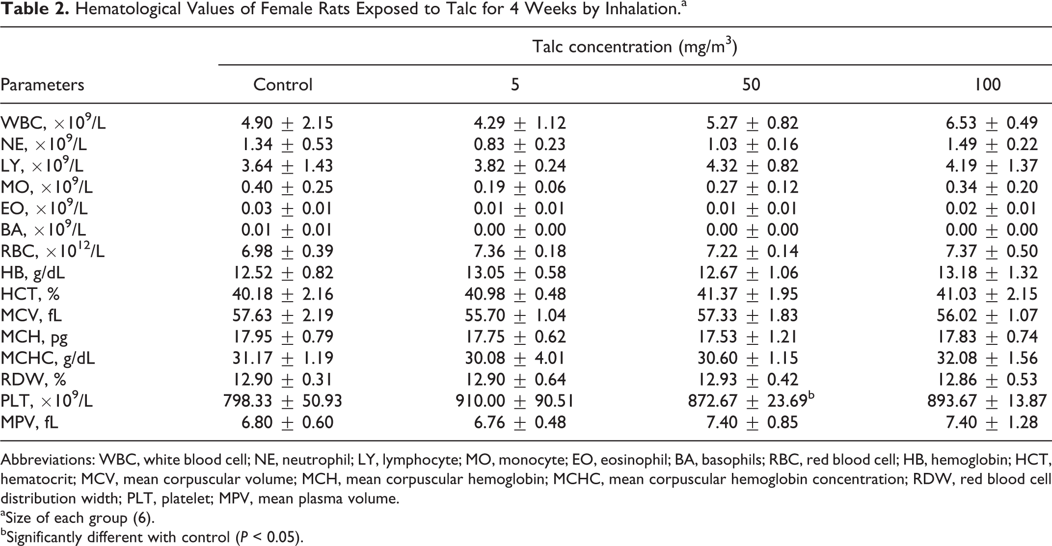
Abbreviations: WBC, white blood cell; NE, neutrophil; LY, lymphocyte; MO, monocyte; EO, eosinophil; BA, basophils; RBC, red blood cell; HB, hemoglobin; HCT, hematocrit; MCV, mean corpuscular volume; MCH, mean corpuscular hemoglobin; MCHC, mean corpuscular hemoglobin concentration; RDW, red blood cell distribution width; PLT, platelet; MPV, mean plasma volume.
aSize of each group (6).
bSignificantly different with control (P < 0.05).
Biochemical Serum Values in Male Rats Exposed to Talc for 4 Weeks by Inhalation.a
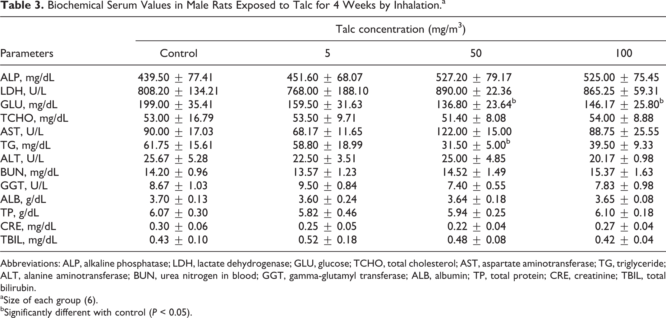
Abbreviations: ALP, alkaline phosphatase; LDH, lactate dehydrogenase; GLU, glucose; TCHO, total cholesterol; AST, aspartate aminotransferase; TG, triglyceride; ALT, alanine aminotransferase; BUN, urea nitrogen in blood; GGT, gamma-glutamyl transferase; ALB, albumin; TP, total protein; CRE, creatinine; TBIL, total bilirubin.
aSize of each group (6).
bSignificantly different with control (P < 0.05).
Biochemical Serum Values in Female Rats Exposed to Talc for 4 Weeks by Inhalation.a
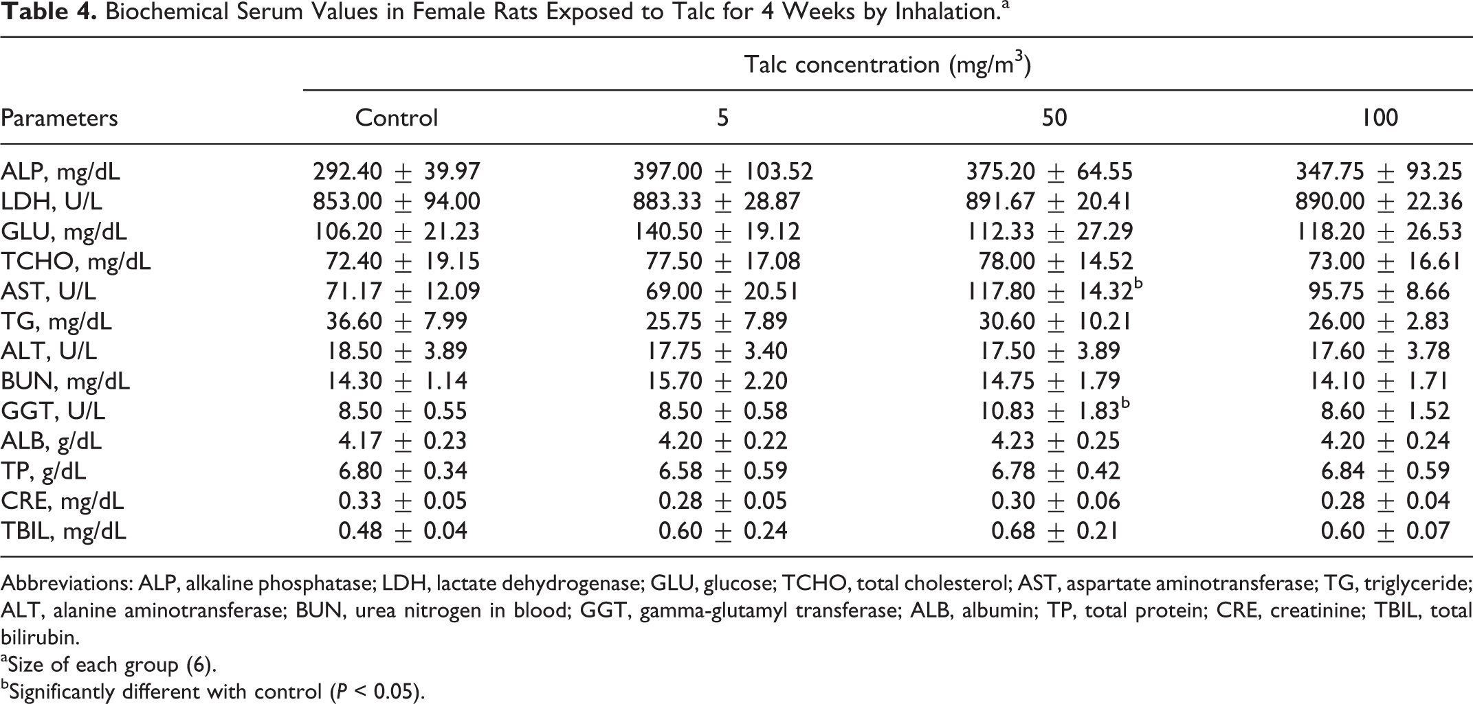
Abbreviations: ALP, alkaline phosphatase; LDH, lactate dehydrogenase; GLU, glucose; TCHO, total cholesterol; AST, aspartate aminotransferase; TG, triglyceride; ALT, alanine aminotransferase; BUN, urea nitrogen in blood; GGT, gamma-glutamyl transferase; ALB, albumin; TP, total protein; CRE, creatinine; TBIL, total bilirubin.
aSize of each group (6).
bSignificantly different with control (P < 0.05).
Lung Histopathology
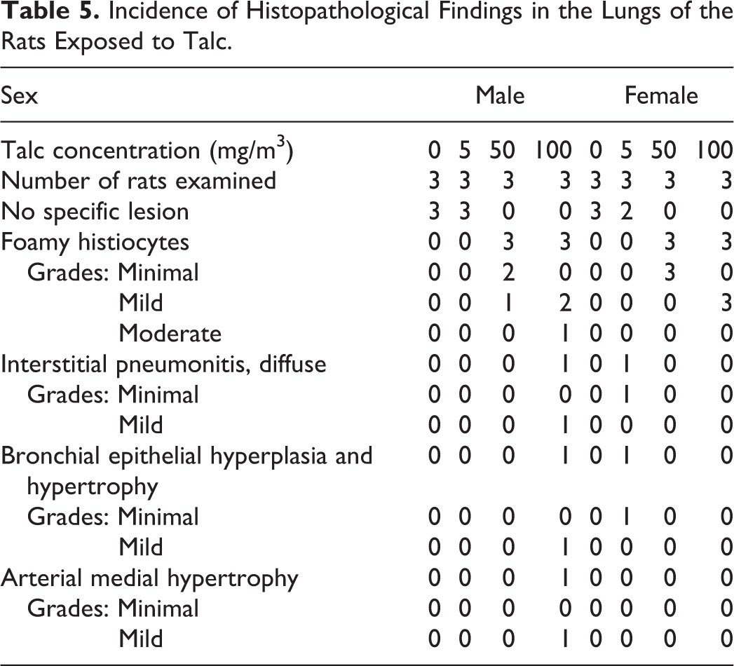
In male and female rats exposed to 50 and 100 mg/m3 of talc, infiltration of foamy macrophages on the alveolar walls and spaces near the terminal and respiratory bronchioles occurred in a concentration-dependent manner (Figures 2 and 3). Interstitial pneumonitis, bronchial epithelial hyperplasia and hypertrophy, and arterial medial hypertrophy were observed in 1 male and female rat, respectively, with no relation to the exposure concentration (Table 5).
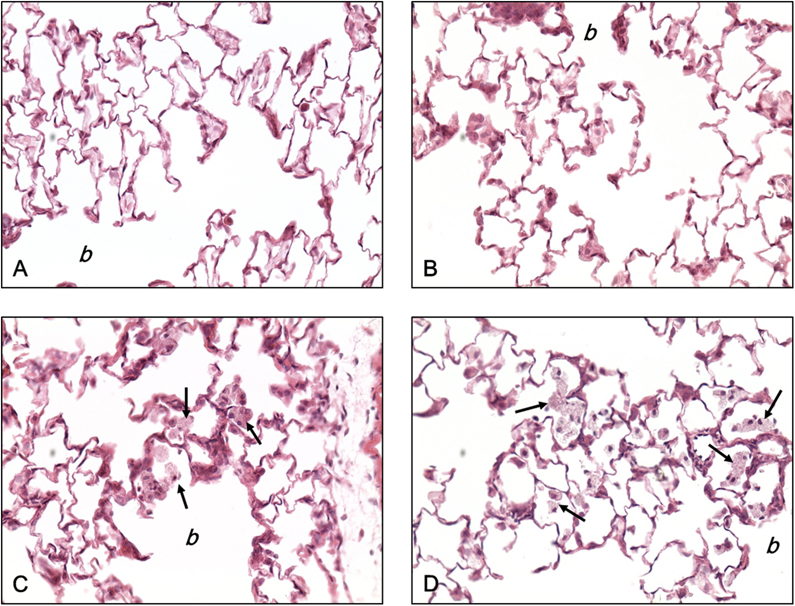
Histopathological findings in the lungs of male rats after exposure to talc for 4 weeks. A, Control and (B) low (50 mg/m3) exposure group. In the middle (C, 50 mg/m3) and high (D, 100 mg/m3) exposure group, the foamy macrophages (arrows) were infiltrated on the alveolar walls and spaces near the terminal bronchioles (b). After hematoxylin and eosin (H&E) staining, the image was taken at 400× magnification.
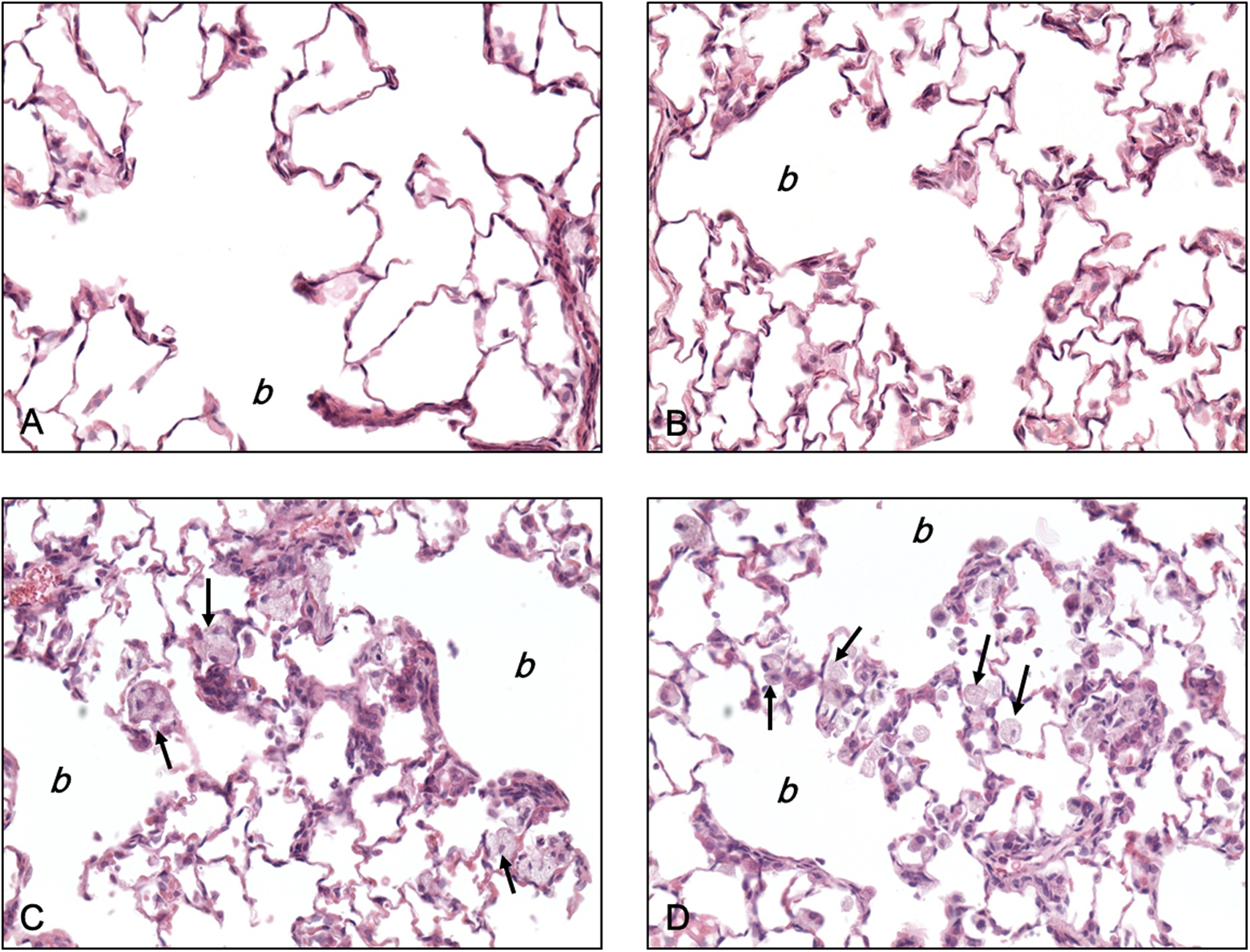
Histopathological findings in the lungs of female rats after exposure to talc for 4 weeks. A, Control and (B) low (50 mg/m3) exposure group. Note the foamy macrophages (arrows) infiltrated on the alveolar walls and spaces near the terminal bronchioles (b) in the middle (C, 50 mg/m3) and high (D, 100 mg/m3) exposure group. After hematoxylin and eosin (H&E) staining, the image was taken at 400× magnification.
Incidence of Histopathological Findings in the Lungs of the Rats Exposed to Talc.
Expression of SOD2 and GPx1 in Lung
A significant fold-induction of SOD2 was evident between the control group and both the low and the high talc inhalation groups of male rats (P < 0.05). Expression of SOD2 in lungs of male rats exposed to the highest concentration of talc was more than twice the level in control rats (Figure 4). Expression of SOD2 in the female rats who inhaled the highest concentration of talc was 4 times that of the control group. Changes in the expression of GPx1 in male rats who inhaled the lowest and highest concentrations of talc were more than double those of control rats. The difference between the control group and low exposed group was significant. Glutathione peroxidase 1 tended to be increased in all females exposed to talc, albeit without statistical significance. Overall, SOD2 was a more significant oxidative stress marker than GPx1 in the lungs of rats exposed to talc.
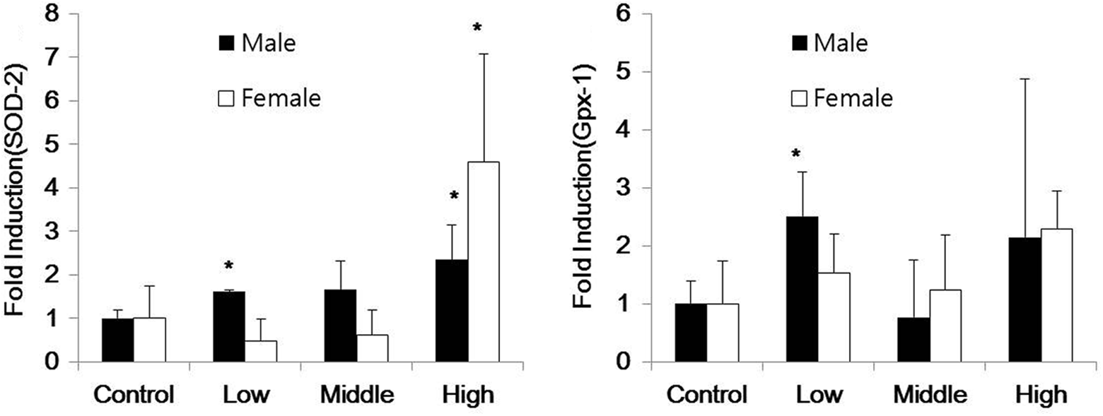
Expression of superoxide dismutase 2 (SOD2) and glutathione peroxidase (GPx1) in the lungs of male and female rats after exposure to talc for 4 weeks. *P < 0.05 versus control group.
Discussion
Talc is a white or gray fine powder that is acid- and temperature-resistant and water insoluble. A great amount of talc has been used occupationally, for example, in the rubber industry and could be exposed to human in our daily life when people use cosmetic products such as baby powder. 35 In addition, exposure of hamsters to an aerosol of cosmetic talc led to deposition of particles in the lung. 36
The present study was performed to evaluate 4-week pulmonary toxicity of inhaled talc including the expression of antioxidant enzymes in lung. Male and female rats were exposed to different concentrations of talc for 4 weeks. The composition of talc used in this study was determined, and hematological, biochemical, and histopathological comparisons with control rats were performed. In addition, the effects of inhaled talc on antioxidant enzymes in lung were evaluated.
Analysis of chemical composition of talc by ICP-AES showed that the talc sample used in this study contained mainly SiO2 and MgO. This finding is consistent with the prior report revealing that talc comprised 63.4% SiO2, 31.9% MgO, and 4.8% water. 37 SiO2 was highly toxic to alveolar macrophages in humans and rats, generating free radical-mediated oxidative stress and inducing tumor necrosis factor, one of cytokine. 38 Numerous studies on SiO2, especially nano-sized particle, revealed that it caused various pulmonary diseases, oxidative stress, and lipid peroxidation. 39
Particles deposited in the alveolar region are phagocytized by alveolar macrophages. The present histopathological examination revealed foamy macrophage infiltrates in the lungs of male and female rats exposed to 50 and 100 mg/m3 talc, indicating that macrophage infiltration was a direct consequence of the inhalation of talc. Macrophage infiltration was found on the alveolar walls and spaces near the terminal bronchioles, which is the route of inhaled particles. Pickrell et al reported that repeated inhalation exposure of rats to talc increased the number of intra-alveolar macrophages. 24 Another talc inhalation study using 4-week-old hamsters revealed lung interstitial pneumonia, alveolar emphysema, alveolar and bronchiolar calcification, alveolar hyperplasia, and alveolar histiocytosis. 36
Exposure to inhaled particles in experimental animals could lead to lung burdens and alveolar inflammation. The inflammatory response might give rise to synthesis of reactive oxygen species and cell injuries. 40 In biological systems, antioxidant enzymes defend cells against various oxidative stresses. We examined the protein expression changes of SOD2 (also called manganese SOD) and GPx1 (also called cytosolic GPx), which are oxidative stress responsive enzymes. Superoxide dismutases catalyze superoxide anions into hydrogen peroxide efficiently. 41 In the present study, SOD2 was induced significantly in male rats exposed to low and high concentrations of inhaled talc and in female rats exposed to the high inhaled concentration. These results were consistent with the previous research, revealing that inhaled cristobalite and TiO2 particles increased SOD2 immunoreactive protein. 42 Another previous study showed that SOD2 and GPx1, representative enzymes associated with oxidative stress response, were significantly upregulated when rats exposed to manufactured ultrafine particles. 43 Glutathione peroxidase 1 detoxifies peroxides with reduced form of GSH, thiol-containing tripeptide, acting as an electron donor, with the production of glutathione disulfide. 44 In the present study, GPx1 tended to increase in rat lungs exposed to talc, albeit not with appreciable statistical significance. Superoxide dismutase 2 was induced significantly more than GPx1 did in rat lungs exposed to talc; therefore, we concluded that SOD2 was more sensitive oxidative stress indicator than GPx1 in this study. Lung tissues of only 3 rats in each male and female group were used in this study on the induction of enzyme, therefore, we might have the experimental limitation for getting significant results of concentration-dependent induction of antioxidant enzymes. It is well known that cellular defense against free radicals is related to various antioxidant enzymes such as catalase, glutathione reductase, and glutamate-cysteine ligase, in addition to GPx and SOD. 45 Further studies on induction of all these enzymes are necessary to understand the antioxidant activities in rats exposed to talc comprehensively.
The decrease of GLU in the male rats might be related to the decreased body weight. There were no treatment-related symptoms or mortality in any of the rats treated with talc during the study period. Histopathological examination revealed that macrophage infiltration was observed on the alveolar walls and spaces near the terminal and respiratory in the 50 mg/m3 and 100 mg/m3 exposure groups. In addition, in male and female rats exposed to 100 mg/m3 talc, SOD2 expression was significantly increased (P < 0.05). Taken together, these results demonstrate that 4 weeks repeated inhalation of talc in rats could induce macrophage aggregations around the terminal airways and oxidative damage in the lung.
Footnotes
Acknowledgments
This study was supported by the National Institute of Environmental Research, Republic of Korea.
Author Contribution
Shim, I. contributed to acquisition, analysis, and interpretation and drafted manuscript; Kim, H. contributed to conception and drafted manuscript; Yang, S. contributed to acquisition and interpretation and critically revised manuscript; Choi, M. contributed to acquisition and analysis and critically revised manuscript; Seo, G. contributed to acquisition and analysis and critically revised manuscript; Kim, P. contributed to conception and critically revised manuscript; Choi, K. contributed to conception and critically revised manuscript. All authors gave final approval and agree to be accountable for all aspects of work ensuring integrity and accuracy.
Declaration of Conflicting Interests
The author(s) declared no potential conflicts of interest with respect to the research, authorship, and/or publication of this article.
Funding
The author(s) received no financial support for the research, authorship, and/or publication of this article.