
Abstract
The health hazard of microwave radiation (MWR) has become a recent subject of interest as a result of the enormous increase in mobile phone usage. The present study aimed to investigate the effects of chronic low-intensity microwave exposure on cognitive function, heat shock protein 70 (HSP70), and DNA damage in rat brain. Experiments were performed on male Fischer rats exposed to MWR for 180 days at 3 different frequencies, namely, 900, 1800 MHz, and 2450 MHz. Animals were divided into 4 groups: group I: sham exposed; group II: exposed to MWR at 900 MHz, specific absorption rate (SAR) 5.953 × 10⁻⁴ W/kg; group III: exposed to 1800 MHz, SAR 5.835×10⁻⁴ W/kg; and group IV: exposed to 2450 MHz, SAR 6.672 × 10⁻⁴ W/kg. All the rats were tested for cognitive function at the end of the exposure period and were subsequently sacrificed to collect brain. Level of HSP70 was estimated by enzyme-linked immunotarget assay and DNA damage was assessed using alkaline comet assay in all the groups. The results showed declined cognitive function, elevated HSP70 level, and DNA damage in the brain of microwave-exposed animals. The results indicated that, chronic low-intensity microwave exposure in the frequency range of 900 to 2450 MHz may cause hazardous effects on the brain.
Introduction
The rapid development in cell phone technology and exposure to microwave radiation (MWR) emitted by cell phones has raised major public concern about the possibility of associated health effects. An association between microwave exposure and its adverse effects is of high impact on public health and it is questionable.
Exposure to MWR is leading to major concern about its effect on cognitive impairment as reduced learning ability. 1 However studies reported on these effects are inconsistent and controversial. It is reported that cognitive impairment, loss of mental concentration, and reduced learning ability occurs due to chronic MWR exposure. 2 Heat shock proteins (HSP) act as molecular chaperones that bind partially damaged or denatured proteins and assist in their removal. Changes in protein conformation in terms of folding and unfolding processes can result in either an increase or a decrease in their biological activity. 3 The accumulation of DNA adduct is the result of imbalance in DNA damage and its repair, and this may lead to cell death or cancer. 4,5
The effect of MWR depends on the energy absorbed by biological tissue and how it is delivered in space and time. The effects also depend on electromagnetic characteristics such as frequency, intensity, and exposure duration. Therefore, it is necessary to evaluate and understand its potential health hazards.
It is observed that the inconsistent findings on the potential health hazards of MWR exposure might be due to differences in experimental exposure setup, protocols followed, and experimental models (biological system) used for investigations. To address this concern, we used specially designed Gigahertz Transverse Electromagnetic (GTEM) cell to expose experimental animals to MWR and study the effects. 6 To best of our knowledge, this is the first study which investigates and compares the effect of low-intensity MWR exposure using 3 different frequencies (900, 1800, and 2450 MHz). Therefore, to investigate the effects of chronic low-intensity MWR, this in vivo study was undertaken using 3 different frequencies used in mobile telecommunication. This study was focused on how chronic low-intensity MWR affects cognitive function, HSP, and DNA damage in rat brain.
Materials and Methods
Microwave Exposure Setup and Dosimetry
The GTEM cell was designed with the help of the Center for Applied Research in Electronics (Microwave Laboratory), Indian Institute of Technology, New Delhi, and Amitech Electronics Ltd Sahibabad, Ghaziabad (UP) to estimate biological effects of MWR (Figure 1A and B). The GTEM cell is a pyramidal tapered, a dual terminated section with its outer cell dimension, length: 220 cm × breath 120 cm × height 80 cm. The MWR generated through microwave generator SMC 100 (Rhode & Schwarz GmbH & Co, Germany). The MWR source consists of a signal generator operating at frequency range from 9 KHz to 3.2 GHz, amplifier, DC regulator, and power meter. The microwave chamber is lined with absorbers that minimize the possibility of any reflections. Electric field was experimentally checked using an E-field probe inserted into the Transverse Electromagnetic cell through a slit wall. Preexposure validation was conducted using spectrum analyzer to ensure the uniformity of the field strength across the volume of GTEM cell. The GTEM cell was placed in a temperature-controlled room (22°C ± 2°C) under constant lighting conditions. Specific absorption rate (SAR) distribution was calculated by the power balance method using the following equation:
7
A, Schematic diagram of microwave exposure setup. B, Gigahertz Transverse Electromagnetic (GTEM) cell.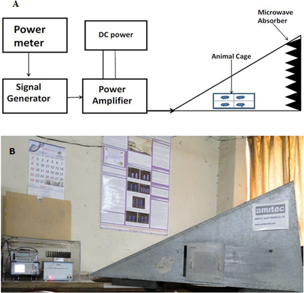

Where, Pabs = RF (radio-frequency) power absorbed per animal (Watt), n = number of animals within the cell, Pin = input power (Watt), Pout = output power (Watt), and Prefl = reflected power (Watt).
Animal Exposure
Male Fischer-344 rats (60 days old and weighing 150-200 g) were obtained from the central animal house facility of the institute and placed in individual-raised, galvanized-wired cages. They were acclimatized to laboratory conditions for 5 days and were kept under standard conditions (temperature 22°C ± 2°C with constant humidity 40%-50%) with alternating 12-hour light and dark cycle. Animals were provided with nutritionally adequate standard diet obtained from Nutrilab (Bangalore, India) and water ad libitum. A total of 24 rats were divided into 4 groups (6 rats in each group): group I (Sham exposed) animals were maintained under the same conditions as that of other groups except microwave generator was kept on switch off mode; group II animals were exposed to MWR at 900 MHz, SAR 5.953 × 10⁻⁴ W/kg; group III animals were exposed to 1800 MHz, SAR 5.835 × 10⁻⁴ W/kg; and group IV animals were exposed to 2450 MHz, SAR 6.672 × 10⁻⁴ W/kg. During the exposure rats were restrained in closed boxes with dimension as length:30 cm × breadth:15 cm × height:20 cm divided into 4 compartments with few holes of 1-cm diameter to facilitate easy movement and breathing, respectively, kept at a distance of 100 cm from the source. At a time 1 group (6 rats) was exposed to whole-body MWR in GTEM cell (Amitech Electronics Ltd, India) at power level of 0.00 dBm for 2 h/day during light period for 5 days/week at the same time for 180 days. Animals had no access to food and water during exposure. All animal procedures were performed in accordance with protocols approved by the Institutional Animal Ethics Committee , University College of Medical Sciences, Delhi, and care of the animals was undertaken as per guidelines of the committee for the Purpose of Control and Supervision of Experiments on Animals, India for laboratory animal facilities. Body temperature of rats was noted by rectal measurements immediately before and after the MWR exposure in all the groups.
Assessment of Cognitive Function
Elevated plus maze paradigm
The Elevated plus maze
Morris Water Maze
The acquisition and retention of a spatial navigation task were examined using a Morris water maze. 6 Animals received a training session consisting of 4 trials in a day for 4 days prior to microwave exposure in Morris water maze (180 cm diameter × 60 cm) filled with water. An escape platform was hidden 2 cm below the surface of water in a fixed location in 1 of the 4 quadrants halfway between the wall and the middle of the pool. The water was made opaque during the task with a nontoxic dye. Each trial consisted of releasing a rat into the water facing the wall of the pool, at 1 of 4 starting compass positions (North, South, East, and West) so that each position could be explored well. The time to reach the escape platform (latency in seconds) was recorded up to a maximum of 3 minutes. The animal which could not find the platform up to 3 minutes were deliberately placed on the platform and allowed to sit for 30 seconds. The time taken by a rat to reach the platform on the fourth day was recorded as initial acquisition latency (IAL). Following 24 h after IAL, a probe test was done, where there was no platform and each rat was randomly released from any one of the positions and tested for the retention of acquired memory. During retention, the time taken by each rat to locate the target quadrant (quadrant in which platform was placed during training) and time spent in the target quadrant for four 15-second interval over 60 seconds was recorded.
Preparation of Brain Samples and Quantification of HSP70 Level
Rats from each group were anesthetized and then decapitated to isolate brain. Whole brain was washed 3 times with phosphate buffer solution (PBS), pH 7.4, and subsequently hippocampus was homogenized with the appropriate amount of PBS at 4°C with protease inhibitors, centrifuged, and the supernatant was stored at −80°C until use. The levels of HSP70 were determined using a commercially available enzyme-linked immunotarget assay Kit (Assay design, New york) according to manufacturer’s instructions.
DNA Damage Analysis Using Alkaline Comet Assay
DNA damage was evaluated using the alkaline comet assay. 9,10 Slides were prepared in duplicates per sample. Remaining brain sample from the HSP 70 assay were used for the comet assay. Briefly, brain tissue was placed in 1 mL chilled mincing solution (Hank balanced salt solution, with 20 mmol/L EDTA and 10% dimethyl sulfoxide [DMSO]) in a Petri dish and chopped into small pieces with a pair of scissors to get a uniform cell suspension. Slides were precoated with 600 µL of low melting agarose (LMA, 1.0%) prepared in PBS. The diluted sample of 600 µL (50 µL cell suspension mixed with 600 µL of 0.75% LMA) was loaded on precoated slide to form the second layer. The slides were kept on ice for 5 minutes to allow the gel to solidify. The slides were immersed in freshly prepared chilled lysing solution containing 2.5 mol/L NaCl, 100 mmol/L EDTA, and 10 mmol/L Tris (pH 10) with 10% DMSO, and 1% Triton X-100 was added just before use. The slides kept in the lysing solution for 1 hour at 4°C, followed by electrophoresis in a horizontal gel electrophoresis tank with agarose ends nearest to the anode. Fresh chilled electrophoresis buffer (1 mmol/L Na2EDTA and 300 mmol/L NaOH, pH >13) was poured into the tank approximately 2.5 mm above the slides. The slides were left in this solution for 25 minutes to allow DNA unwinding and expression of alkali-labile sites as DNA strand breaks. Electrophoresis was conducted at 0.9 V/cm for 60 minutes at 4°C. All these steps were performed under dim light. The electrophoresis tank was covered with black paper to avoid additional DNA damage due to stray light. After electrophoresis slides were drained and placed horizontally in a tray. Tris buffer (0.4 mol/L; pH 7.5) was added drop-wise and left for 5 minutes to neutralize the excess alkali. Neutralization of slides was repeated 3 times and subsequently slides were dried and stored.
Dried slides were rehydrated and each slide was stained on the next day with 100 µL of ethidium bromide (20 mg/mL) for 5 min. Slides were randomized and coded to blind the scorer. All slides were scored by 1 person to avoid interscorer variability. Scoring done using an image-analysis system (Kinetic Imaging, Liverpool, United Kingdom) attached to a fluorescent microscope (BX51, Olympus, Japan). The microscope was connected to a computer through a charge-coupled device camera to transport images to software (Komet 5.0) for analysis. Images from 100 cells (50 from each replicate slide) were analyzed. Undamaged cells had an intact nucleus without a tail and damaged cells had the appearance of a comet. To quantify DNA damage, following parameters were evaluated: percentage of DNA content in the head and tail, olive tail moment (OTM), tail extent moment, and tail length (TL) using Komet 5.0 software (Kinetic Imaging, Liverpool, United Kingdom).
Statistical Analysis
Statistical analysis was performed with SPSS (version 16.0). All values were expressed as mean ± standard deviation. Significance of differences among groups was determined by 1-way analysis of variance followed by Tukey test. Statistical significance was accepted at P value <.05.
Results
Microwave exposure resulted in no change in body temperature.
Effect of MWR on Cognitive Function
The influence of MWR on cognitive function is shown in Figure 2, where all the MWR-exposed groups showed higher TL (transfer latency) when compared to sham-exposed group, but when TL was compared between the microwaves-exposed groups, that is, 900 MHz, 1800 MHz, and 2450 MHz, no significant difference was found. TL of first the day (on 180th day; end of exposure duration) indicated the acquisition of learning behavior of animals, whereas TL of next day (24 hours after 180 days of microwave exposure) indicated retention of information or memory. A significant difference was observed in Transfer latency between sham-exposed and microwave-exposed groups (Figure 2 A and B). Rats exposed to MWR took more time to enter one of the closed arms of EPM when compared to sham-exposed animals following MWR exposure. This increase in transfer latency indicates impairment in learning and memory.
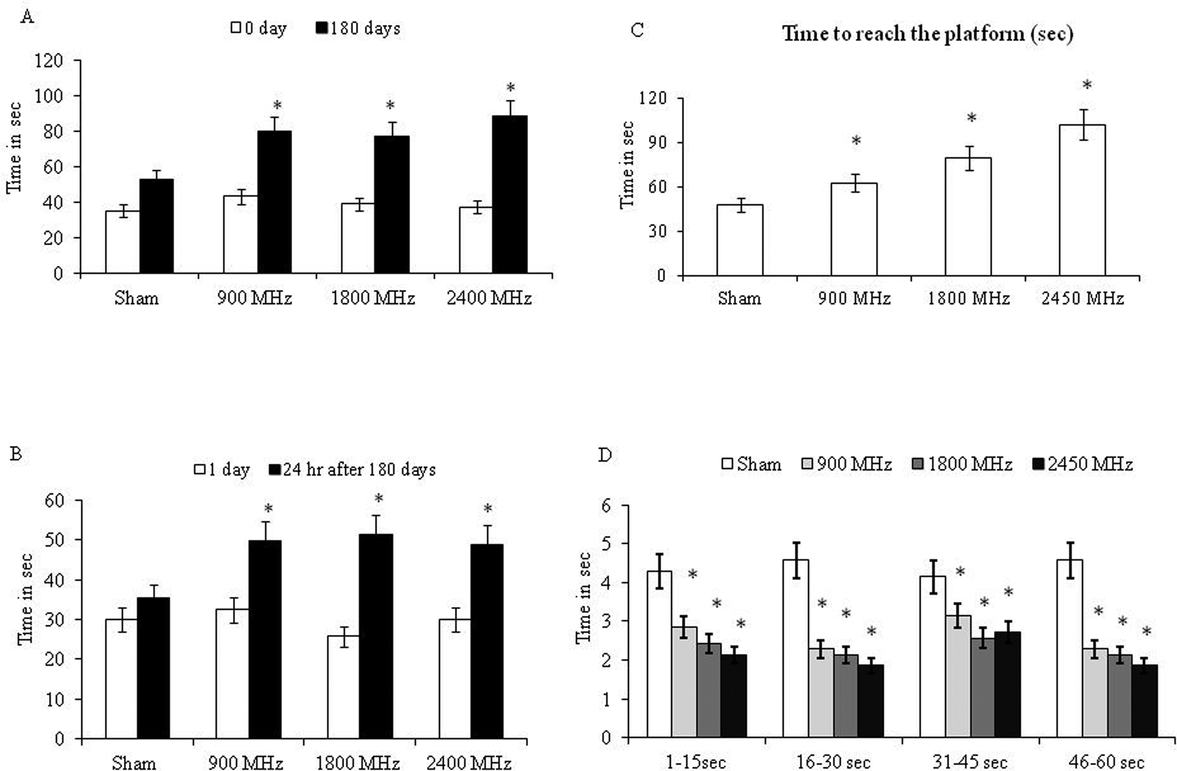
Effect of microwave radiation exposure on rat behavior. A, Time taken by rats to enter 1 of the closed arms during Elevated plus maze (EPM; acquisition) B, Time taken by rats to enter one of the closed arms during EPM (retention). C, Escape latency time (ELT) of rats during Water maze test to locate hidden platform. D, Time spent in target quadrant.*shows significant difference from Sham-exposed group (P < .05). Values are expressed as mean ± standard deviation (SD; 6 animals/group).
Spatial memory performance was evaluated using the Morris water maze in all the experimental groups. Significant difference with respect to escape time was observed between microwave-exposed groups and sham-exposed groups. During the probe trial (with the removed platform) microwave-exposed rats took longer time to locate the place where the platform was placed (Figure 2 C and D). The time to reach the target quadrant was significantly longer in microwave-exposed group and the time spent in the target quadrant was significantly shorter when compared to the sham-exposed groups.
Effect of MWR on HSP70 Level
Microwave radiation exposure for 180 days showed a significant increase in the level of HSP70 in all the microwave-exposed groups (900 MHz, 1800 MHz, and 2450 MHz), when compared with sham-exposed group (P < .05; Figure 3).
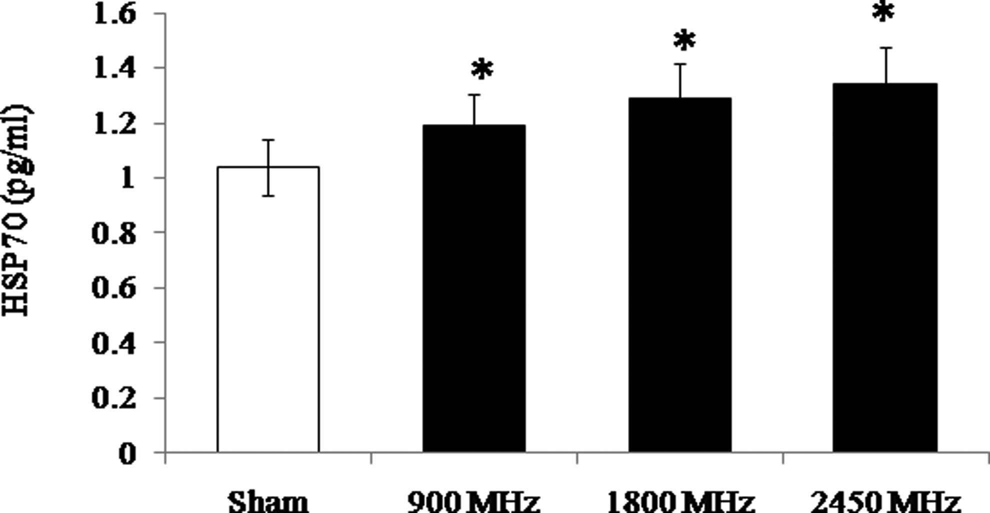
Effect of microwave radiation exposure after 180 days on level of heat shock protein 70 (HSP70) protein (pg/mL) in rat brain.* P < .05 when compared with sham-exposed group. Values are expressed as mean ± standard deviation (SD;6 animals/group).
Effect of MWR on DNA Damage
Comet assay performed on brain tissue following exposure to MWR showed a significant increase in the percentage of DNA in tail, tail extent moment, OTM and TL in brain cells all microwave-exposed animals when compared to sham-exposed animals (Figure 4). The percentage of DNA migrating into the tail region was significantly enhanced in all the 3 groups, that is, 900 MHz, 1800 MHz, and 2450 MHz when compared to sham-exposed group (P < .05). Correspondingly, the percentage of DNA in the head was significantly reduced in all the microwave-exposed groups, that is, 900 MHz, 1800 MHz, and 2450 MHz (P < .05; Figure 5A). The head/tail DNA in 1800 MHz- and 2450 MHz-exposed group showed significant difference (P < .05) when compared with the 900 MHz-exposed group. The OTM was also increased significantly (P < .05) in all the microwave-exposed groups (Figure 5 B) when compared to sham-exposed group. The OTM in 1800 MHz- and 2450 MHz-exposed group were significantly (P < .05) increased when compared to 900 MHz-exposed group.
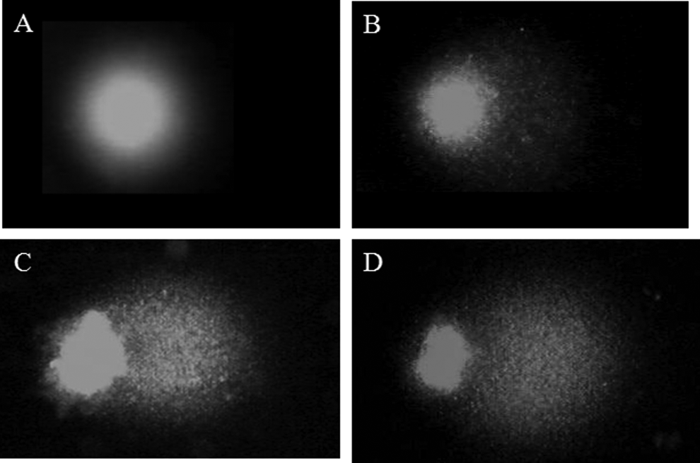
Representative picture of comet (DNA damage) at different frequencies (A) Sham exposed, (B) 900 MHz exposed, (C) 1800 MHz exposed, and (D) 2450 MHz exposed.
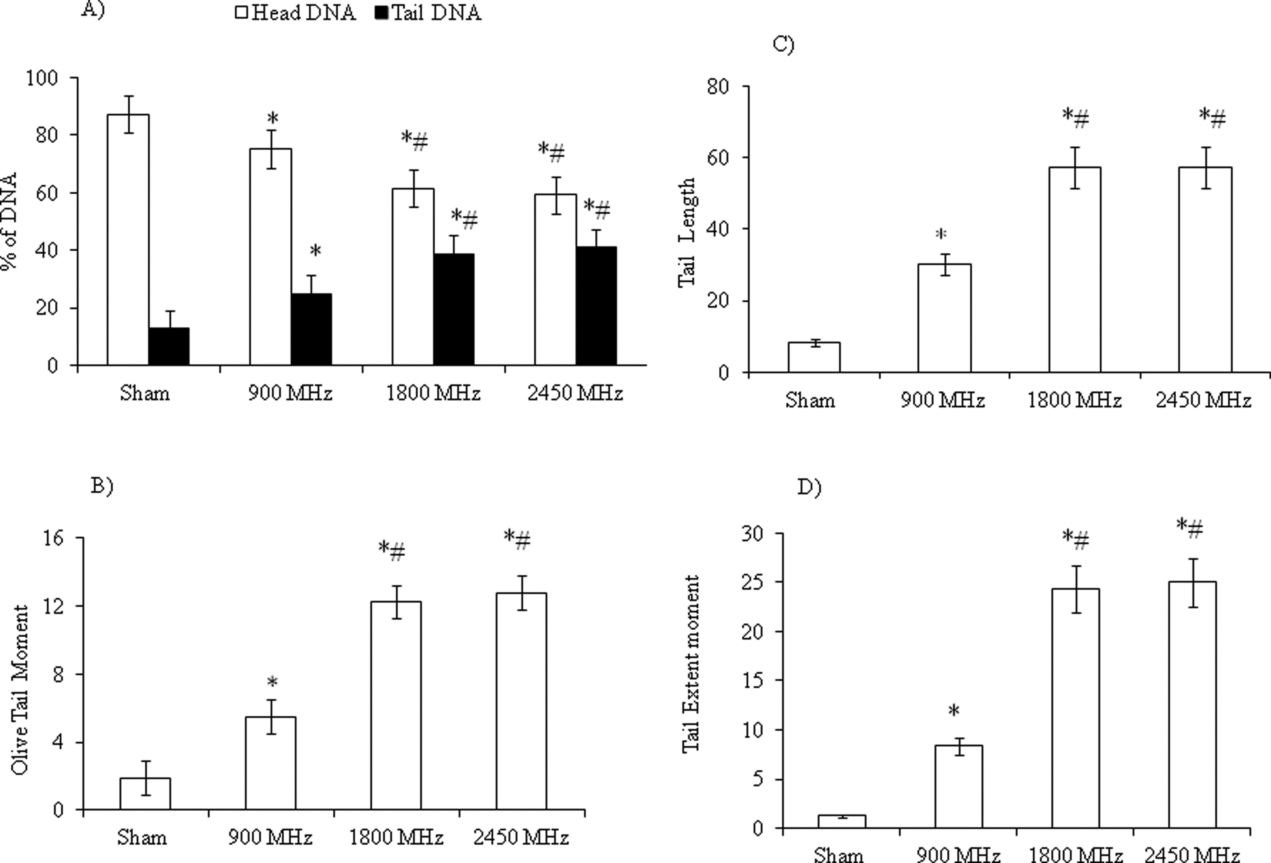
Effect of microwave exposure for 180 days on DNA in rat brain. (A) Percentage of DNA in head and tail, (B) Olive tail moment (arbitrary unit), (C) tail length (µm), and (D) tail extent moment.*P < .05 when compared with sham-exposed group; # P < .05 when compared with 900 MHz-exposed group. Values are expressed as mean ± standard deviation (SD; 6 animals/group).
Similarly, the TL of comet was increased significantly in animals exposed to 900, 1800, and 2450 MHz (P < .05) in comparison with sham-exposed group. The 1800 MHz-and 2450 MHz-exposed group showed significant (P < .05) increase in TL when compared to 900 MHz-exposed group (Figure 5C). Significant (P < .05) increment in tail extent moment was noted in all microwave-exposed group when compared to sham-exposed group. Similarly, the tail extent moment in 1800 MHz- and 2450 MHz-exposed group showed significant differences (P < .05) when compared to 900 MHz-exposed group (Figure 5D).
Discussion
Public concerns are increasing on possible adverse effects of MWR used in mobile telephony on health. The human body is like an electrochemical instrument of exquisite sensitivity whose orderly functioning and control are underpinned by oscillatory electrical processes of various kinds, each characterized by a specific frequency, some of which happen to be close to those used in Global system for Mobile Communication (GSM). The exact mechanism behind the biological action of MWR exposure is still unknown. The present study provides evidence that low-intensity microwave exposure results in cognitive impairment, elevation in HSP level and DNA damage in rat brain.
In the present study, it is demonstrated that low-intensity chronic microwave exposure upto 180 days at 3 frequencies, that is, 900, 1800 and 2450 MHz causes impairment in learning and memory as tested by EPM and Morris water maze test. Our earlier study also suggested that low-intensity microwave exposure for 30 days at 900 MHz at 5.953 × 10⁻⁴ W/kg, 1800 MHz at 5.835 ×10⁻⁴ W/kg, and 2450 MHz at 6.672 ×10⁻⁴ W/kg, causes impairment in learning and memory. 11 Earlier we reported that MWR exposure upto 30 days at 900 MHz even at low intensity affects the cognitive function. 6 This could be due to direct or indirect interaction of microwaves in brain of experimental rats. 12 Nittby et al 13 reported significant impairment in cognitive function after 55 weeks of exposure in rats exposed to MWR with whole-body SAR value of 0.6 and 60 mW/kg. Narayanan et al 14 reported that animals exposed to the GSM mobile phone at (900/1800 MHz) with 50 missed calls/day for 4 weeks showed alterations in the acquisition of learning response in the Morris water maze test. Decline in the cognitive function may be a result of damage in the blood–brain barrier and the cells of the brain, that is, hippocampus which is concerned with learning, memory, and movement. 15,16
In the present study, MWR triggered an increase in the levels of HSP70 in all the microwave-exposed groups, that is, groups exposed to 900 MHz, 1800 MHz, and 2450 MHz (SAR 5.953 × 10⁻⁴ W/kg, 5.835 × 10⁻⁴ W/kg, and 6.672 × 10⁻⁴ W/kg, respectively) that might be related to nonthermal effects of the electromagnetic field (EMF) produced by the MWR exposure. Heat shock protein 70 is one of the most studied HSPs and is the central component of the cellular network of molecular chaperones, folding catalyst, and protect cells against a variety of environmental stressors and MWR is one of them. 17 It is reported that nonthermal radiofrequency energy induces heat shock response in various cellular targets and observed different results in regard to the cell sensitivity to EMFs. 18 –20 In our earlier study, we observed that low-intensity microwave exposure leads to elevation in the level of HSP70. 11 The elevation in the level of HSP70 in hippocampus, which controls behavioral and cognitive functions including spatial and working memory, may be the possible reason for cognitive decline in the microwave-exposed rats. A study by Jorge et al 21 observed that acute exposure to 2.45 GHz EMFs triggered an imbalance in anatomical HSP levels on exposure of MWR.
In the present study, DNA damage was also observed in brain after chronic low-intensity MWR exposure. Earlier in our study, we found that low-intensity MWR exposure for 30 days is capable of interacting with DNA by unknown mechanism and causes single-strand DNA breaks. 11 It is apparent from our study that at such low level of microwave exposure and the range of frequency from 900 to 2450 MHz could be the genotoxic by indirect mechanism. The biochemical compounds in living cells are composed of charges and dipoles that can interact with electric and magnetic fields by various mechanisms. The high frequency EMF (2.45 GHz and 50 Hz modulated) exerts their genotoxic effects in male Wistar rats as evidenced by a significant increase in DNA strand breaks after 2 hours exposure per day to EMF for 35 days with whole-body SAR of 0.11 W/kg. 22 Campisi et al 23 reported an increase in oxygen radicals accompanied by an increase in DNA strand breaks in primary rat glia cells after exposure to high frequency field (900 MHz). Xu et al 24 reported that DNA adduct rate caused by oxygen radicals in the mitochondria of primary cultured neurons (nerve cells) significantly increased after 24 hours GSM exposure. The increase in single- and double-strand DNA breaks was observed in brain cells of rats exposed to 2450 MHz for 2 hours at whole-body SAR 0.6 W/kg. 25,26 Usikalu M et al 27 reported that low SAR and 2.45 GHz MWR exposure can induce a single-strand break in brain cells of rats. Thus, we are confronted with the question whether or not the same deleterious alteration may also occur in brain from regular microwave exposure. Therefore, it would be necessary to further explore the differential effects of different exposure parameters such as frequency, duration of exposure, and pattern.
Conclusion
The present study demonstrates that exposure to low-intensity MWR leads to harmful effects on rat brain as evidenced by declined cognitive function, increased HSP70 and DNA damage. To better understand the molecular mechanism of action on cognitive function further studies with different intensities and durations of microwave exposure are needed.
Footnotes
Acknowledgments
One of the authors Pravin Suryakantrao Deshmukh is grateful to ICMR for senior research fellowship (SRF) support.
Declaration of Conflicting Interests
The author(s) declared no potential conflicts of interest with respect to the research, authorship, and/or publication of this article.
Funding
Authors are grateful to Indian Council of Medical Research (ICMR), New Delhi for the grant; in the form of the Extramural Research Project vide sanction letter No. 5/8/4-4(env) 07-NCD-I dated 3-08-09.