
Abstract
In this study, an in vivo toxicological safety assessment of Bacillus licheniformis Me1, a native isolate from milk, was performed. An acute toxicity study in male albino Wistar rats demonstrated no treatment-related illness or mortality. A 90-day subchronic oral toxicity study using 2 doses (1.1 × 10¹⁰ and 1.1 × 10¹¹ colony-forming unit [CFU]/kg body weight [BW], respectively) failed to show dose-dependent illness or mortality. Moreover, neither significant differences in serum biochemical and hematological analyses nor histopathological changes in organs or tissues were found when compared to the control groups. The no-observed-adverse-effect level (NOAEL) was found to be greater than 1.1 × 10¹¹ CFU/kg BW. The in vivo micronucleus assay in mice did not reveal any signs of genotoxic effect at any of the doses tested. Furthermore, dermal and acute eye irritation tests conducted in rabbits showed no edema or erythema and ocular lesions. These results suggest that B licheniformis Me1 can be considered safe for food industry applications.
Introduction
Probiotics are “beneficial live microorganisms which, when administered in adequate amounts, confer a health benefit to the host.” 1 Within the last decade, the supplementation of probiotics to various food products and as therapeutic agents against several infectious diseases has increased tremendously. 2 The health benefits of probiotics include inhibition of gastrointestinal pathogens; aid in digestion and nutrient absorption; prevention of infectious diarrhea, irritable bowel syndrome, constipation, gas, or lactose intolerance; and contribution to immune function. Furthermore, other clinical investigations document further benefits such as lowering cholesterol levels, preventing dental caries, delaying of allergies in children, and treatment and prevention of vaginal and urinary infections in women. 3 Probiotics can also serve as an alternative to commonly used therapeutic and prophylactic avenues for increasing incidence of antibiotic resistance and a growing prevalence of cancer and allergic conditions in an aging population.
The majority of the probiotics studied and commercialized are lactic acid bacteria (LAB) and bifidobacteria. 4,5 In contrast, only very few species of the spore-forming Bacillus genus are used3,6 ; although, there are several advantages of spore-forming Bacillus spp over LAB as probiotics. The underlying rationale for this perception is that Bacillus can survive in food products requiring harsh processing conditions such as high temperature and pressure, they survive better under gastrointestinal tract (GIT) conditions, possess a long shelf life, and remain viable throughout the shelf life at room temperature and refrigerated conditions. 7 Due to its better survivability, the effective dose required for Bacillus as probiotic supplements is less, as compared to LAB. 8
Bacillus spp which are commonly available for human and animal consumption as probiotics include Bacillus subtilis, B. licheniformis, B. clausii, B. coagulans, B. cereus, B. pumilus, and B. laterosporus. 9 In humans, Bacillus probiotics are used as health food supplements, for prophylaxis of gastrointestinal disorders, 10 and as therapeutic agents for treatment of urinary tract infections. 11 B. subtilis and B. licheniformis are probiotic components of Biosporin, which is licensed as a medicine in Ukraine and Russia for the treatment of GI disorders. 12,13 Recently, GanedenBC30 (B. coagulans) received its generally recognized as safe (GRAS) status by an independent scientific panel. 14 Presently, it is marketed in various food products and claims to have several health benefits. 8 In animals, probiotics are being used as growth-promoting and prophylactic agents (eg, Toyerocin 15 ) after antibiotic growth supplements were banned by the European Union (EU) in 2006. 7 Bacillus probiotics are also broadly used in aquaculture, particularly in shellfish, to enhance growth and resistance to disease. 16
Some of the Bacillus spp, such as B. anthracis, B. cereus, B. thuringiensis, B. pseudomycoides, and B. weihenstephanesis, are known for their ability to produce toxins. B. cereus produces enterotoxins and emetic toxins and has been recognized as the major cause of food poisoning and other nongastrointestinal infections. However, not all strains of B. cereus are pathogenic and their pathogenicity varies with strains. 17 In addition, B. thuriengiensis, commonly used as a biopesticide, produces enterotoxins and causes gastroenteritis. 18 The B. subtilis group (including B. subtilis, B. licheniformis, B. pumilus, and B. amyloliquefaciens) is considered relatively safe but is also reported to be involved in food borne illness. 19 –22 Overall, the involvement of strains of the B. subtilis group has been considered of little significance in food poisoning incidents. Another concern about the application of Bacillus in food products is that certain strains of Bacillus can cause opportunistic infections. Oggioni et al 23 reported B. subtilis, used for probiotic preparations, caused septicemia in immunocompromised patients. The probability for the presence of transmissible antibiotic resistance genes in the probiotic candidates should also be considered in any probiotic applications. A recent report proved the presence of macrocide-resistant erm gene in a probiotic B. claussi strain. 24 Therefore, the use of these spore-forming bacteria as dietary supplements, functional foods, and for incorporation in pharmaceutical products requires careful safety assessment using suitable models.
Only limited information is available about the in vivo as well as in vitro safety evaluation of Bacillus spp, viz, B. subtilis, B. coagulans, B. indicus, and B. licheniformis, and these cultures are described to be safe for use without having any adverse effects. 6,25 –27 Several health benefits of Bacillus, in addition to inherent resistance of its spores to environmental stress, emphasize the scope of exploring various Bacillus spp with respect to its use as commercial probiotics. Thus, in the present study, we have evaluated the safety of the bacterial culture, B. licheniformis Me1, a native isolate from milk, for its potential applications in food industry as a probiotic supplement.
Materials and Methods
Chemicals and Reagent Kits
Assay kits for determination of urea, creatinine, bilirubin, aspartate aminotransferase (SGOT), alanine amino transferase (SGPT), lactate dehydrogenase (LDH), creatinine kinase (CK Nac), and alkaline phosphatase (ALP) were obtained from Aspen Laboratories Pvt Ltd, India. Assay kit for analysis of glucose, cholesterol, and triglycerides (TG) were from Span Diagnostics Ltd, India. Kits for determination of sodium, potassium, and chloride were obtained from Coreal Clinical Systems, India. All other chemicals and solvents used for this study were of the highest purity obtained from Merck Pvt Ltd, India.
Preparation of Test Article and Diet
The culture B. licheniformis Me1 isolated from milk was used as the test article in this study. This culture was initially screened based on its potent antibacterial activity and subsequently identified using several biochemical and molecular techniques. (Nithya and Halami, unpublished data). The test article was cultivated in Luria Bertani (LB) broth for 36 hours at 37°C under shaking (150 rpm) condition. Subsequently, cells were harvested by centrifugation. The test article was prepared at a concentration of 1.1 × 10 11 colony-forming unit (CFU)/g of cell pellet for experimental feed preparation. The diet was prepared according to Reeves et al. 28 For acute and subchronic toxicity studies, AIN93M and AIN93G diets were used, respectively. For the subchronic study, diet was prepared once every 2 days with the test article. According to the body weight (BW) changes in the rat and feed intake changes, adjustments for the addition of test article in the diet were made. The experimental diet was stored at 4°C to ensure viability.
Experimental Rats
Animal care and handling conformed to the guidelines of the Committee for the purpose of control and supervision of experiments on animals (CPCSEA), Government of India, and the protocols were approved by the Institutional Animal Ethical Committee (IAEC). The albino Wistar rats bred in the Central Food Technological Research Institute (CFTRI) animal house facility were housed in stainless steel cages (1 rat/cage) with a 12-hour light/dark cycle in a controlled atmosphere viz temperature 22°C ± 3°C and a relative humidity of 60% to 70%. The animals were fed ad libitum and had free access to drinking water during the experimental period. The drinking water was periodically analyzed and was determined to be free of contaminants. The animals were acclimatized to above experimental conditions for 5 days prior to the start of dosing for any toxicity study.
Acute Toxicity Study
An acute toxicity study was performed in accordance with Organization for Economic Cooperation and Development (OECD) Guideline for the Testing of Chemicals No. 423; Acute Oral Toxicity—Acute Toxic Class Method, adopted December 17, 2001. Twelve healthy adult male albino Wistar rats (220 ± 10 g) were randomly assigned into 2 groups (6 per group), namely the control group which received the vehicle alone and the probiotic-treated group which received test article of 1000 mg/kg BW of rat, as a single dose. This dose level corresponds to 1.1 × 10¹¹ CFU/kg BW of rat and was selected based on previous references that have evaluated the in vivo safety of probiotic bacteria. 25,26,29 No female rats were used for acute toxicity study. Following the initial dose, the animals were observed with respect to general behavior, signs of toxicity, and mortality for 4 continuous hours and then twice per day over a period of 14 consecutive days. The general behavior, signs of toxicity include changes in the skin and fur, eyes and mucous membranes, and also respiratory, circulatory, digestive, autonomic, and central nervous systems. Observations were also made on behavior patterns, such as tremors, convulsions, salivation, stool consistency, lethargy, sleep and changes in gait, posture, and response to handling. Daily feed and water intake was recorded throughout the experimental period and the BW of the rats were documented on days 0, 4, 7, 10, and 14. On the 15th day of the experiment, following an overnight fast, the animals were weighed and sacrificed with ether anesthesia. The blood samples were collected in sterile conditions by cardiac puncture in ethylenediaminetetraacetic acid (EDTA-2K) containing tubes for hematological analysis and in nonanticoagulant tubes for serum biochemical investigations. The vital organs were weighed and relative organ weights (g/100 g BW) were calculated. A gross pathological organ examination was performed. Histopatholological analysis (if necessary) was done for any treatment-related abnormalities.
Subchronic Toxicity Study
Male and female albino Wistar rats bred in the CFTRI animal house facility were selected and randomly split into 3 groups, each comprising 6 males and 6 females, weighing 36 ± 1 g. The first group was kept as control and fed only the basal diet. Experimental diet consisting of basal diet supplemented with test article at 2 dose levels 100 and 1000 mg/kg BW/d (which corresponds to 1.1 × 10¹⁰ and 1.1 × 10¹¹ CFU) was fed to groups 2 and 3, respectively, for 13 weeks. Test diet and uncontaminated water were available ad libitum throughout the experimental period. At the end of the experiment, all surviving animals were fasted overnight before anesthetization with diethyl ether and sacrificed. The results were analyzed according to the OECD Guideline for the Testing of Chemicals No. 408; Repeated Dose 90-Day Oral Toxicity Study in Rodents, adopted September 21, 1998.
Feed and water intake, clinical observations, and BW
The feed intake was recorded daily and from this, the mean daily feed intake for each week was calculated. The water intake by the rats was also noted. All the animals were examined for general behavior, signs of toxicity, and mortality, twice daily, as in the case of acute toxicity study. Body weights were measured at the initiation of the experiment and then at weekly intervals. Before sacrifice, the final BW of the rats was also recorded following overnight fasting.
Viability of B licheniformis Me1
Fresh fecal samples were collected from each test group along with the control group on randomly selected days (10, 30, 50, and 80). This was done to confirm that the administered test article survived the stress within the GIT. The feces samples were homogenized in normal saline (0.85% NaCl) and serially diluted. The diluted homogenates (0.1 mL) were spread plated on LB agar plates for the enumeration of B. licheniformis Me1. After incubation at 37°C for 24 hours, the numbers of colonies were counted based on the colony appearance and recorded accordingly. A control group of untreated mice was also analyzed.
Hematology and biochemical studies
At the end of the experiment, all the surviving animals were fasted overnight before anesthetization and necropsy. Blood samples were collected in 2 centrifuge tubes: one pre-filled with EDTA-2K as an anticoagulant and other without any supplement. The hematological parameters determined with an automated hematology analyzer, K-4500 (Sysmex Corp, Japan), included white blood cells (WBCs), red blood cells (RBCs), hemoglobin (HGB), mean corpuscular volume (MCV), mean corpuscular hemoglobin (MCH), MCH concentration (MCHC), platelet count (PLT), and differential leukocyte count. For measuring the differential leukocyte count, blood samples were mixed with 1/4 volume of 5.0% EDTA-2K and analyzed with a Microx HEG-120A (Omron Tateishi Electronics Co, Ltd, Tokyo, Japan). The non-EDTA-2K-treated blood samples were used for a biochemical examination of the serum. These samples were used to determine the levels of glucose, triglycerides, cholesterol, urea, creatinine, and for the activity of ALP, SGPT, SGOT, and LDH by using standard kits. Blood biochemistry determinations were performed manually with a spectrophotometer (Shimadzu, Japan).
Relative organ weights and histopathological analysis
Gross observations were made at necropsy and recorded. Before further histopathological examinations, the organ weights of liver, lungs, kidneys, heart, spleen, testis/ovary, epididymis/uterus, brain, and adrenals were weighed. The relative organ weights were calculated based on the final BW of the rats. At necropsy the vital organs were surgically removed from the rats, washed with normal saline, fixed, and preserved in 10% neutral phosphate-buffered formalin. Collected tissues were grossly and microscopically examined during histopathological examination according to OECD guidelines. All the major tissues were further processed and trimmed, embedded in paraffin, sectioned to a thickness of 4 µm and stained with hematoxylin and eosin, 30 and analyzed by light microscopy.
Micronucleus Assay in Mice
Adult male and female mice (Swiss Albino, Mus musculas, CFT strain) weighing 36 ± 2 g were obtained from our animal house facility. The study was conducted according to OECD Guideline for the Testing of Chemicals No. 474; Mammalian micronucleus test, Adopted July 21, 1997. Mice of each gender were randomly assigned into 4 groups and housed (5 animals in a single cage). The environmental condition in the experimental room was maintained viz temperature 22°C ± 3°C, relative humidity 60% to 70%, at a 12-hour light/dark cycle. The animals had free access to the standard diet 31 and water, which were available ad libitum. Animals were acclimatized to the experimental conditions for 5 days before the start of the study. The animals were weighed and observed for signs of illness or other abnormalities at the start of the study. Ethane methane sulfonate (EMS; 5 μg/mL) and water were used as positive and negative controls, respectively. The test article, B licheniformis Me1 was administrated to 2 different mice groups at concentrations, namely, 1.1 × 10¹⁰ CFU/kg BW and 1.1 × 10¹¹ CFU/kg BW of mice, using water as a vehicle. The test article was given by oral route for 2 consecutive days at 24-hour interval. The positive and negative controls were given to the control groups mice only once by intraperitoneal injection on the last test article administration day. Thirty-six hours after the last day of the test article administration, blood samples from the mice were collected by tail trimming. Peripheral blood samples were smeared on acridine orange coded slides and observed under a fluorescent microscope. A minimum of 2000 reticulocytes were scored for the presence of micronuclei. The proportion of immature erythrocytes among total erythrocytes was determined for each test group and compared with control.
Acute Eye Irritation Study in Rabbits
Male albino rabbits weighing 2500 ± 250 g from our animal house facility were used for the study. Animals were housed individually in metal cages at 20°C ± 3°C, relative humidity of 40% to 70% and at a 12-h light/dark cycle. Animals were fed ad libitum with conventional laboratory diet. Tap water was routinely analyzed for contaminants and was also available ad libitum. Three healthy male albino rabbits were used for acute eye irritation study. Both eyes of the animals were examined 24 hours prior to the start of the study. Only animals not showing any ocular defects or preexisting corneal injury were used in the experiment. The dosage of test article (0.1 g of the undiluted B. licheniformis Me1 cell mass at a concentration of 1.1 × 10¹¹ CFU/g) was placed into the conjunctival sac of the left eye of each rabbit. The untreated right eye served as the control. Eyes were not washed after the application. Eyes were examined for any lesions at 1, 24, 48, and 72 hours after the test article application. Any clinical signs of toxicity or signs of ill health of the animals were recorded during the study. At the end of the experiment, the weight of animals was determined. The study was performed in accordance with the OECD Guidelines for Testing of Chemicals No. 405; Acute Eye Irritation/Corrosion, adopted April 24, 2002. Eye irritation scores were evaluated according to the Draize (1977) and the OECD 405 (April 24, 2002) scoring systems.
Acute Skin Irritation Study in Rabbits
The conditions for housing, feeding, and drinking of the rabbits were the same as described above. For the acute skin irritation experiment, 3 male albino rabbits were used. The fur was removed from a small area of the animal’s trunk, approximately 24 hours before the test. An undiluted dose of 0.5 g of the test article (corresponding to a concentration of 1.1 × 10¹³ CFU/g) was moistened sufficiently with water to ensure good contact with the skin and applied to a small skin area (approximately 6 cm2) of 3 animals and covered with sterile gauze patches, held in place with nonirritating tape. Untreated skin areas of each animal served as the control. After 4 hours of exposure, the test article was removed from the animal’s skin by washing with water. Animals were examined for erythema and edema at 1, 24, 48, and 72 hours after the removal of the test article. The test article was evaluated according to the Draize (1959) method (OECD 404, 2002) for any skin irritant effect. The study was performed in accordance with the OECD Guidelines for Testing of Chemicals No. 404; Acute dermal irritation/Corrosion, adopted April 24, 2002.
Statistical Analysis
Statistical analysis of the data was performed with SPSS Software (version 16.0). Comparison of results between control and treatment groups of male and female groups were separately carried out by 1-way analysis of variance (ANOVA), and a post hoc analysis of individual pair difference was performed by Duncan multiple range tests. All the data are presented as mean ± standard error of the mean (SEM). A P value of < .05 was taken as statistically significant.
Results
Acute Oral Toxicity Study
A 14-day oral acute toxicity study in adult male albino Wistar rats was performed to investigate the short-term effect of the test article, B. licheniformis Me1 administration. The results of this study provide useful preliminary toxicity data to determine appropriate dose levels for repeated-dose toxicity studies as well as for determining possible target organs to be examined, more closely in toxicity studies of a longer duration. No clinically related signs or any mortality were observed during the 14 consecutive days in the treated male rats. A single dose of 1.1 × 10¹¹ CFU/kg BW produced no treatment-related signs in behavior or changes in locomotor activity, respiratory, digestive, and circulatory or central nervous system, and autonomic activity. No abnormality was observed in skin, fur, and eyes and also no obvious signs of toxicity or any changes in other physiological activities immediately after administration or during the posttreatment period in any of the animals. Feed and water uptake were normal and there was no loss or gain in BW when compared to the control group. At necropsy, neither significant difference in the relative organ’s weight nor gross pathological alternations in the internal organs were found in all the treated and the control rats and hence, according to the OECD Guideline No. 423, histopathological examinations of organs were not carried out.
Subchronic Oral Toxicity in Rats
Mortality and clinical symptoms
A 13-week repeated-dose toxicity study was performed in rats for determination of a no-observed-adverse-effect level (NOAEL) and used to establish a safe chronic oral dose for humans. During this experimental period, all the animals survived the test article administration. Animals of both treated and control groups appeared and behaved normal in their cages throughout the experimental period, with no clinical signs of toxicity or allergic reactions. Neither treatment-related incidence of diarrhea, colonic effects, or stomach irritation, constipation, or other gastrointestinal disorders nor changes in locomotors activity, respiratory, circulatory, autonomic and central nervous system were observed.
Feed and water intake and BW
Table 1 shows the daily feed intake of male and female rats over 13 weeks. Male rat group fed with a test dose of 1.1 × 10¹¹ CFU/kg BW showed a significantly higher (P < .05) feed intake at the end of the experimental period. In all other cases, feed intake was normal for both male and female rats without any significant difference between the control and experimental groups, respectively. Also, throughout the study, no statistically significant changes concerning the BW of both male and female Wistar rats fed with test article were observed, when compared with control groups (Figures 1 and 2). The water intake was normal for all the rat groups during the entire experimental period.
Daily Feed Intake of Male and Female Wistar Rats Fed With Test Article for 13 Weeksa,b
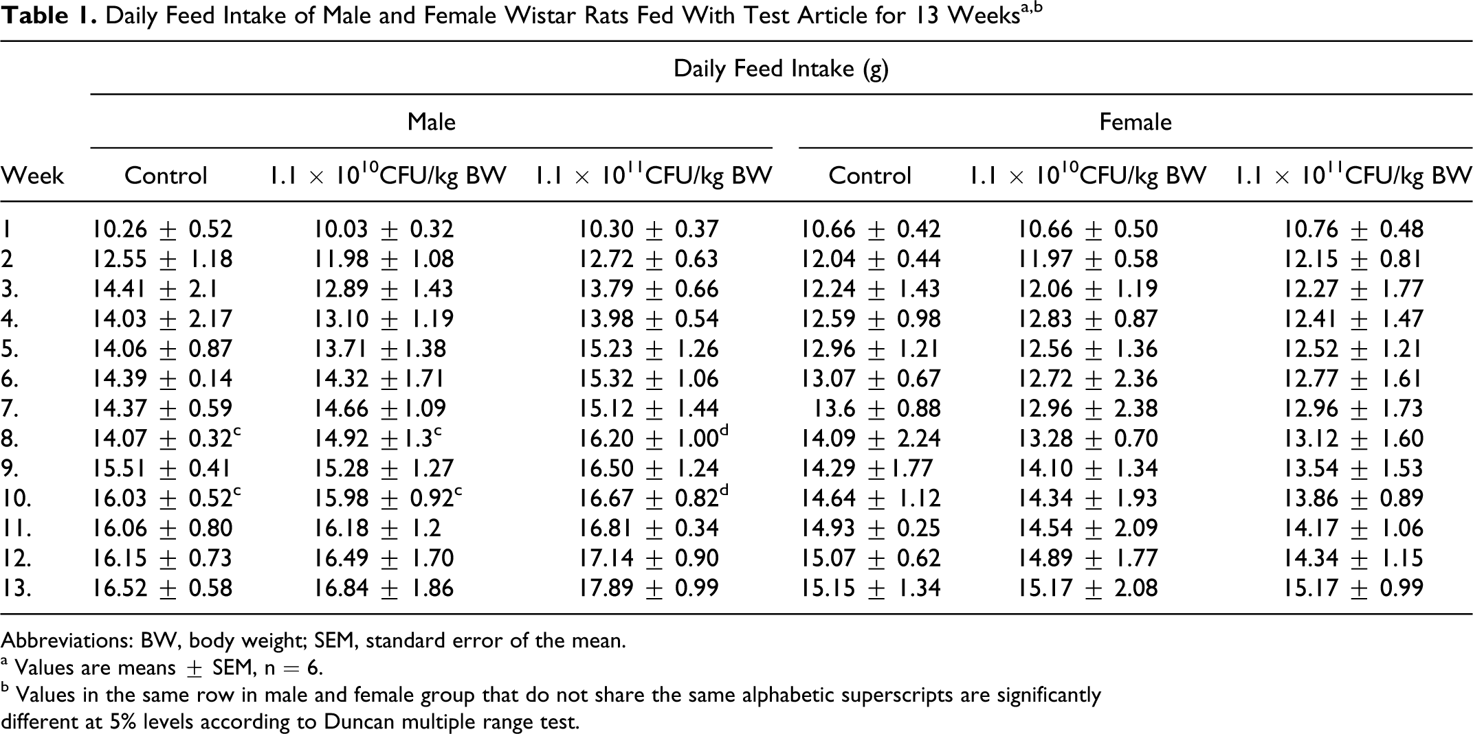
Abbreviations: BW, body weight; SEM, standard error of the mean.
a Values are means ± SEM, n = 6.
b Values in the same row in male and female group that do not share the same alphabetic superscripts are significantly
different at 5% levels according to Duncan multiple range test.
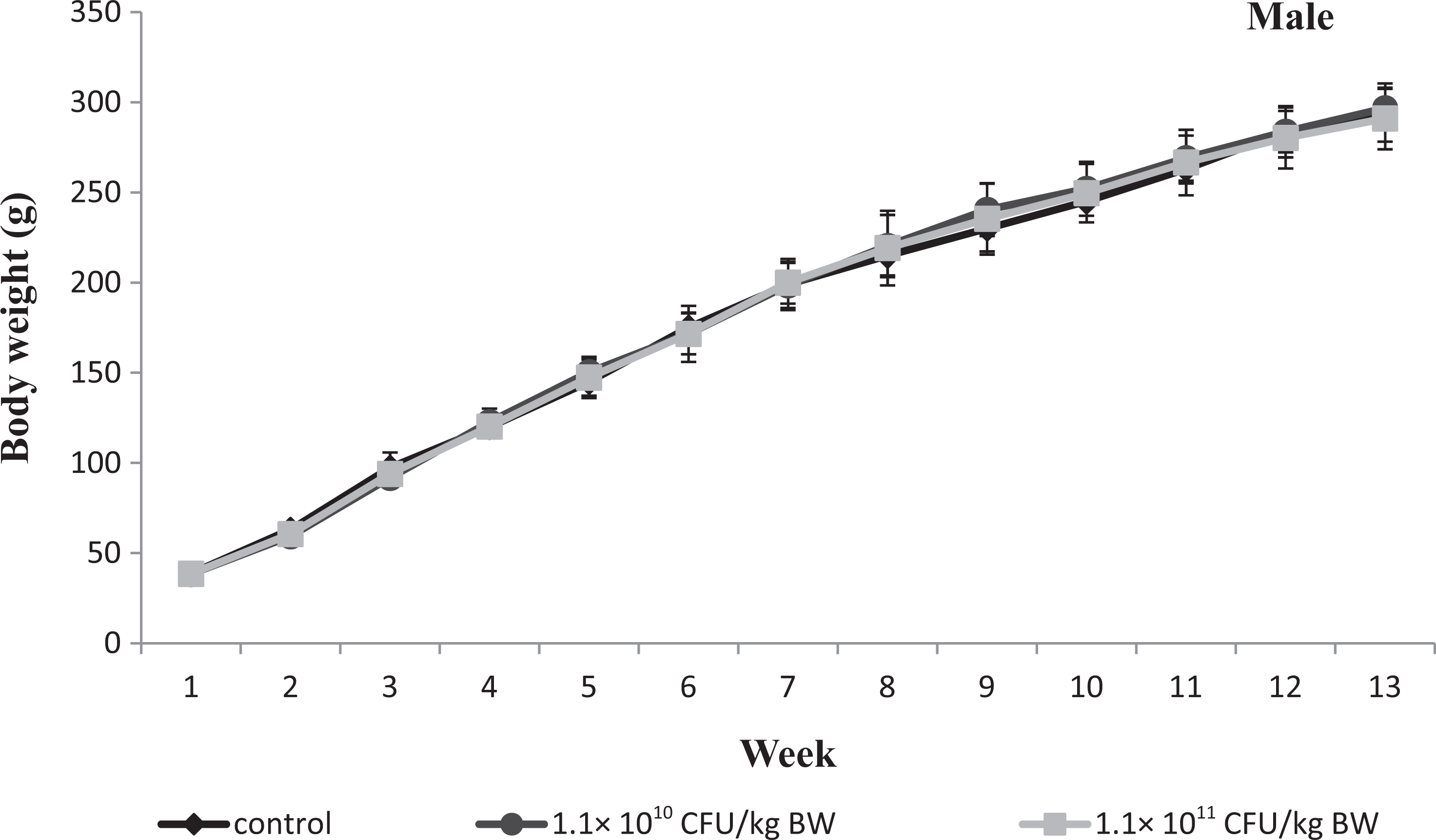
Graph showing body weight of male Wistar rats given test article for 13 weeks. Values are means ± standard error of the mean (SEM) of 6 rats.
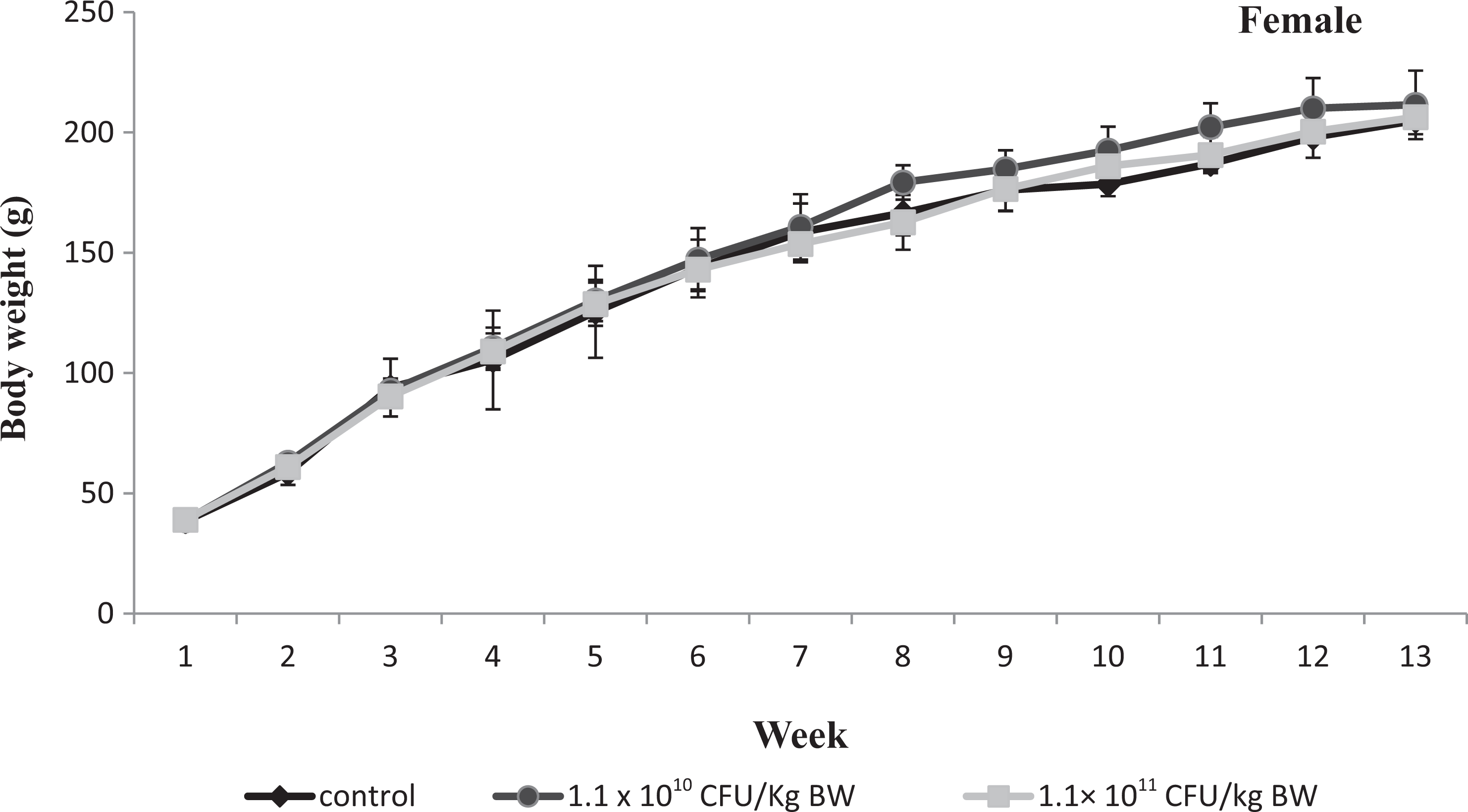
Graph showing body weight of female Wistar rats given test article for 13 weeks. Values are means ± standard error of the mean (SEM) of 6 rats.
Viability of B. licheniformis Me1
Viability of the test article in the rat GIT was determined by monitoring the presence of B licheniformis Me1 in the feces of the treated rats during the experimental period (on days 10, 30, 50, and 80). An average of ∼1 × 10⁶ CFU of B. licheniformis Me1 per gram of the rat’s faeces was observed. There was no marked difference in the numbers of B. licheniformis in the feces across test groups on any of the days tested. From this, it can be concluded that Me1 was able to survive the GIT conditions and not all fed test article was excreted out.
Relative organ weights and histopathology
At necropsy, macroscopic observation of the organs revealed no treatment-related damages or differences. The relative organ weights of adrenals, spleen, kidney, and heart did not show any significant deviation from that of control in any of the treated animal groups (Supplemental Tables 1 and 2). However, a marginal, but statistically nonsignificant decrease in relative organ weight of lungs in females was noticed (Supplemental Table 2).
Histopathological examination of the tissues of the control and treated groups revealed no treatment-related abnormalities in morphology or toxicity of the organs. No swelling of epithelium and occlusion of intestinal lumen were noticed even in the highest dose group. The villi pattern of the small intestine was well preserved in all the rats treated with B. licheniformis Me1. Other vital organs like heart and brain also showed a normal structure. There were no microscopic or macroscopic lesions in any organs that could be attributed due to the treatments.
Hematology and serum biochemical studies
No significant differences were found in any of the analyzed aspects in hematology for both male and female test groups when compared to that of the control groups (Supplemental Table 3). On serum biochemical analysis, no statistically significant dose-dependent alterations were detected either in the levels of glucose, cholesterol, triglyceride, urea, or in the activity of ALP, LDH, AST, and ALT in both the sexes in comparison to the control groups (Supplemental Table 4).
Micronucleus Assay in Mice
The micronucleus test was conducted to investigate the formation of micronuclei containing chromosome fragments or whole chromosomes and is considered the most reliable assay for cytogenetic damage. In the micronucleus assay, neither any differences in BW between the treatment groups compared to the control group nor any signs of toxicity were noted in clinical observations following administration of the test article at doses of 1.1 × 10¹¹ and 1.1 × 10¹⁰ CFU/kg BW. The ratio of reticulocytes to total erythrocytes was used an indicator for the evaluation of bone marrow toxicity. The results clearly demonstrate that the number of immature erythrocytes in each dose did not significantly increase above the concurrent negative (water) control frequencies. Additionally, it was always within the historical negative control range. As expected, animals in the EMS-treated positive control group showed a significant increase in the frequency of micronuclei compared to the negative controls. In contrast, no statistically significant induction of micronuclei in the test article fed groups was observed. The ratio of reticulocytes to total erythrocytes in the treated groups did not show any significant decrease as compared to the negative control group. For female mice, the average reticulocyte to total erythrocyte ratio in the negative control group was 2.17%. Also, the treated groups 1.1 × 10¹¹ and 1.1 × 10¹⁰ CFU/kg BW per day showed 2.07% and 1.93% of reticulocyte to total erythrocyte ratio, respectively, a reduction of 25.9% from the positive control group. In male mice, the negative control group showed a ratio of 1.85% and the 2 dose groups 1.1 × 10¹¹ and 1.1 × 10¹⁰ CFU/kg BW/d exhibited 1.51% and 1.10% reticulocytes to erythrocytes ratio. In the male group, the positive control showed 27.12% decrease in the ratio. These results clearly show that Me1 did not cause any signs of bone marrow cytotoxicity of mice in the range of the doses tested.
In the negative control groups, the incidence of micronucleated reticulocytes in the peripheral blood per 1000 reticulocytes was 1.18 ± 0.5 in males and 0.98 ± 1.1 in females. These results were within the historical reference range. 27 The positive control group had a statistically increased mean frequency of 26.14 ± 3 in males and 23.2 ± 5 in females as compared to the negative control group. The micronucleated reticulocytes per 1000 reticulocytes were found to be 1.03 ± 0.7 and 0.89 ± 1.2 in males and 1.2 ± 1 and 1.03 ± 0.9 in females at the test article dose levels of 1.1 × 10¹¹ and 1.1 × 10¹⁰ CFU/kg BW per day, respectively. Since the mice peripheral blood micronucleus assay did not show any statistically significant changes according to the OECD Guideline for the Testing of Chemicals No. 474, there is no indication that B. licheniformis Me1 administration caused any genocytotoxicity.
Acute Eye Irritation Study in Rabbits
B. licheniformis Me1 cell mass application to the conjunctival mucosa of the eyes did not result in any test article–related irritant effect at any time during the experimental period. Furthermore, we did not observe any negative symptoms in either the cornea or the iris. According to the European Commission (EC) criteria of 2001/59/EEC for the classification and labeling requirements for dangerous substances and preparations, the test article is not required to be classified or labeled as an irritant to the eye.
Skin Irritation Study in Rabbits
B. licheniformis Me1 cell mass application to the skin did not reveal any clinical signs of erythema and edema at 1 hour after removal of the patch and until 72 hours later. According to the EC directive 2001/59/EEC, it is not required to classify or label the test article as a skin irritant.
Discussion
As the demand for probiotic foods among consumers continues to grow, several new foods are likely to include probiotics in the future. It is of obvious importance that novel probiotic cultures for human and animal consumption are evaluated carefully and precisely for safety and efficacy before commercialization. 26 Many reports have shown that selected strains of Bacillus, with a history for safe use in the food industry, are increasingly being incorporated with health-promoting “functional foods” to provide digestive and immune health benefits. Moreover, Bacillus can also serve as an alternative to commonly used LAB for therapeutic, prophylactic, and growth supplements for animals and humans. 8,10,32,33
The ability of B. licheniformis to produce a wide array of antimicrobial substances with a broad inhibitory spectrum 34 and other promising probiotic characteristics 35,36 makes it as a suitable and potential candidate for application in probiotic food products. B. licheniformis Me1 used in this study was a native isolate from milk. The isolate is gram-positive, catalase-positive, nonhemolytic and nonphospholytic, and able to hydrolyse starch, phytate, casein, and produce exopolysaccharides. The isolate was identified as B. licheniformis by 16S rDNA sequencing in combination with morphological, physiological, and biochemical characteristics and phylogenetic studies (Nithya and Halami, unpublished data). The gene sequence has been deposited at GenBank under the accession number HM564028 (http:/www.ncbi.nlm.nih.gov/Genbank). B. licheniformis Me1 produces an antibacterial peptide that can inhibit various food-borne pathogens, including both gram-positive (Bacillus cereus F4433, Staphylococcus aureus FR1722, Listeria innocua FB21, L monocytogenes Scott A, L murrayi FB69, and Micrococcus luteus ATCC9341) and gram-negative bacteria (Salmonella paratyphi FB254, S typhimurium MTCC1254, Escherichia coli CFR02, and Yersinia enterocolitica MTCC859). Furthermore, our previous study revealed prominent probiotic properties of the culture, such as acid and bile tolerance, sensitiveness to antibiotics, antioxidant property, and ability to grow under anaerobic conditions, and so on 37 .
To assess the safety of B. licheniformis Me1 strain, an in vivo acute and subchronic toxicity study was conducted. Studies, including acute (single administration) toxicity and a repeated administration (subchronic) toxicity assessment have been recommended for assessing the safety of probiotics. 1,38 To study the acute toxicity, a limit test was conducted with a maximum dose level of 1.1 × 10¹¹ CFU/kg BW of rat. Since, no treatment-related mortality, morbidity, or clinical symptoms resulted in this acute oral toxicity study, B. licheniformis Me1 can be considered nontoxic per the OECD guidelines. The result indicated that the oral LD50 for the test article was ≥1.1 × 10¹¹ CFU/kg BW. Similar results were reported for the safety analysis of other probiotic strains of Bacillus such as B. subtilis and B. licheniformis 26 and B coagulans. 27
Organ weight changes have long been accepted as a sensitive indicator of chemically induced changes to organs. Therefore, in toxicological experiments, the comparison of organ weights between control and treated groups have conventionally been used to predict the toxic effect of a test article. 39 The absence of significant changes in the vital organs of the treated groups during the subchronic oral toxicity study shows that the ingestion of B. licheniformis Me1 did not induce any anomalous lesions or inflammation of these organs. Similar observations were found by Sorokulova et al 35 in a safety assessment of probiotic biosporin, but the administration of B. coagulans resulted in difference in organ weight of liver, brain, and hemorrhages in the lungs. 27
Hematological values were not significantly affected by the administration of B. licheniformis Me1 between the control and the treated groups for all the parameters analyzed. These findings suggest that the test article B. licheniformis Me1 may not be toxic as they do not significantly affect the circulating RBC nor the haematopoiesis or leucopoiesis. Furthermore, the normal metabolism of the treated animals was not affected by the test article fortification. Hong et al 6 showed similar findings in in vivo toxicity studies of B. subtilis and B. indicus conducted in rabbits. Absence of any significant changes in the activity of diagnostic marker enzymes along with no histological alternations, namely inflammation, lesion, and cellular infiltration in the vital organs, emphasize the safety of test article augmentation at levels used in the present study. Results obtained are in accordance with the data for Lactobacillus 40 and Bacillus 27 probiotics.
The viability test result indicates the survivability of the test article B. licheniformis Me1 within the rat GIT and points the fact that the ingested dose actually reaches the region of the GIT where it should exert its effect. 41 Having reached the small intestine, a proportion of the ingested test article would have colonized in the nutrient-rich region and would have established itself alone or in association with other gut microbes in the small intestine. Moreover, our previous in vitro probiotic studies have shown that this Bacillus culture grows efficiently in anaerobic environment, is tolerant of GIT conditions, and also has efficient adherence to hydrocarbons, an attribute related to the hydrophobicity of the culture.37 Furthermore, several other authors have documented the persistence of Bacillus spp in the GIT of the experimental animal models. 25,42,43 The Scientific Committee on Animal Nutrition (SCAN) 44 reported the presence of approximately 80% of the fed B. licheniformis NCTC13123 in the digestive tract of pigs by doing a microbiological examination of feces collected from piglets during a tolerance test.
The in vivo micronucleus test conducted in mice to assay the cytogenotoxic effect of B. licheniformis Me1 administration showed that there was no statistically significant dose-related increase in the incidence of micronucleated immature erythrocytes for the treatment groups compared with the concurrent control group. Also, there were no substantial and statistically significant dose-related decreases in the proportion of immature erythrocytes. These findings were similar to that observed for B. coagulans. 27 Acute eye and skin irritation tests conducted in rabbits failed to show any signs of eye irritation and skin erythema or edema. A similar result for skin irritation test was reported on application of B. licheniformis NCTC13123; while in eye irritation study, a diffuse corneal opacification and evidence of conjunctivitis were observed. 44 Endres et al 27 also reported a slight conjunctiva irritation and erythema on the treated skin surface within 1 hour of B. coagulans application. However, they reported the reaction was fully reversible later and thus, it can be assumed that these reactions might be due to the technical manipulation of administration.
The concentration of B. licheniformis Me1 (1.1 × 10¹¹ CFUs/kg BW of rat) used in this study corresponds to 77 × 1011 CFUs for an average 70 kg human being. Thus, the concentration used can be considered 2566 to 77 000 times safe for human consumption, as the suggested human dose is in the range of 1 × 10⁸ to 3 × 10⁹ CFUs.
Conclusion
The test article B. licheniformis Me1 administered to rats at a dose of 1.1 × 10¹¹ CFU/kg BW did not show any sign of toxicity or mortality using in vivo oral acute and subchronic assessments. Hence, the NOAEL for both males and females is considered to be higher than 1.1 × 10¹¹ CFU/kg BW per d, which was the highest dose tested. Furthermore, a micronucleus assay carried out in mice using B. licheniformis Me1 did not demonstrate any evidence for cytogenotoxicity. In addition, applications of B. licheniformis Me1 did not produce any signs of skin or eye irritation. Based on these results and according to the highest dose levels required by OECD guidelines for materials of low toxicity, it can be concluded that the test article B. licheniformis Me1 is safe in rodents and can be potentially considered a safe probiotic for future use.
Footnotes
Acknowledgments
The authors are grateful to the Director, CFTRI, Mysore and Dr S Umesh Kumar, Head, Food Microbiology Department, CFTRI, and the staff of Animal house, B&N Department for providing all facilities to carry out this experiment successfully. V.N. acknowledges CSIR for the Fellowships. Work was carried out under the Institute project MLP83.
The author(s) declared no potential conflicts of interest with respect to the research, authorship, and/or publication of this article.
The author(s) received no financial support for the research, authorship, and/or publication of this article