
Abstract
Sprague Dawley rats (10/sex/group) were given a single intravenous (iv) dose of CUMI-101 to determine acute toxicity of CUMI-101 and radiation dosimetry estimations were conducted in baboons with [11C]CUMI-101. Intravenous administration of CUMI-101 did not produce overt biologically or toxicologically significant adverse effects except transient hypoactivity immediately after dose in the mid- and high-dose groups, which is not considered to be a dose-limiting toxic effect. No adverse effects were observed in the low-dose group. The no observed adverse effect level (NOAEL) is considered to be 44.05 µg/kg for a single iv dose administration in rats. The maximum tolerated dose (MTD) was estimated to be 881 µg/kg for a single iv dose administration. The Medical Internal Radiation Dose (MIRDOSE) estimates indicate the maximum permissible single-study dosage of [11C]CUMI-101 in humans is 52 mCi with testes and urinary bladder as the critical organ for males and females, respectively.
Keywords
Introduction
Serotonin1A (5-HT1A) receptors belong to the family of G-protein-coupled receptors (GPCRs) and contribute to serotonin transmission in brain. 1 Serotonin1A receptors have been implicated in the pathophysiology of mood and anxiety disorders, sexual function, eating disorders, neurodegenerative diseases, and in the mechanism of action of antidepressants. 2–9 The density and number of 5-HT1A receptors enables its quantification in living participants using positron emission tomography (PET). 10,11 Serotonin1A receptors occur in high- (HA) and low-affinity (LA) agonist-binding states. In contrast, antagonists bind to the HA and LA conformations of 5-HT1A receptors with comparable affinity. It is the HA state of the receptor which is coupled to G-proteins and therefore agonists provide a measure of functional 5-HT1A receptors. 12,13 Among the successful antagonist tracers, [11C]WAY-100635 is the most commonly used 5-HT1A ligand for in vivo studies in human participants. 14–16 Several efforts have been made to develop a 5-HT1A receptor agonist imaging agent in vivo. 17–19 [O-methyl-11C]2-(4-(4-(2-methoxyphenyl)piperazin-1-yl)butyl)-4-methyl-1,2,4-triazine-3,5(2H,4H)dione ([11C]CUMI-101) is the only radiotracer available at present (Ki = 0.15 nM; Emax = 80%; EC50 = 0.1 nM; Figure 1). 13 [11C]CUMI-101 can image HA sites in baboons and human participants. 12,20 [11C]CUMI-101 is also sensitive to large changes in endogenous 5-HT produced by the administration of citalopram and fenfluramine to baboons. 21 According to the Food and Drug Administration (FDA), the extended single-dose toxicity studies in animals can be used to support single-dose studies in humans. Specifically, this guidance states “The study should be designed to establish a dose inducing a minimal toxic effect, or alternatively, establishing a margin of safety. To establish a margin of safety, the sponsor should demonstrate that a large multiple (eg, 100X) of the proposed human dose does not induce adverse effects in the experimental animals. Scaling from animals to humans based on body surface area can be used to select the dose for use in the clinical trial. Scaling based on pharmacokinetic and pharmacodynamic modeling would also be appropriate if such data are available” (Guidance for Industry, Investigators, and Reviewers Exploratory Investigational Drug Application [E-IND], FDA). Since these preclinical studies are needed to demonstrate to the Radioactive Drug Research Committee (RDRC) and FDA that a candidate imaging agent is understood well enough for designing appropriate clinical experiments, most of the work to be conducted to achieve these objectives must be performed and the resulting data analyzed and reported in strict compliance with the FDA’s good laboratory practice (GLP) regulations for nonclinical laboratory studies (21 CFR 58). We now report toxicology and dosimetry determinations prior to performing human studies with [11C]CUMI1-101. We report toxicology evaluation of CUMI-101 in rats and dosimetry estimations for [11C]CUMI-101 in baboons.
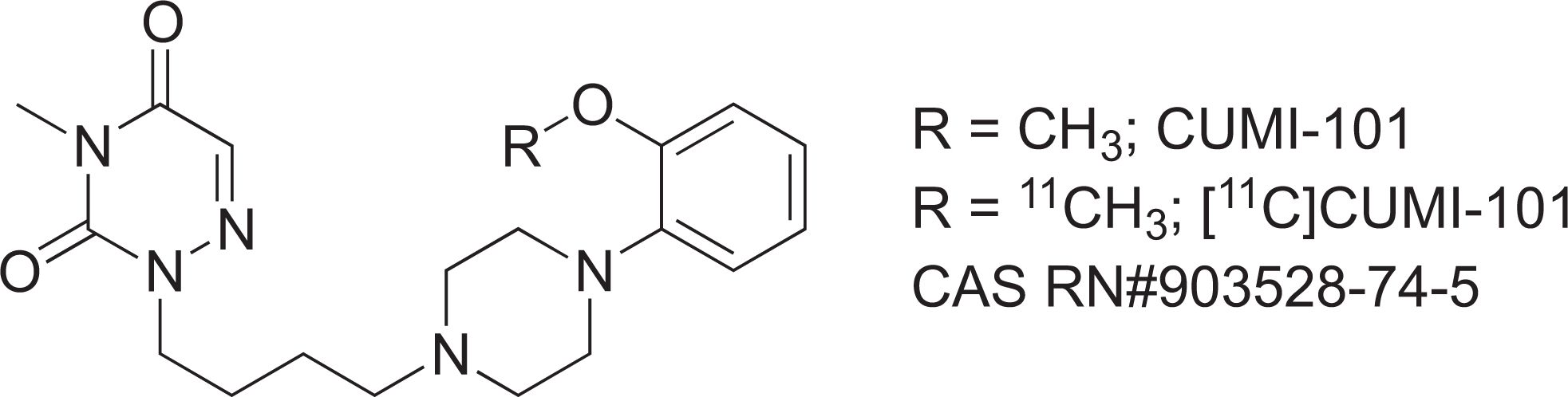
Chemical structures of CUMI-101 and [11C]CUMI-101.
Materials and Methods
Synthesis of CUMI-101 and [11C]CUMI-101
The syntheses of CUMI-101 and [11C]CUMI-101 (CAS RN# 903528-74-5) were performed based on a procedure reported previously by us. 13 In brief, coupling of 2-(4-chlorobutyl)-4-methyl-1,2,4-triazine-3,5(2H,4H)dione with 2-(piperazin-1-yl)phenol and 1-(2-methoxyphenyl)piperazine provided desmethyl-CUMI-101 and CUMI-101, respectively. Radiosynthesis of [11C]CUMI-101 was performed by [11C]methylation of desmethyl-CUMI-101 using [11C]MeOTf followed by purification using reverse phase high-performance liquid chromatography (RP-HPLC) and C-18 Sep-Pak.
Rat Toxicity Study
The toxicity study was carried out in male and female Sprague Dawley rats by SRI International, Menlo Park, California, in accordance with the FDA “Good Laboratory Practice for Nonclinical Laboratory Studies,” as described in 21 CFR Part 58. All animal studies were approved by SRI’s Institutional Animal Care and Use Committee (IACUC) and were performed in animal facilities that are accredited by the Association for Assessment and Accreditation of Laboratory Animal Care, International (AAALAC). SRI files assurances with the Public Health Service (PHS) Office of Laboratory Animal Welfare (OLAW) and adheres to PHS standards and practices.
Maximum human dose of CUMI-101 proposed for a single dose is 10 µg/70 kg person. 10 µg/70 kg = 0.143 µg/kg × 37 (human surface area conversion) = 5.286 µg/m2. Scaling for the rat gives 5.286 µg/m2/6 (rat surface area conversion) = 0.881 µg/kg as an equivalent human dose; 1000 × 0.881 = 881 µg/kg as the 1000× dose in this study.
Fifty male (7-9 weeks old, 191-264 g) and 50 female (8-10 weeks old, 178-224 g) Sprague Dawley rats supplied from Charles River Laboratories (Hollister, CA) were used in this study. Upon arrival, the rats were quarantined for 3 days. The rats were housed (5 per cage) in polycarbonate hanging cages with Sanichip bedding. The animal room was monitored for temperature (65°F-79°F, 18°C–26°C) and humidity (30%-70%) with a light cycle of 12-h light/12-h dark. The rats were fed with Purina Certified Rodent Chow (#5002; Richmond, Indiana) ad libitum. Water (purified, reverse osmosis) was provided ad libitum during quarantine and study periods. Male and female rats (10 males and 10 females per group) were given CUMI-101 iv on day 1 as a single dose of 881 μg/kg (5286 µg/m2; 1000 times the human dose; group 2) or as 2 daily doses of 440.5 μg/kg per day (2643 µg/m2; 500 times the human dose; group 3) or 88.1 μg/kg (528.6 µg/m2; 100 times the human dose; group 4) or 44.05 μg/kg (264.3 µg/m2; 50 times the human dose; group 5). Control rats (10 males and 10 females) were given a single iv dose of vehicle (at an equivalent volume 5 mL/kg on day 1 [Group 1]).
Preparation of Dose Formulations
The test article CUMI-101 (also known as CUNBID-101 or MMP) formulations were prepared by dissolving the appropriate amount of test article in ethanol (Pharmco, Brooksfield, Connecticutt). Sterile saline was then added to achieve the target concentration in final vehicle formulation of 5% ethanol and 95% saline. Serial dilution using 5% ethanol/95% saline from the stock solution was used to achieve the mid and low doses. The pH was adjusted using 0.1 N HCl (JT Baker, Phillipsburg, New Jersey; Lot# T40429) to achieve a final pH = 7. Formulations were stored refrigerated protected from light for 3 days at 3.3°C to 5.0°C before use. Formulations were brought to room temperature prior to administration to the animals. All dosing formulations were confirmed to be within ±10% of the target concentration for all groups. All dose levels were scaled to surface area for comparison with proposed human dosages.
Observation Protocol
All animals were individually identified by ear punch and were observed at least once daily for signs of mortality, morbidity, injury, and availability of food and water. All clinical observations were recorded daily (2-4 hours post dose on the days of dose administration) and more often as clinical effects warranted. Individual body weights were measured and recorded for each animal on day 1 prior to dosing and at necropsy (days 3 and 15 for groups 1-5). Food consumption was measured for a period of about 24 hours per cage, twice weekly throughout the study.
Clinical Pathology
Blood for clinical chemistry and hematology evaluations was collected on days 3 and 15. Hematology samples were collected using EDTA as anticoagulant. No anticoagulants were used for serum chemistry samples. Animals were killed on days 3 and 15 (interim and terminal necropsy of groups 1-5) necropsy of group 3) by overdose of sodium pentobarbital (250 mg/kg) administered by intraperitoneal (ip) injection. External examination of all body orifices and surfaces and an examination of all cranial, thoracic, and abdominal organs were recorded. All retained tissues and organs were fixed in phosphate-buffered 10% formalin. Organ weights recorded for all survived animals at scheduled sacrifice. These include adrenal glands, brain, heart, kidneys, liver, spleen, thymus, testes, and ovaries. Paired organs were combined and weighed together, except kidneys.
Histopathology
Histopathology were performed on group 1 (control) and 2 (1000× human dose). Sections of the tissues from animals to be evaluated were embedded in paraffin (5 μm thick) and stained with hematoxylin and eosin by Biopathology Sciences Inc (Redwood City, CA). Each lesion was listed and coded by the most specific topographic and morphologic diagnoses, severity, and distribution using Systemized Nomenclature of Medicine (SNOMED) and National Toxicology Program (NTP) codes as guide. Four-step grading system (minimal, mild, moderate, and marked) was used to define gradable lesions for comparison between treated and control groups.
Data Analyses
Mean and standard deviation were calculated for body weight, food consumption, organ weights, and clinical pathology data. One-way analysis of variance (ANOVA) was performed on clinical pathology (LABCAT module, HE v. 4.42, Princeton, NJ) and organ weights data (LABCAT module OW v3.24). Body weights (LABCAT In-Life v. 6.2) for groups 4 and 5 were not compared with those for groups 1 and 3 since dosing of groups 4 and 5 animals was initiated on different days because of protocol changes in amendments. The values reported as individual food consumption data were calculated from cage totals rather than individual measurements. No statistical analysis was performed on food consumption. In all cases Criteria for Null Hypothesis Rejection was defined as P < .05.
Dosimetry Studies of [11C]CUMI-101 in Baboons
Dosimetry estimations in baboons were performed based on protocol developed by us.22–24
In brief, fasted animals were immobilized with ketamine (10 mg/kg intramuscularly [im]) and anesthetized with 1.8% isoflurane via an endotracheal tube. Temperature was kept constant at 37
Results
Mortality/Morbidity and Clinical Observations
All animals survived until their scheduled sacrifice. Almost all male and female rats in the mid- and high-dose groups (groups 2-4) displayed slight-to-moderate hypoactivity (severity 1-on a 0-4 scale) on day 1 immediately after dose administration. The incidence of the hypoactivity appeared to be dose dependent, with 19 of 20 animals in the high-dose group (group 2) having moderate hypoactivity. The animals recovered from the hypoactivity within a few hours after dose administration and appeared normal by the last time point at which clinical observations were performed on day 1. All animals, except 1 female in group 3, in the mid and high dose (groups 2-4) also displayed tachypnea (rapid breathing) immediately after dosing on day 1; some animals also had shallow breathing. These animals recovered by the next day. Rats in the low-dose group at 44.05 μg/kg (50× human dose) and the control group did not exhibit hypoactivity or tachypnea after dose administration.
Body Weight and Food Consumption
No test article-related changes in body weights and food consumption were observed in the treated groups compared with the control group.
Clinical Pathology
Clinical pathology (hematology and clinical chemistry) was evaluated on days 3 and 15 (2 and 14 days after dose administration, respectively). All the changes were sporadic, were not clearly dose related, and generally represented small changes within the normal ranges for rats. Therefore, none of these changes were considered to be of toxicological significance.
Organ Weights
Brain weight in the males of group 2 at the terminal sacrifice on day 15 was slightly decreased compared with the control rats. The change was not correlated with microscopic evaluation and is considered to be a sporadic variation.
Histopathology
All tissues (∼45 per animal) were examined histopathologically from all rats in groups 1 (the controls) and 2 (high dose, 1000× human dose). Minor differences in type, incidence, and severity of findings exist between and within these 2 study groups, but no finding varies consistently and no variation is attributed to the test compound. The findings are generally similar to those commonly encountered as background findings for male and female rats of this strain and the general age group. The gross stomach mass in group 1 female #11 is histologically a hematoma on the serosal aspect of the stomach, an incidental finding. Inflammation in the heart is compatible with murine progressive cardiomyopathy, a commonly encountered background lesion in young rats of this strain. No histopathology examination was made of any tissues from rats in group 3, 4, or 5 because lesions at the highest dose were not considered test article related.
Biodistribution of [11C]CUMI-101 in Baboons
All animals tolerated the study and there were no discernable effects of the radiotracer injections on systolic or diastolic blood pressure, heart rate, respiratory rate, or rectal temperature. The whole body biodistribution of [11C]CUMI-101 is shown in Figure 2.
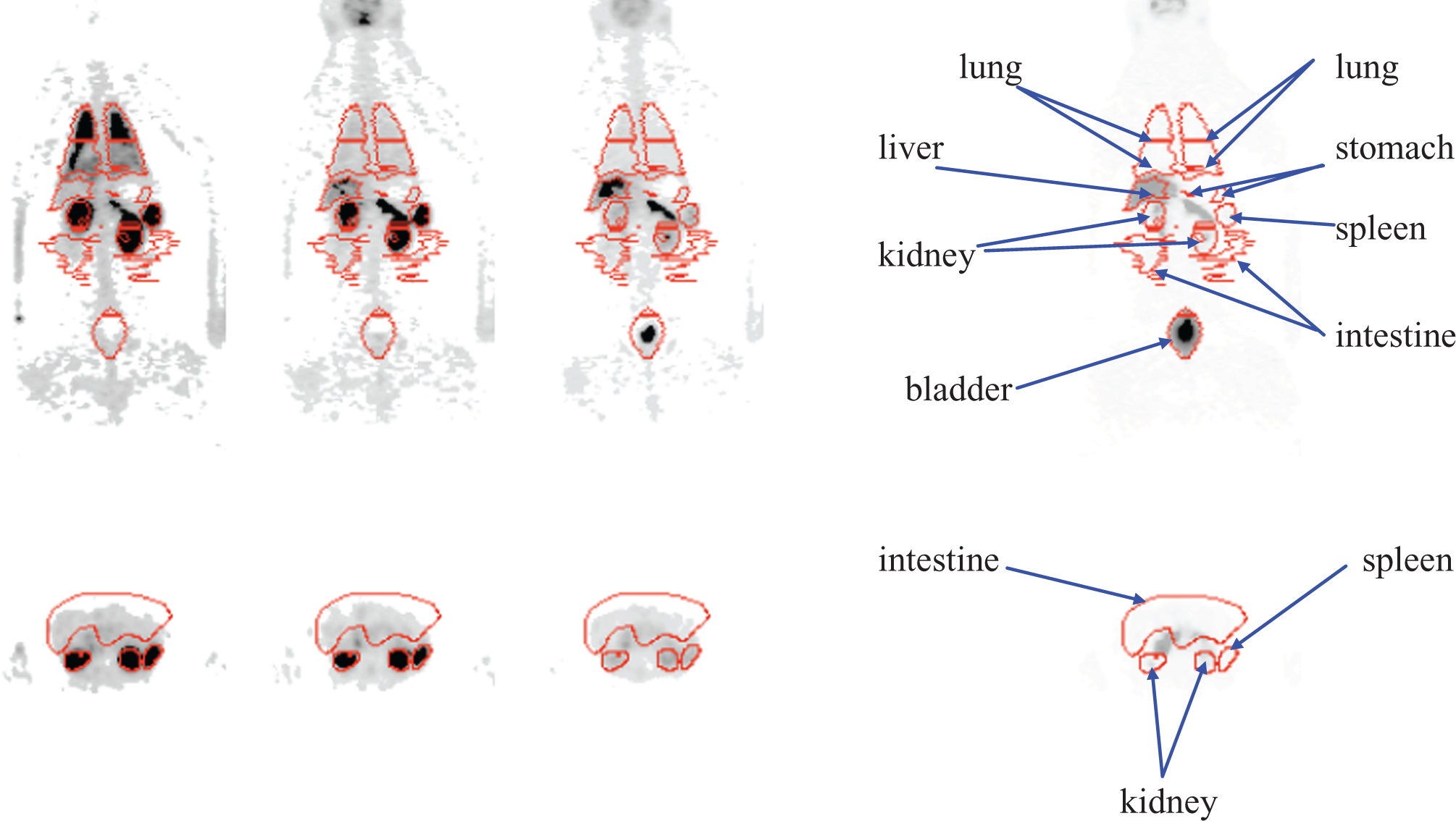
Whole body images of [11C]CUMI-101 in baboons. Note: Left to right: frame 1, 2, 4, 8 (Midtime 0.75, 4.38, 13.65 and 80.18 minutes), top: coronal images, bottom: transaxial images.
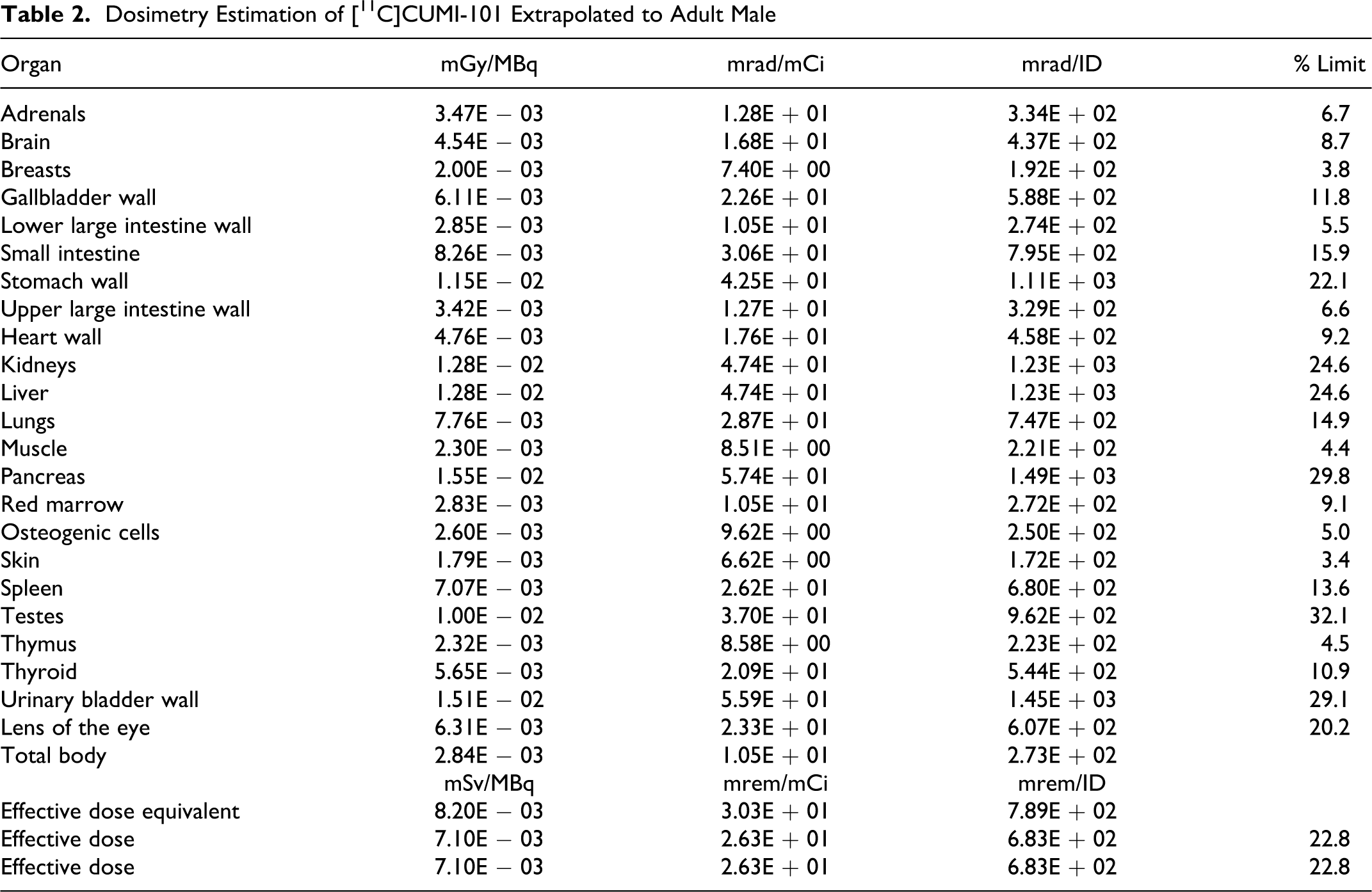
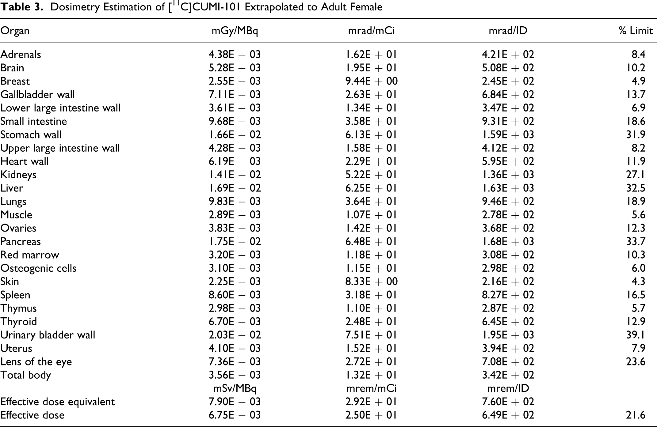
Several organs were clearly identifiable based on location by the administration of [11C]CUMI-101. A TAC generated from some of the regions is illustrated in Figure 3. The area under the curve was calculated as the sum of the trapezoidal integration and the integral of the exponential functions from time zero to infinity. The residence times are shown in Table 1 with the liver having the highest residence time. Residence times were then used as input to the MIRDOSE3 program. Tables 2 and 3 give the dosimetry estimates for the 70-kg adult male and adult female (57 kg), respectively. The data indicate elimination of [11C]CUMI-101 via the hepatobiliary and renal systems. Testes for adult male and urinary bladder for female received the highest estimated radiation dose and were the critical organ (1.00E − 02 mGy/MBq and 2.03E − 02 rad/mCi, respectively) estimates for adult human males and females and validate the baboon as a substitute for human participants.
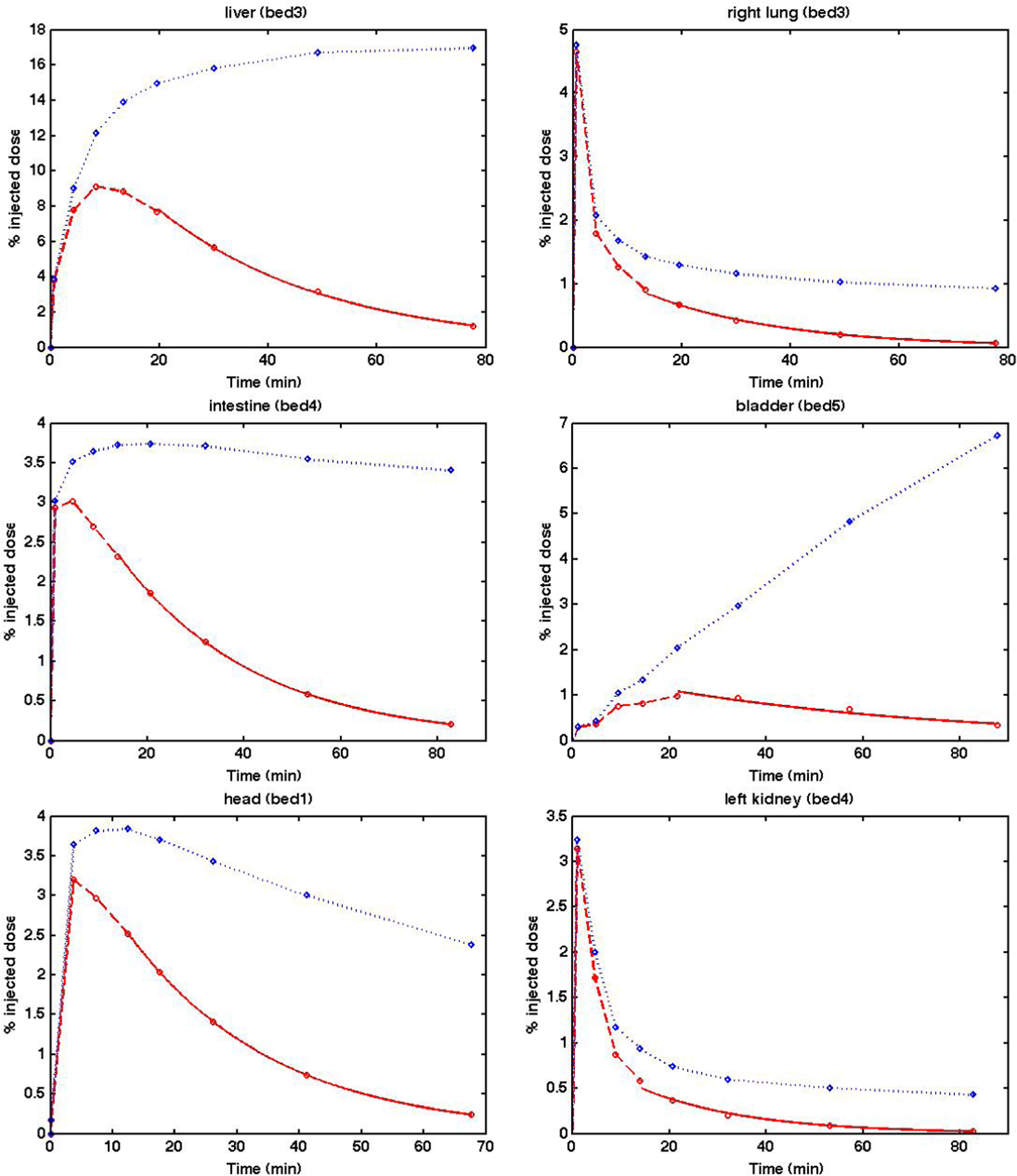
Time–activity curves of [11C]CUMI-101 in various organs. Note: The dotted lines represent decay corrected and the dashed and solid lines are non-decay-corrected TACs for each organ.
Average Residence Time of [11C]CUMI-101 in Baboonsa
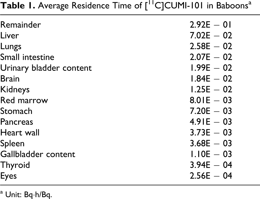
a Unit: Bq·h/Bq.
Dosimetry Estimation of [11C]CUMI-101 Extrapolated to Adult Male
Dosimetry Estimation of [11C]CUMI-101 Extrapolated to Adult Female
Discussion
All animals evaluated in the rat toxicity study survived until their scheduled necropsy. Male and female rats in the mid- and high-dose groups (groups 2-4) displayed slight-to-moderate hypoactivity on day 1 immediately after dose administration. The severity of the hypoactivity appeared to be dose dependent, with the highest severity observed in the high-dose group (group 2). The animals recovered from the hypoactivity within a few hours and appeared normal by the last time point at which clinical observations were performed on day 1. Rats in the low-dose group at 44.05 μg/kg (264.3 μg/m2, 50× human dose) and control group did not exhibit hypoactivity after dose administration. No drug-related effects were found for clinical pathology, body weights, food consumption, organ weights, and macroscopic and microscopic evaluations. In conclusion, iv administration of CUMI-101 to male and female rats once or twice a day did not produce overt biologically or toxicologically significant adverse effects other than hypoactivity in the mid- and high-dose groups. The hypoactivity is not considered to be a dose-limiting toxic effect. No adverse effects were observed in the low-dose group. The no observed adverse effect level (NOAEL) is considered to be 44.05 μg/kg (264.3 μg/m2) for administration of a single iv dose. The maximum tolerated dose (MTD) is considered to be at least 881 μg/kg (5286 μg/m2) for administration of a single iv dose.
The [11C]CUMI-101 was well tolerated in the animals, with no meaningful changes in any vital signs. Our study shows the single study maximal radiation exposure calculated by our group in male baboon studies involving [11C]CUMI-101 will remain below the 21 CFR 361.1 dose limit for research participants which is 26 mCi in males and 26 mCi in females (ie, calculation based upon the testes and the bladder as the critical organs, respectively; 3 rads per single study for the whole body, active blood forming organs, lens of the eyes, and gonads; 5 rads for other organ per single study limit). The maximum annual dose limit (15 rads) is 135 mCi in males and 170 mCi in females. Thus, the dose of 52 mCi in males and 52 mCi in females per year (dosage for 2 PET sessions) is below this limit. The favorable toxicology and dosimetry findings make [11C]CUMI-101 suitable for human use. Dosimetry findings will need to be validated in human participants.
Footnotes
The author(s) declared no potential conflicts of interest with respect to the research, authorship, and/or publication of this article.
This work was partially supported by a research grant from the National Institutes of Health (K08 MH76258-01A1). We thank the National Institute of Mental Health Toxicology screening program for financial support for toxicology studies (Contract number: NO1-MH-32001).