
Abstract
Fertility and early embryonic developmental toxicity in rats were evaluated by intravenously administering astragaloside IV (AS-IV) daily at 0.25, 0.5, and 1.0 mg/kg for 4 weeks before mating, throughout the mating period, and continuing to day 6 of gestation period in females. Perinatal toxicity in rats was evaluated on gestational days (GD) 15 to 21 and lactational days LD (LD) 1 to 21. Astragaloside IV had no maternal toxicity at 0.25 to 1.0 mg/kg in rats. Although it has an inhibitory effect on female fertility in F0/F1 rats, AS-IV was devoid of early embryonic developmental toxicity in F0/F1 rats and in the survival parameters of F1 postnatal rats. Maternal AS-IV exposure at the dose of 1.0 mg/kg per d resulted in a significant delay in time for fur development, eye opening, and cliff parry reflex of pups compared to control group (P < .05), whereas it did not affect the memory and learning of F1 pups.
Keywords
Radix Astragali (Huangqi), the dried root of Astragalus membranaceus (Fisch.) Bge. var. mongholicus (Bge.) Hsiao, or A membranaceus (Fisch.) Bge., is one of the most widely prescribed Chinese herbs in many formulas. Astragaloside IV ([AS-IV]; 3-0-β-D-xylopyranosyl-6-0-β-D-glucopyrano-sylcycloastra-genol), a purified small molecular saponin (molecular weight [MW] 784) of this herb, is one of the main active ingredients of Radix Astragali, which is an herbal remedy widely used in traditional Chinese medicine for the treatment of type 2 diabetes 1 and cerebrovascular 2,3 and cardiovascular diseases. 4 Astragaloside IV has been shown to possess many pharmacologic activities such as antihypertension, 4 positive inotropic action, 5 anti-inflammation, 6 antinociception, 7 anti-infarction, 2 antiviral activity, 8 neuroprotective activity, 9 and hepatoprotective activity. 10 In addition, AS-IV has been demonstrated to increase T and B lymphocyte proliferation and antibody production in vivo and in vitro and to inhibit the production of interleukin 1 (IL-1) and tumor necrosis factor α (TNF-α) from peritoneal macrophages in vitro. 11 Astragaloside IV improved TNF-α-induced insulin resistance in 3T3-L1 adipocytes. The antilipolytic action of AS-IV in adipocytes may allow this agent to decrease the circulating free fatty acid (FFA) levels, which would thus increase insulin sensitivity and treat cardiovascular diseases. 12 Regulation of tight junctional proteins in the endothelial cells may be one mechanism for blood−brain barrier protection in ischemia/reperfusion rats. 3 However, there is little information about the reproductive and developmental toxicity after intravenous administration of AS-IV in animals. In our previous embryo-fetal development toxicity studies, 13 the results indicated that AS-IV was maternally toxic at 1.0 mg/kg in rats and fetotoxic at a dose higher than 0.5 mg/kg, but devoid of teratogenic effects in rats and rabbits. To see whether AS-IV had any reproductive toxicity, we performed fertility and early embryonic developmental toxicity and peritoxicity study of AS-IV in rats. The current study was designed to evaluate potential reproductive toxicity of AS-IV using the standard segment I (general reproductive toxicity test) and segment III (peripartum reproductive toxicity test) recommended by the State Food and Drug Administration (SFDA) of China and in accordance with the regulations for good laboratory practice of China.
Materials and methods
Animals and Housing Conditions
Animals used in this study were experimentally naive, nulliparous females weighing 205 ± 25 g (180-230 g) and males weighing 225 ± 25 g (200-250 g) Sprague-Dawley (SD) rats (Sino-British Sippr/BK Lab Animal Ltd, Shanghai, China; Permit No rats: SCXK Shanghai 2003-0002). Animals were reared on a basal diet (Shi-lin Tech Ltd, Shanghai, China) and filtered tap water ad libitum and maintained in an air-conditioned room at 22-24°C, with a relative humidity of 60%, a 12-hour light (8:00-20:00)/dark (20:00-8:00) cycle, and ventilation at 10 to 15 changes/h. The males and females were acclimated to the laboratory for 8 days prior to the start of the experiment. Five rats were housed in a cage with free access to food and water. No animal had been exposed previously to any test material. The animal studies were approved by the Second Military Medical University Animal Ethics Committee (Shanghai, China). The experimental procedures were carried out in accordance with the Guidelines for Animal Experimentation of Second Military Medical University (Shanghai, China). The studies were carried out under Good Laboratory Practice regulations for nonclinical laboratory studies.
Chemical
The AS-IV (Lot no 060310, College of Pharmacy of the Second Military Medical University, Shanghai, China) used in this study was of high purity (99.2%) as determined by high-performance liquid chromatography (HPLC) analysis. Astragaloside IV was extracted daily (according to the document 14 ) and prepared as a dosing solution in ethanol–propylene glycol (50:50, v/v) vehicle. As the compound will be proposed for intravenous use in humans in the future, AS-IV was administered intravenously in this study. Astragaloside IV was dissolved directly in the vehicle and injected daily and the injected volume was adjusted to 1 mL/kg.
Experimental Design
Experimental rats were administered intravenously daily with 0.25, 0.5, and 1.0 mg/kg AS-IV, which is approximately 1/20 to 1/80 of rat maximum tolerated dose (MTD) and was conjectured not be able to cause a toxic effect in 0.25 dose mg/kg. The anticipated clinical dose for humans is 10 mg/60 kg per d compared to the doses administered in our previous embryo-fetal development toxicity studies.
Segment I: Fertility and Early Embryonic Development Toxicity
Exposure regimen
Young adult male rats (n = 21) were administered intravenously daily with 0.25, 0.5, and 1.0 mg/kg AS-IV for 4 weeks before mating and throughout the mating period. Young adult female rats (n = 21) were dosed once daily from 14 days prior to mating and throughout the mating until day 6 of gestation. Animals in the control group were administered with the same amount of the vehicle only. The day of the first dosing was designated as day 0 of the administration/premating period. Each female was mated with a single male of the same dosage group until copulation occurred or the mating period of 10 days had elapsed. During the mating period, vaginal smears were examined daily for the presence of sperm, and the presence of sperm in the vaginal smear and/or a vaginal plug were considered evidence of successful mating. The day of successful mating was designated as day 0 of gestation.
Clinical observations, body weight, and food consumption
All animals were observed daily in the early morning as well as in the afternoon on working days, namely from Monday to Friday, for abnormalities, signs of ill health, and reaction to treatment. Animals were weighed once when allocated to groups and at sacrifice. Body weight of males was recorded on days 0, 7, 14, 21, and 28 of the dosing period. Body weight of females was recorded on days 0, 7, and 14 of the premating period and on days 0, 7, and 14 of the gestation period. Weight gain in males was calculated by subtracting their weight on day 0 from their weight on day 28. Food consumption was recorded on days 0, 7, 14, 21, and 28 of the dosing period in males, and on days 0, 7, and 14 of the premating period, on days 0, 7, and 14 of the gestation period in females.
Fertility and reproductive performance of F0 rats
For each group, the number of females placed with males, males mated with females, successful copulations (number of females mated), males that became sires, pregnant females, the number and location of implantation sites, resorptions, and live fetuses were recorded. Fertility and reproductive performance parameters were calculated on mating index and female fertility index.
Organ weights, gross necropsy, and histopathology
After the mating period of 10 days, all males were necropsied and body weights were measured. The livers, kidneys, testes, and epididymides were removed and weighed. All female rats showing successful reproductive performance were necropsied on day 14 of gestation, and body weights were measured. The livers, kidneys, and both ovaries in females were removed and weighed. Females that did not copulate were euthanized on the sixth day after the 10-day mating period; the abdomen and thoracic cavity were opened, and a gross internal examination was performed. Histopathological evaluations were performed on the testes, epididymides, and ovaries of all animals in the control and highest dose groups. For the histopathological examination, the target organs were fixed in 10% neutral-buffered formalin (following Bouin fixation for the testes and epididymides) and processed routinely for embedding in paraffin, and sections were prepared for staining with hematoxylin–eosin.
Sperm parameters
Sperm parameters were determined for all live F0 male adults on the day of the scheduled terminal sacrifice. The right testis was used to count testicular homogenization-resistant spermatid heads. The right cauda epididymis was weighed and used for sperm analysis. Sperm motility was analyzed using a computer-assisted cell motion analyzer (TOX IVOS, Hamilton Thorne Biosciences, Beverly, Massachusetts). The percentage of motile sperm was determined. Sperm activity was classified by 4 grades according to sperm swimming state in accordance with World Health Organization (WHO) recommendation 15 (sperm activity = [I + II + III]/[I + II + III + IV] × 100). After the recording of sperm motion, the cauda epididymal fluid was diluted and sperm were enumerated using a hemacytometer under a light microscope. A sperm count per gram of epididymal tissue was obtained by dividing the total count by the weight of the cauda epididymis. The sperms were stained with eosin and mounted on a glass slide. Two hundred sperms in each sample were examined under a light microscope, and the percentage of morphologically abnormal sperm was calculated. Total abnormal sperm ratio (%) = the number of morphologically abnormal sperm/total number of sperm observed × 100.
Segment III: Perinatal and Postnatal Toxicity
Exposure regimen
Dose levels of 0.25, 0.5, and 1.0 mg/kg were administered daily at a dose volume of 1 mL/kg on gestational days (GD) 15 to 21 and on lactational days (LD) 1 to 21. Animals in the control groups were administered with the same amount of the vehicle only.
Animals
Each female was co-housed (1:1) with a male until copulation occurred or the mating period of 10 days had elapsed. During the mating period, vaginal smears were examined daily for the presence of sperm, and the presence of sperm in the vaginal smear and/or a vaginal plug were considered evidence of successful mating. The day of successful mating was designated as day 0 of gestation. After mating, all males were necropsied. Eighty-four timed-pregnant female Sprague-Dawley rats were randomly assigned to 3 dose groups and a control group by a computer-generated body weight–sorting program using the GD0 body weights to ensure that mean body weights were similar among treatment groups. This resulted with 21 individuals in each of the 4 groups. All mated rats were housed individually throughout gestation and lactation in polycarbonate shoe box cages with hardwood sawdust bedding. Pups were housed with their dams in shoe box cages until weaning on postnatal day (PND) 21. When the dam was removed, F1 pups remained in shoe box cages with all of their littermates until approximately PND 28, at which time they were moved to hanging wire cages where they were housed 2 per cage with littermates of the same gender. On approximately PND 35, F1 pups were housed individually in hanging wire cages. Food (Lab Diet certified rodent diet; Shi-lin Tech Ltd, Shanghai, China) and drinking water were provided ad libitum.
Mating and termination procedures and general observations
All animals were observed daily in the early morning for abnormalities, signs of ill health, and reaction to treatment, as well as in the afternoon on weekdays. Maternal body weights were measured on days 0, 9, 15, and 18 of gestation and 1, 4, 7, 14, and 21 of lactation. Maternal food consumption was recorded on days 15 and 21 of the gestation period and on days 3, 7, 14, and 21 of the lactation period. F0-mated females were allowed to deliver the litters. Litters born during the day were designated as being on PND 0, and litters born overnight were designated as being on PND 1. The dams were housed with their offspring in these cages until PND 21 (weaning). After weaning, the mated females were necropsied.
Each surviving litter was examined twice a day and deaths were recorded. Until PND 4, those litters with 9 or more pups were culled to a total of 8 pups by random selection of 4 males and 4 females, when available. If a litter had fewer than 4 pups of one gender, more pups of the opposite gender were selected to have a total of 8 pups. The remaining offspring were weighed and euthanized. Each 8-pup litter (4 males and 4 females, if possible) was weighed on PND 4, 7, and then weekly. The weights were recorded as total litter weight up through PND 21, then divided by the number of pups for average body weights.
All F1 pups were observed for physical and reflex development. Daily observations continued until PND 65 and weekly thereafter until scheduled necropsy. From each treatment group, 10 males and 10 females were randomly chosen to be tested for Y-maze tests after 4 weeks. From each treatment group, 21 males and 21 females were randomly selected after PND 100. Female rats were transferred to the home cage with a male of the same group, and cohabited on a 1:1 basis until successful copulation occurred or the mating period of 10 days had elapsed. During the mating period, vaginal smears were examined daily for the presence of sperm, and the presence of sperm in the vaginal smear and/or a vaginal plug were considered evidence of successful mating. The day of successful mating was designated as day 0 of pregnancy. The F1 female rats were mated to non-littermate males. F1 female were sacrificed by CO2 asphyxiation followed by exsanguination on GD14. The number and locations of implantation sites were recorded. Resorptions and live and dead fetuses were recorded.
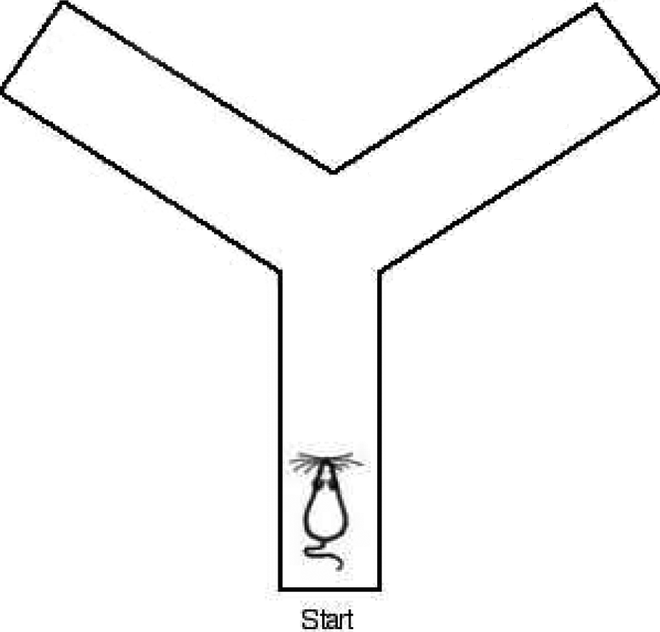
The schematic diagram of the Y-maze. Y-maze has three identical arms. The rat starts at the end of one arm, then chooses one of the other two.
Survival parameters of F1 postnatal rats and the reproductive ability of F1 rats
The following survival parameters of F1 postnatal rats were calculated: Live birth index, pup mortality on day 1, and viability index on day 4. For each group, the numbers of females placed with males, males mated with females, successful copulations (number of females mated), males that became sires, pregnant females, the number and locations of implantation sites, and resorptions and live fetuses were recorded. The following fertility and reproductive performance parameters in F1 rats were calculated: mating index, male fertility index, female fertility index, and female fecundity index.
Physical and reflex development of F1 rats
Developmental and neurobehavioral effects in the offspring were investigated using a test battery 16,17 including assessment of physical development, reflex ontogeny, motor function, motor activity, and learning and memory.
The body weights as well as the state of postnatal development of the F1 pups were monitored during the lactation period. At birth, the following data were recorded: number of live and dead pups, gross malformation, and individual pup weight. After these measures were taken, litter sizes were randomly reduced to 8 pups. Body weight was again measured on PND 4, PND 7, and then every week. Physical and reflex development parameters are evaluated following the articles by Iezhitsa 18 and Vorhees. 19 The day of pinnae detachment, incisor eruption, fur development, eye opening, and ear opening were also monitored until all pups in a given litter were positive for all developmental parameters. Offspring were assessed for indicators of sexual maturation during the post-weaning period. Female offspring were examined daily for the presence of an open vagina, beginning on PND 30. Male offspring were examined daily for balanopreputial separation, beginning on PND 25. Throughout the lactation period, the pups were evaluated in terms of reflex development and neuromuscular maturation. The day of occurrence for surface righting reflex, 20 writhing (swivel by means of the concertina movement of 1 drawleg), creeping (moving ahead slowly with its abdomen close to the floor, not raising its trunk), locomotor activity, mid-air righting reflex, 21 cliff parry, and negative geotaxis reflex were also monitored until all pups in a given litter were positive for all developmental parameters beginning on PND 1, PND 6, PND 7, PND 17, PND 15, PND 5, and PND 7. Consequently, the frequency of animals showing each of the above parameters was recorded for each day of the observational period. To accomplish that, each litter was considered an experiment unit.
The ability of learning and memory were measured by Y-maze
Recording the spontaneous behavior in a Y-maze assessed spatial working memory performance. 22 From each treatment group, 10 males and 10 females were selected randomly for assessment of learning and memory.
The Y-maze is similar to the T-maze; however, it has 3 identical lanes rather than 2. The rat starts from the end of 1 lane and end in any of the other 2 lanes. Y-maze is easier for rats to learn because of the less sharp turns compared to T-maze. Structure and mechanism of Y-maze: Also known as trisection radiant maze, Y-maze is composed of 3 lanes (1, 2, and 3) of the same length joined to a center. Each lane is covered with copper wires for the electric shock. These wires are 14 cm in length and 0.2 cm in diameter. On the top of each lane’s end, there is a 15-W lamp for signal. The control panel of this Y-maze has voltage-controlling switch, delay-controlling switch, and 4 switches for 1, 2, 3, and 0 separately. The lamp will be turned on once the corresponding switch is closed, and we define this as the signal for “safe zone” or “red light zone”; the other 2 lanes with lamps off are “nonsafe zone” or “electric shock zone.” There is no electric shock when the 0 switch is closed. Before starting the “electric shock at the crossing area” experiment, we let the rat stay in the starting district (where the rat starts) 3 to 5 minutes for adaptation. Then we will change the safe zone and nonsafe zone in a certain sequence or randomly observe the rat’s learning procedure: get out of the nonsafe zone and get into the safe zone. Rats may still get into the nonsafe zone under the electric shock until they get trained after several times of training. It is defined as passive avoidance response when electric shock is needed to stimulate the rat to find the safe zone. After several times of training, the rat will run to the safe zone once the signal lamp is turned on and the electric shock is still off; we define this as conditioned reflex to distinguish light and dark (active avoidance response). Method: The experiment is carried out in a quiet dark Y-maze at the same time every day. There is a lamp at the end of each lane and the floor of each lane is covered with electric wires. Once the lamp of the lane is on, there is no current, that is, this lane is the safe zone; the other 2 lanes without lamps turned on are nonsafe zone because current is passing through the wire. Safe zone and nonsafe zone can be exchanged with each other randomly. Once the rat has stayed in the safe zone for 30 seconds, the safe zone will be changed to the other 2 lanes randomly. Five seconds after the change, current will pass through the nonsafe zone and stimulate the rat to run to the safe zone. It is defined as positive when the rats get to the safe zone in 10 seconds at one time and vice versa. Learning study: Procedures A and B—Procedure A: The directions of the rats' learning experiment are 1-2, 2-3, and 3-1. After a rat gets to the safe zone and stays there for 30 seconds, put the rat back to the lane it started and give it a electric shock 1 minute later. Repeat the above procedure till the rat can find the safe zone at one time and take a confirmative trial. Then start all over again from the new safe zone. This procedure will be carried out in the direction mentioned above. Once the rat can find the safe zone at one time no matter which lane it is in, we will go to the procedure B. Procedure B—The directions of the rats' learning experiment are 1-2-3, 2-3-1, and 3-1-2. Repeat the electric shock training as mentioned in procedure A. There is a 1-minute rest time between every trial. Every rat will be trained for 30 times and the right times are recorded. The qualified standards are 10/30 and 15/30 for A and B separately. The screenings are carried out every 2 days. Memory study: Pick out the qualified rats, give them electric shock test as in procedure B, 24 hours later. Memory index: training times needed to get 9 correct responses out of 10 consecutivetrials. Index for observing: 1. The amount of qualified rats; 2. Training times needed to get qualified: the less training time needed to get qualified in the learning experiment and memory experiment, the better learning ability or the better learning efficiency of rats and vice versa.
Gross necropsy and histopathology
Organs or tissues showing macroscopic abnormalities were preserved in a neutral, phosphate-buffered 4% formaldehyde solution (the testes were preserved in Bouin fixative). All stillborn pups and pups found dead during the study were examined macroscopically for structural abnormalities or pathological changes. All collected samples of the high-dose and control groups, as well as the organs and tissues showing macroscopic abnormalities, were subjected to microscopic examination.
Statistica Evaluation
Body weight, body weight gain, food consumption, numbers of implantations and pups delivered, delivery index, sperm parameters, organ weight, organ/body weight ratio (relative organ weight), and the viability of pups were analyzed for statistical significance as follows: Bartlett test of homogeneity of variance was used to determine whether the groups had equivalent variances. If the variances were equivalent, the groups were compared by 1-way analysis of variance (ANOVA) with Tukey posttest. The incidence of females with normal estrous cycles, copulation index, fertility index, gestation index, neonatal sex ratio, and completion rate of the reflex response were analyzed by Fisher exact test. The probability of .05 was used as the criterion for significance.
Results
Segment I: Fertility and Early Embryonic Development Toxicity
Clinical observations, body weight, and food consumption during the premating, mating, and gestation periods (F0)
There were no compound-related clinical signs of toxicity in either male or female F0 rats during the premating, mating, or gestation periods.
Table 1 shows the body weights of F0 males and females during the dosing period. The body weight and body weight gain of F0 males and females exhibited no significant differences between the control and treatment groups. No significant changes in food consumption were observed in F0 rats of either sex (data not shown). All pregnant animals appeared healthy at sacrifice. There was no significant difference in gravid uterus weight between the control and treatment groups or among the treated groups.
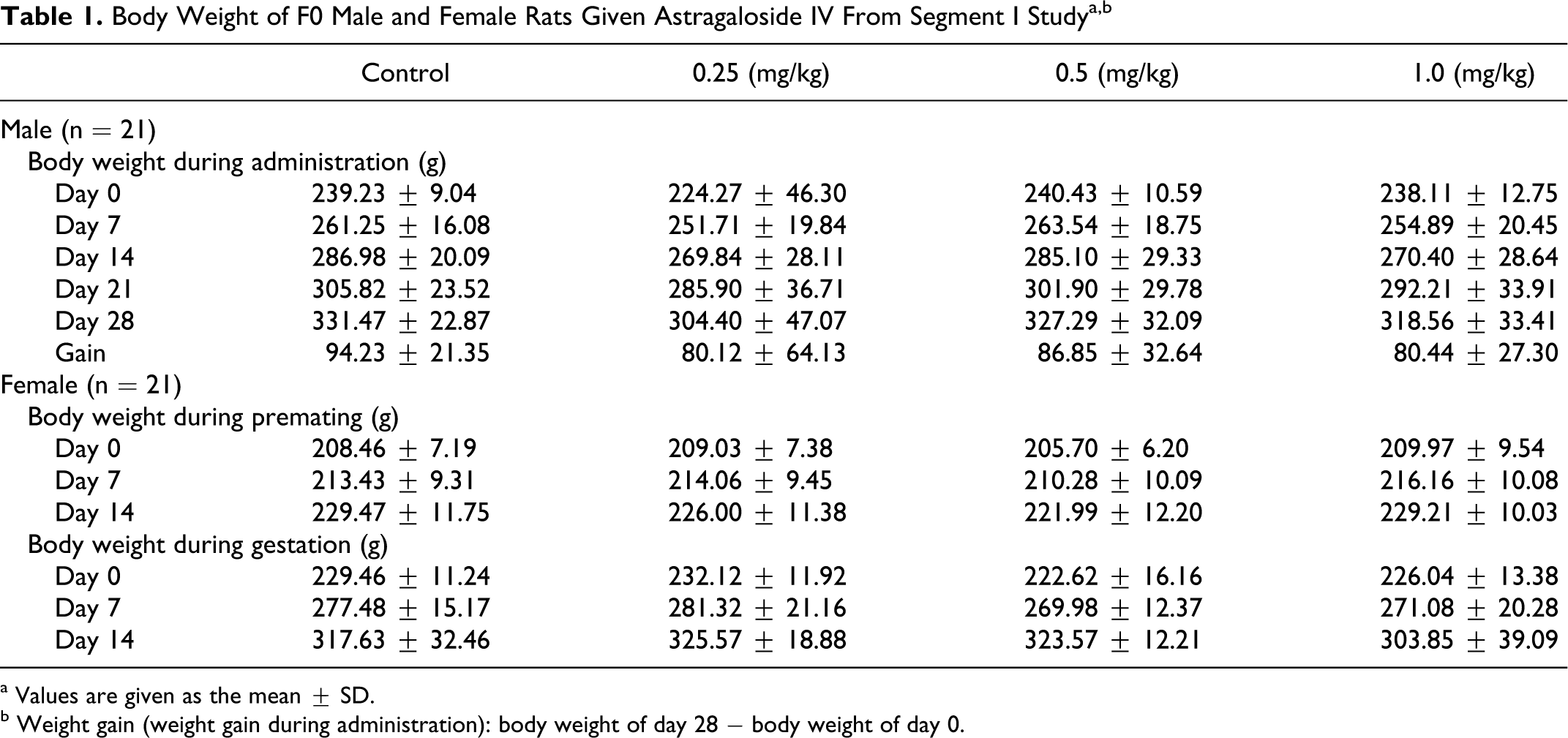
a Values are given as the mean ± SD.
b Weight gain (weight gain during administration): body weight of day 28 − body weight of day 0.
Fertility and reproductive performance of F0 rats
The reproductive and developmental parameters for F0 parent are presented in Table 2. In F0 parent animals, there were no significant differences in the fertility index, the proportion of resorptions, or dead fetuses per litter between the treatment and control groups. The copulation index was lower throughout the mating period at 1.0 mg/kg per d (1.0 mg/kg group vs control, 71.43% vs 90.48%, χ2 = 3.30, .05 < P < .10).
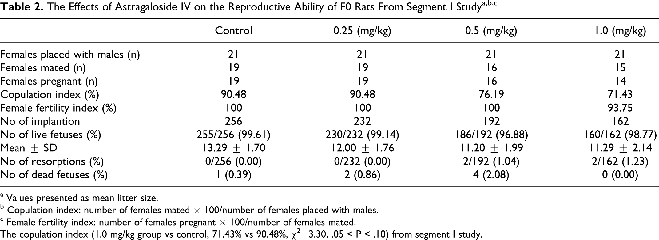
a Values presented as mean litter size.
b Copulation index: number of females mated × 100/number of females placed with males.
c Female fertility index: number of females pregnant × 100/number of females mated.
The copulation index (1.0 mg/kg group vs control, 71.43% vs 90.48%, χ2=3.30, .05 < P < .10) from segment I study.
Organ weights (F0 adults), gross necropsy, and histopathology
There were no significant differences in liver, kidney, epididymides, and testes weights between the control and treatment group males (Table 3); additionally, there were no differences in the weights of body, liver, kidneys, or ovaries of pregnant females. There were no compound-related gross lesions or microscopic alterations in the reproductive organs of F0 males and females. No reproductive difficulty was observed.
Organ Weight of Male and Female F0 Rats Given Astragaloside IV From Segment I Study a
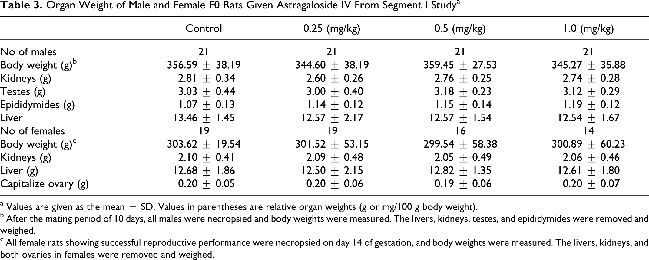
a Values are given as the mean ± SD. Values in parentheses are relative organ weights (g or mg/100 g body weight).
b After the mating period of 10 days, all males were necropsied and body weights were measured. The livers, kidneys, testes, and epididymides were removed and weighed.
c All female rats showing successful reproductive performance were necropsied on day 14 of gestation, and body weights were measured. The livers, kidneys, and both ovaries in females were removed and weighed.
Sperm parameters (F0 adults)
Table 4 shows the sperm parameters in F0 adult males. No significant changes in sperm counts, percentage of motile sperm, or total abnormal sperm ratios were recorded in F0 adults between the control and treated groups.
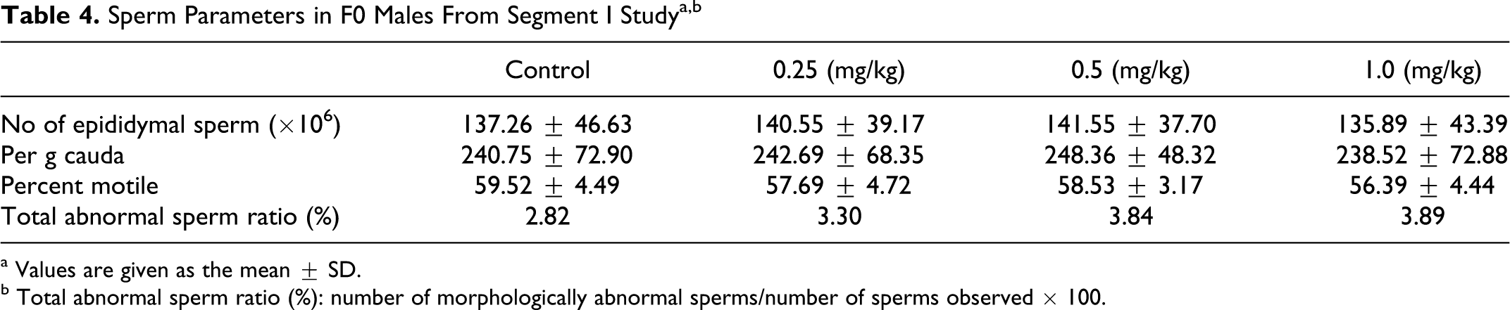
a Values are given as the mean ± SD.
b Total abnormal sperm ratio (%): number of morphologically abnormal sperms/number of sperms observed × 100.
Segment III: Perinatal and Postnatal Toxicity
Clinical observations, body weight, and food consumption (F0/F1)
There were no compound-related clinical signs of toxicity in mated female F0 rats during the gestation or lactation periods. The body weights of F0 females during dosing are shown in Table 5. Body weights of F1 (male and female) rats are shown in Table 6. The body weights and body weight gain of F0 females exhibited no significant difference between the control and treatment groups. The body weights of F1 (male and female) rats per litter exhibited no significant difference between the control and treatment groups. Food consumption of F0 females was not significantly reduced during pregnancy. Food consumption in the 0.25 to 1.0 mg/kg groups was significantly less than that of the control group on days 3 and 7 of lactation, whereas it is not in a dose-dependent relationship. Food consumption of F0 females in the 0.5 and 1.0 mg/kg groups was not significantly different on days 14 and 21 of lactation.
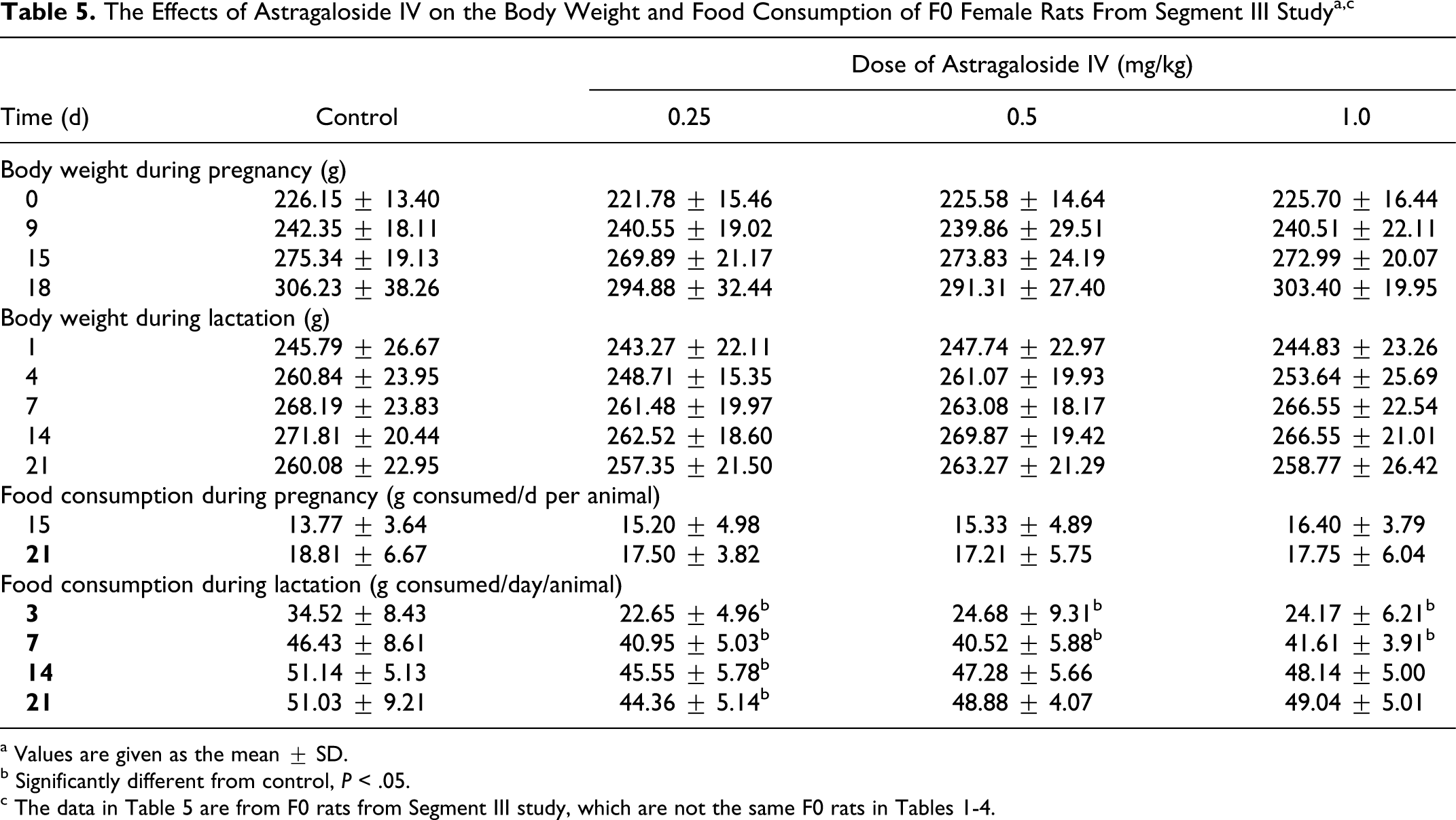
a Values are given as the mean ± SD.
b Significantly different from control, P < .05.
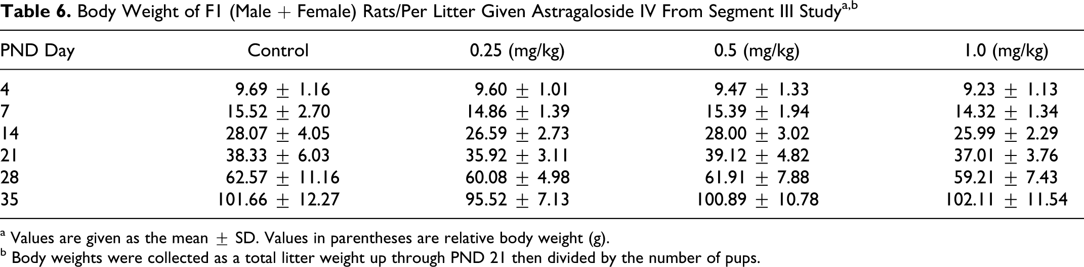
a Values are given as the mean ± SD. Values in parentheses are relative body weight (g).
b Body weights were collected as a total litter weight up through PND 21 then divided by the number of pups.
The survival parameters of F1 postnatal rats and the reproductive ability of F1 rats
No malformed F1 pup was found in any groups. Live litter size, number of live born, stillborn, live birth index, viability index day 4, and viability index days 4 to 21 were comparable among all groups (Table 7). Pup mortality from PND1 in the 0.5 mg/kg per d group was significantly greater than that of the control group, but there were no significant differences (0.5 mg/kg group vs control, 3.39% vs 0.46%, χ2 = 3.26, P > .05; Table 7). No difference in clinical observations was recorded on PND 4, 7, and 14. Macroscopic examination of pups did not reveal gross malformations or findings that would indicate abnormal development (data not shown).
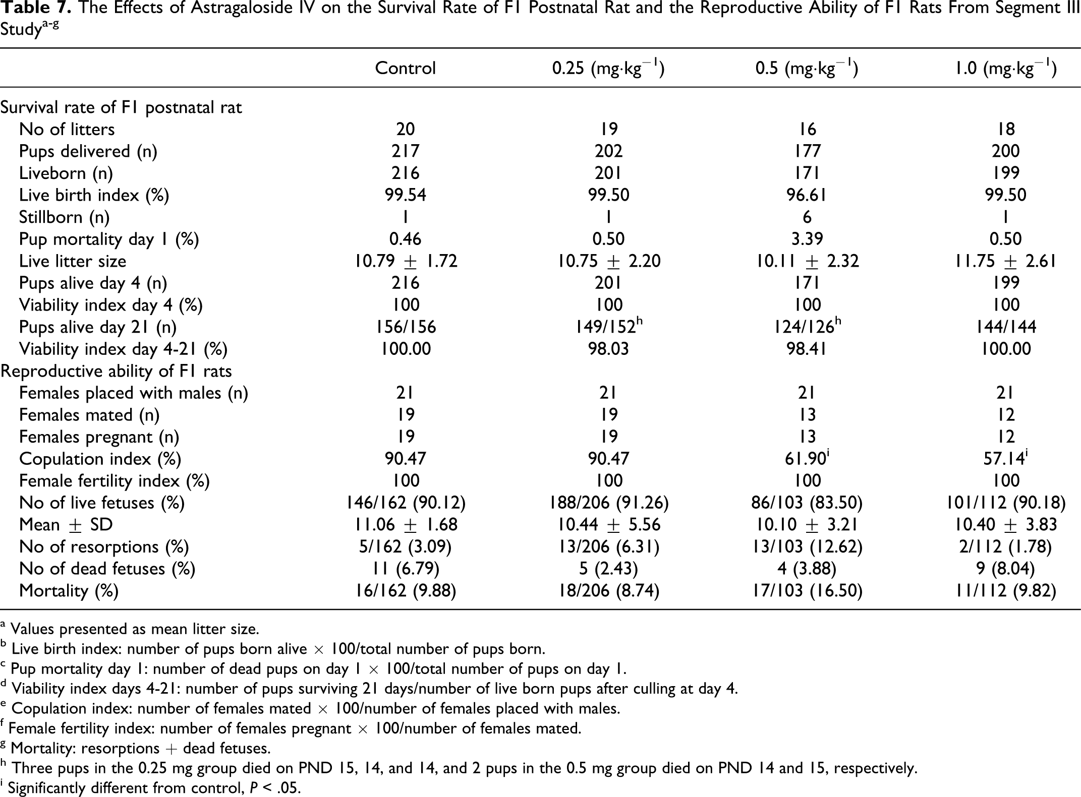
a Values presented as mean litter size.
b Live birth index: number of pups born alive × 100/total number of pups born.
c Pup mortality day 1: number of dead pups on day 1 × 100/total number of pups on day 1.
d Viability index days 4-21: number of pups surviving 21 days/number of live born pups after culling at day 4.
e Copulation index: number of females mated × 100/number of females placed with males.
f Female fertility index: number of females pregnant × 100/number of females mated.
g Mortality: resorptions + dead fetuses.
h Three pups in the 0.25 mg group died on PND 15, 14, and 14, and 2 pups in the 0.5 mg group died on PND 14 and 15, respectively.
i Significantly different from control, P < .05.
In all, 2 females in the control group, 8 females in the 0.5 mg/kg/d groups, and 9 females in the 1.0 mg/kg per d groups did not mate. All mated females became pregnant. In F1 parent females, there was no significant difference in female fertility index from segment III study between the control and treatment groups or among the treated groups. The copulation index from the segment III study in the 0.5 and 1.0 mg/kg groups was significantly greater than those of the control group (0.5 mg/kg group vs control, 61.90% vs 90.47%, χ2 = 4.73, P < .05; 1.0mg /kg group vs control, 57.14% vs 90.47%, χ2 = 6.04, P < .05; Table 7). There were no significant differences in the proportion of resorptions, dead fetuses, or mortality between the treatment groups and the control group.
Physical and reflex development of F1 rats
Physical development of F1 pups is presented in Table 8. Maternal AS-IV exposure at the dose of 1.0 mg/kg per d resulted in a significant delay in the time of the hair appearance, eye opening, locomotor activity, and cliff parry reflex of pups compared to the control group (P < .05). The completion of hair appearance, ear opening, descent of testicles into the scrotum, cliff parry, and writhing was delayed in male and female F1 pups at 0.5 mg/kg per d. No significant alterations in means of body weight increase of newborns were detected between control and treated groups (Table 6). We did not find a significant difference between groups in tests memory and learning (Y-maze; Table 9).
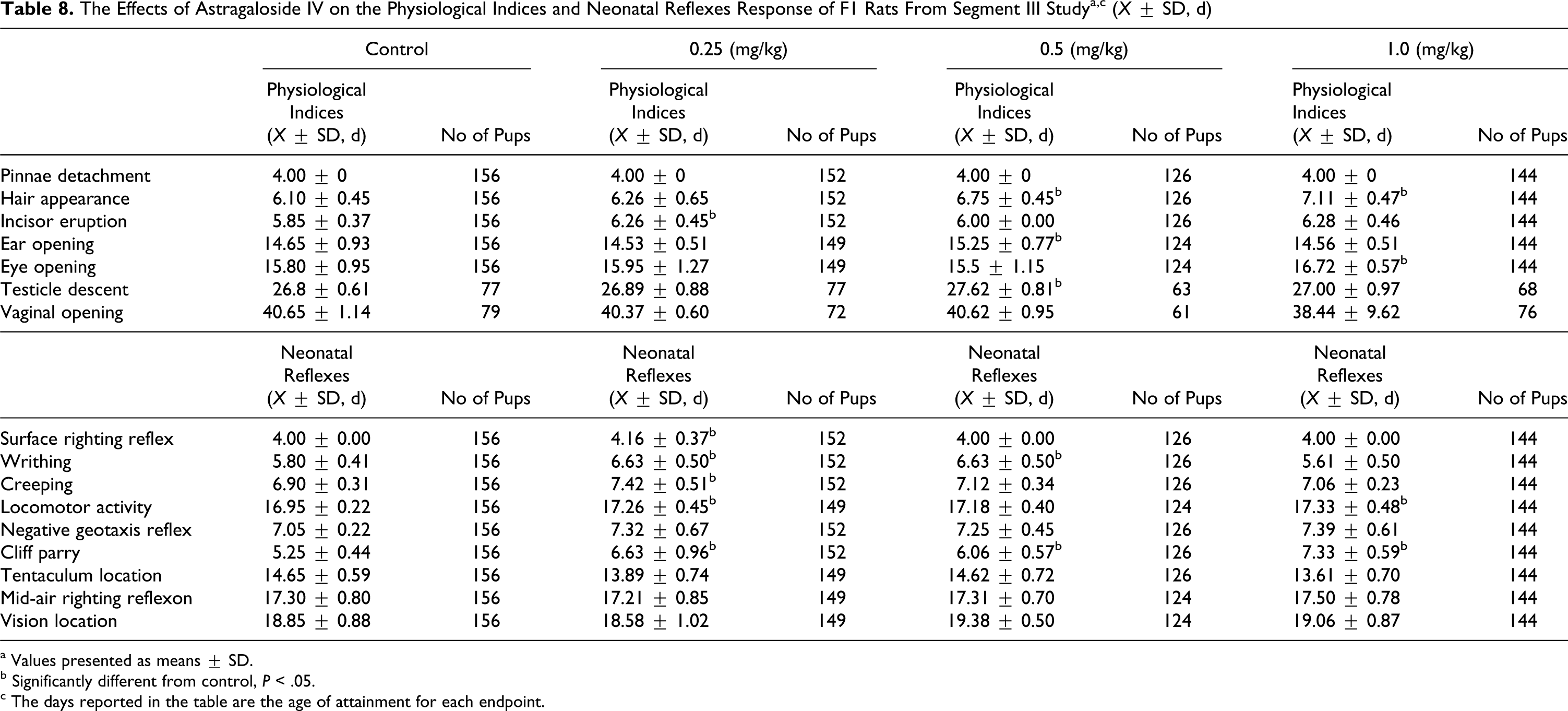
a Values presented as means ± SD.
b Significantly different from control, P < .05.
c The days reported in the table are the age of attainment for each endpoint.
The Effects of Astragaloside IV on the Ability of Learning of F1 Rats a
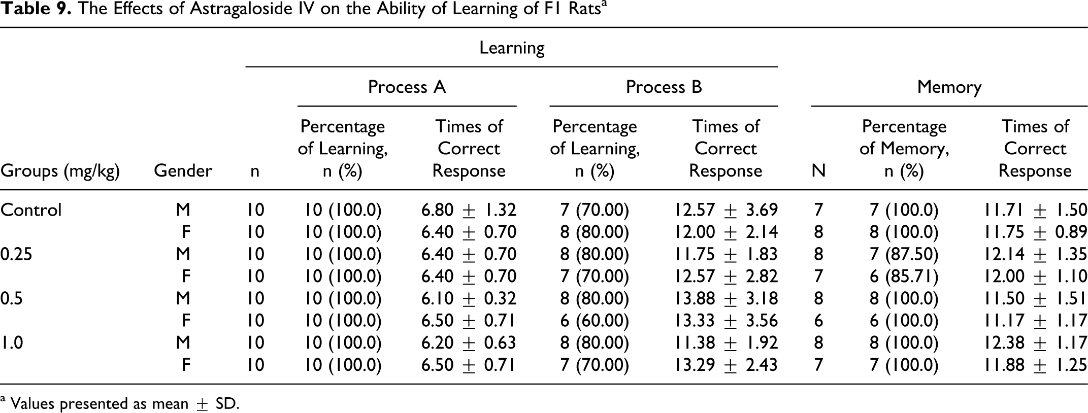
a Values presented as mean ± SD.
Necropsy and histopathology
No compound-related gross lesions or remarkable microscopic alterations of tissues and organs, including the reproductive organs, were recorded in F0 and F1 males and females in the highest dose group and dead animals before the scheduled term. There were no compound-related gross lesions or microscopic alterations observed in male or female F1/F2 pups, including pups that died before weaning (data not shown).
Discussion
Astragaloside IV, a saponin isolated from A membranaceus, exhibits not only potent cardioprotective effects by reducing infarct size, attenuating arrhythmias, and improving ventricular function in ischemic heart disease, 4,5 but also blood−brain barrier protective effects against ischemia/reperfusion injury by reducing infarct size induced by ischemia and decreasing the tight junction proteins, occludin, and zonae occludens-1 (ZO-1) in endothelial cells. 2,3 More recently AS-IV was reported to be capable of decreasing the frequencies of synchronized spontaneous Ca2+oscillations and spontaneous excitatory postsynaptic currents in hippocampal neurons, 23 increasing the rate of peripheral nerve regeneration, enhancing axonal regeneration, and reconstructing of neuronal synapses as well as preventing Ab(25-35)-induced neuronal death. 24,25 Astragaloside IV can also protect dopaminergic neurons against 6-Hydroxydopamine (6-OHDA)-induced degeneration, and promote neurite outgrowth and increased tyrosine hydroxylase (TH) and nitric oxide synthase (NOS) immunoreactivity of dopaminergic neurons. 9 Astragaloside IV is now being developed as a cardioprotective and neuroprotective agent for treating cardio- and cerebrovascular diseases. In this study, we conducted reproductive and developmental toxicity testing in F0 and F1 to observe its maternal toxicity, fertility, and early embryonic development toxicity, as well as peri- and postnatal toxicity 26,27 in rats to support its clinical safety.
In our acute toxicity studies, the MTD in rats is 20 mg/kg (data not published). Considering previous embryo-fetal development toxicity studies and the MTD in rats, rats were administered intravenously daily with 0.25, 0.5, and 1.0 mg/kg AS-IV in this study, which is approximately 1/20 to 1/80 of rat MTD. In this study, AS-IV showed no maternal toxicity at 0.25 to 1.0 mg/kg in rats. Although food consumption in the 0.25 to 1.0 mg/kg groups from segment III study was significantly less than that of the control group on days 3 and 7 of lactation, they are not in a dose-dependent relationship. Food consumption of F0 females in the 0.5 and 1.0 mg/kg groups was not significantly different on days 14 and 21 of lactation between the treatment groups and the control group. These data were not considered indicative of parental toxicity. A membranaceus has been routinely used in China for patients with stroke or chronic debilitating diseases, and even for normal participants who wish to further improve their vital functions. 2 Therefore, AS-IV may support the animals with less food consumption without a decrease in body weight, especially during lactation. All pregnant animals appeared healthy at sacrifice. However, the pups were probably also consuming the food by day 18 of lactation. All of these results indicated that AS-IV has no maternal toxicity at 0.25 to 1.0 mg/kg in rats.
The highest doses, however, did cause a decrease in pregnancy outcome because no decreases in fertility and copulation index were recorded in the F0 animals from the segment I study. The copulation index (1.0 mg/kg group vs control, 71.43% vs 90.48%, χ2 = 3.30, .05 < P < .10) from the segment I study were significantly reduced throughout the mating period at 1.0 mg/kg per d. The differences in copulation index between the 2 groups were not statistically significant (P < .01), but there is dose−response relation. This result showed that AS-IV had an inhibitory effect on the fertility of F0 rat but was devoid of early embryonic developmental toxicity. Meanwhile, in F1 parent females, there was no significant difference in female fertility index in the segment III study between the control and treatment groups or among the treated groups. However, the copulation index from segment III study in the 0.5 and 1.0 mg/kg groups was significantly less than those of the control group (0.5 mg/kg group vs control, 61.90% vs 90.47%, χ2 = 4.73, P < .05; 1.0 mg /kg group vs control, 57.14% vs 90.47 %, χ2 = 6.04, P < .05; Table 7). Although there are no enough data to definitely attribute this to an effect on the male or female, there may be a contributory factor from the males' exposure in that the fecundity index is not changed by treatment.
The body weights of F1 (male and female) rats per litter exhibited no significant differences between the control and treated groups. However, due to lack of data by gender difference, whether males and females may differ in their rate of body weight gain under the influence of the drug is not clear. Maternal AS-IV exposure at the dose of 1.0 mg/kg per d resulted in a significant delay in time of fur appearance, eye opening, locomotor activity, and cliff parry reflex of pups compared to control group (P < .05). Our data demonstrated that maternal exposure of AS-IV resulted in transient delay in reflex maturation in pups at 0.5 mg/kg per d. However, there is not enough data to demonstrate an effect on physical and reflex development in F1 rats. Further research is required on the mediation of AS-IV action during peripartum. We did not find a significant difference between groups in tests of memory and learning. No significant alterations in means of body weight increases of newborns were detected between control and treated groups.
In summary, our study is the first experimental study investigating the effect of fertility and early embryonic development toxicity and peri- and postnatal toxicity of AS-IV in rats. Astragaloside IV was devoid of maternal toxicity at 0.25 to 1.0 mg/kg in rats. Exposure to AS-IV up to concentrations of 1.0 mg/kg per d in F0 and F1 rats appears to have an inhibitory effect on female fertility of F0/F1 rats but was devoid of early embryonic development toxicity in F0/F1 rats. Astragaloside IV did not affect the survival parameters of F1 postnatal rats. Maternal AS-IV exposure at the dose of 1.0 mg/kg per d resulted in a significant delay in reaction time of the fur development, eye opening locomotor activity, and cliff parry reflex of pups. It does not affect the memory and learning of F1 pups. Further research is required on the mediation of AS-IV action during peripartum, and thorough investigation is required for elucidating the action mechanism and the applicability of these results in humans.
In light of these findings, it is prudent to advise caution to women who might use AS-IV to combat cardio- and cerebrovascular sickness during peripartum, as the anticipated clinical dose for humans is 10 mg/60 kg per d, which is comparable to the doses administered in the segments I and III studies.
Footnotes
Notes
The author(s) declared no conflicts of interest with respect to the authorship and/or publication of this article.
The author(s) disclosed receipt of the following financial support for the research and/or authorship of this article: the National Natural Science Foundation of China (30400360).