
Abstract
Liver cancer is one of the most lethal cancer types in humans, but our understanding of the molecular mechanisms underlying this process remains insufficient. Here, we conducted high-content screening of the potential genes involved in liver cancer metastasis, which we selected from the Kyoto Encyclopedia of Genes and Genomes (KEGG) pathway database, based on the SAMcell method and RNA interference technology. We identified two powerful genes in the liver cancer metastasis process, AEG-1 and AKR1C2, both of which proved to be positive regulators in promoting metastasis in liver cancer. Further clinical results verified their roles in liver cancer. In summary, these findings could provide new insight into the liver cancer mechanism and potentially therapeutic novel targets for liver cancer therapies in the future.
Introduction
We currently suffer from a lack of efficient therapies to control most solid tumors because of cancer metastasis. To our knowledge, metastasis is a complex and multifaceted process, consisting of tumor invasion, tumor cell dissemination, colonization of distant organs, and metastatic outgrowth. 1 These processes are mediated by diverse genes in several pathways, including cell proliferation, angiogenesis, motility, invasiveness, and so on. Some of these genes, such as VEGF, SRC, and MMP, have been identified as either promoting genes or suppressing genes.1,2 However, our knowledge of the molecular mechanism underlying metastasis is far from sufficient. We still lack efficient therapies to control metastasis effectively.
Both positive and negative regulatory genes of metastasis have provided us with hints for developing potential diagnostic methods and subsequent clinical drugs. Genes that are positive regulators are usually expressed at high levels and are more likely to act as markers for predicting metastasis. Furthermore, the inhibition of some positive regulatory genes leads to adverse effects on tumor metastasis, such as the disruption of invasion and metastasis, inhibition of cell proliferation, and even induction of cell apoptosis. These inhibitors might be developed into novel drug targets in preclinical and clinical stage research and might even finally become efficient methods to treat cancer. Thus, identifying these positive regulatory genes is of great importance. 3
Liver cancer or hepatocellular carcinoma (HCC) is one of the most deadly diseases. Major risks, such as infection with hepatitis B virus (HBV) or hepatitis C virus (HCV), alcoholic liver disease, and nonalcoholic fatty liver disease, can lead to the development of cirrhosis in most liver cancer patients. 4 Although recent research suggests that the overall survival rate is increasing, we still lack a systematic understanding of the underlying molecular mechanism of liver cancer, especially during the metastatic process.4,5
High-content screening is a powerful tool for identifying the roles of large quantities of genes in a short period of time, and it would probably help us scratch the surface of the metastatic process. 6 Traditional screening methods for cell migration, such as the transwell and wound-healing assays, are not suitable for high-content screening because of low throughput, poor efficiency, and sophisticated operations. Recently, a method called SAMcell (Self-Assembly Cell Microarray) has been successfully used to perform screening for metastasis and to identify novel regulators in this process. 7 This method integrated micro-fabrication technology, reverse-transfection technology, and RNA interference technology, and it investigated cell behavior directly on a cell chip. Here, we screened a library of genes related to liver cancer and identified two powerful genes, AEG-1 and AKR1C2, involved in the liver cancer metastasis process.
Materials and Methods
Small Interfering RNA and Plasmid
Small interfering RNA (siRNA) duplexes were obtained from View-Solid Biotech (Beijing, China). The silencing efficiency of these siRNAs for their targeted messenger RNAs (mRNAs) was tested by quantitative real-time PCR at 24 to 48 h after transfection. All of the reactions were run in triplicate. During the RNA interference (RNAi) experiments, we used two specific siRNAs and a scrambled siRNA as a negative control (siNC).
The human AEG-1 and AKR1C2 complementary DNA (cDNA) clones were constructed into the pcDNA3.1 vector (Addgene, Cambridge, MA). During overexpressing experiments, we used the empty vector as a negative control (Vector NC).
Cell Culture and Transfection
The two types of HCC lines (MHCC-97H and Huh7 cells) were obtained from the American Type Culture Collection (ATCC, Manassas, VA). Cell lines, including MHCC-97H and Huh7, were cultured in Dulbecco’s modified Eagle’s medium containing 10 % fetal bovine serum, 100 U/mL−1 penicillin, and 0.1 mg/mL−1 streptomycin under humidified conditions of 95% air and 5% CO2 at 37 °C. The transfection experiments were followed with the Lipofectamine 3000 (Invitrogen, Carlsbad, CA) transfection reagent protocol.
Fabrication of SAMcell
Glass slides (2.2 × 2.2 cm) were washed with detergent and MilliQ (Millipore, US) water. After drying, the slides were covered with poly(N-isopropylacrylamide) (Sigma-Aldrich, St. Louis, MO) dissolved in ethanol (6% w/v). The slides were etched via a shadow mask by oxygen plasma for 5 min at 200 W of power. The reverse transfection protocol was previously described. In brief, 3 µL OptiMEM (Invitrogen) containing sucrose and 2.5 µL Lipofectamine 3000 (Invitrogen) were transferred to each tube and mixed thoroughly. Then, 2 µg siRNA/1 µg of plasmid/chemicals/proteins were added to each tube, and the mixture was incubated for 20 min at room temperature. Finally, 7.25 µL of a 0.2 % (w/v) gelatin (Type B; Sigma-Aldrich) solution was added to each tube and mixed thoroughly. After UV sterilization, the reverse transfection reagent was printed on the chip using a nanodispenser (Phoenix; Art Robbins Instruments, Sunnyvale, CA). Next, the slides were fixed in a 6-well plate with melted wax. Approximately 3 mL of 37 °C medium containing 5 × 10⁵ cells was transferred to each well. Approximately 24 to 48 h later, the dishes were moved at room temperature for 5 min and were washed with phosphate-buffered saline (PBS) three times to ensure the total removal of the polymer.
Transwell Migration and Invasion Assay
For migration assays, MHCC-97H or Huh7 cells were seeded into the upper chamber of a transwell insert (pore size, 8 µm; Costar [Corning, Corning, NY]) in 100 µL of serum-free medium per well. Medium at a volume of 600 µL, containing 10 % serum, was placed in the lower chamber to act as a chemoattractant. Nonmigratory cells were removed from the upper chamber by scraping with a cotton bud. The cells remaining on the lower surface of the insert were fixed with 4% formaldehyde (Sigma-Aldrich) and were stained by Crystal Violet (C3886; Sigma-Aldrich). For invasion assays, cells were seeded in a Matrigel (Bio-Rad, Hercules, CA)–coated chamber and were incubated at 37 °C.
Cell Proliferation Assay
MHCC-97H or Huh7 cells were transfected with the indicated siRNAs or plasmids. After 1 day, the cells were counted and seeded in a 12-well plate at a density of 5 × 10⁴ cells per well. After 4 days of incubation, the cells were trypsinized for complete detachment and counted by hemocytometer measurement.
Immunofluorescence
Cells were seeded onto sterile cover slides and allowed to attach overnight. The cells were then fixed with 4% formaldehyde, permeabilized with 0.1% Triton X-100, and blocked in 2% bovine serum albumin for 1 h at room temperature. The expression of E-cadherin and vimentin was examined using targeted antibodies (anti–E-cadherin: ab76055; Abcam, Cambridge, MA) (antivimentin: ab8978; Abcam) and visualized using anti–rabbit IgG (H + L), F (ab)2 fragment (Alexa Fluor 488 Conjugate; Cell Signaling Technology, Beverly, MA). Immunofluorescence was examined using a microscope (EcliPSE Ti-U; Nikon, Tokyo, Japan). The antibodies were diluted 1:200.
Immunoblotting
Lysates were resolved by electrophoresis, transferred to a polyvinylidene difluoride membrane (Millipore, Billerica, MA), and probed with antibodies against AEG-1 (ab45338; Abcam) and AKR1C2 (ab131375; Abcam). The antibodies were diluted 1:1000.
Human Liver Samples and Immunohistochemistry
All human liver samples were obtained from the Beijing Youan Hospital. Before surgery at the center, all patients provided written informed consent to allow any excess tissue to be used for research studies.
Immunohistochemistry was performed at the Beijing Youan Hospital. Tissue samples were fixed in 10% buffered formalin for 12 h, followed by washing with PBS, transfer to 70% ethanol, and then embedding in paraffin. Immunohistochemistry using antibodies against AEG-1 (ab45338; Abcam) and AKR1C2 (ab131375; Abcam) was performed on paraffin sections.
The scoring criteria were determined during a preliminary evaluation, using a multiheaded microscope to reach a consensus. The staining results for each antibody were interpreted by two of the authors independently, without prior knowledge of the clinical or pathological parameters. For each sample, at least five fields and >500 cells were analyzed. The number of immune-positive cells was semi-quantitatively estimated. First, a scoring system was determined according to the staining intensity as follows: 0, colorless; 1, light yellow; 2, brown-yellow; and 3, dark brown. Scoring according to the percentage of positive cells was determined as follows: 0, no positive cells; 1, <10% positively stained cells; 2, 11% to 50% positively stained cells; 3, 51% to 75% positively stained cells; and 4, >75% positively stained cells. If the product of multiplication of the staining intensity by the percentage of positive cells was ≥2, the sample was considered immune positive (+).
Statistical Analysis
All of the results are expressed as the means and totals derived from independent experiments. When comparing two groups, Student’s unpaired t test (two-tailed) was used. For all of the tests, a p value <0.05 was considered significant. The Benjamini and Hochberg false discovery rate was used as a correction for multiple testing. * indicates p < 0.05; ** indicates p < 0.01; *** indicates p < 0.001. Error bars represent the standard deviations (SDs) of at least three independent experiments.
Results and Discussion
SAMcell Screening Identifies AEG-1 and AKR1C2 as Positive Regulators of Liver Cancer Metastasis
We used the highly metastatic MHCC-97H cell line to conduct loss-of-function screening. We selected 103 candidate genes related to liver cancer from the Kyoto Encyclopedia of Genes and Genomes (KEGG) pathway, 8 most of which have not been reported clearly during the process of metastasis. Then, we constructed the siRNA library, with three specific siRNAs against one gene as a pool.
We performed cell migration screening with the aforementioned SAMcell method.
7
Briefly, we printed siRNA pools as well as scrambled siRNA on each chip and seeded MHCC-97H cells. After incubation for 48 h, we started to record the images of cell islands. Cell chips were recorded by a microscope system (Image Xpress; Molecular Devices, Sunnyvale, CA). Areas of cell islands were also measured using this system. Four repeats were performed in each group, and we used the mean. The areas of cell islands increased as a linear function of time within a certain period so that the slopes could represent the migratory activities.
7
Consequently, we could use a formula to calculate the cell island’s migration speed, MS =
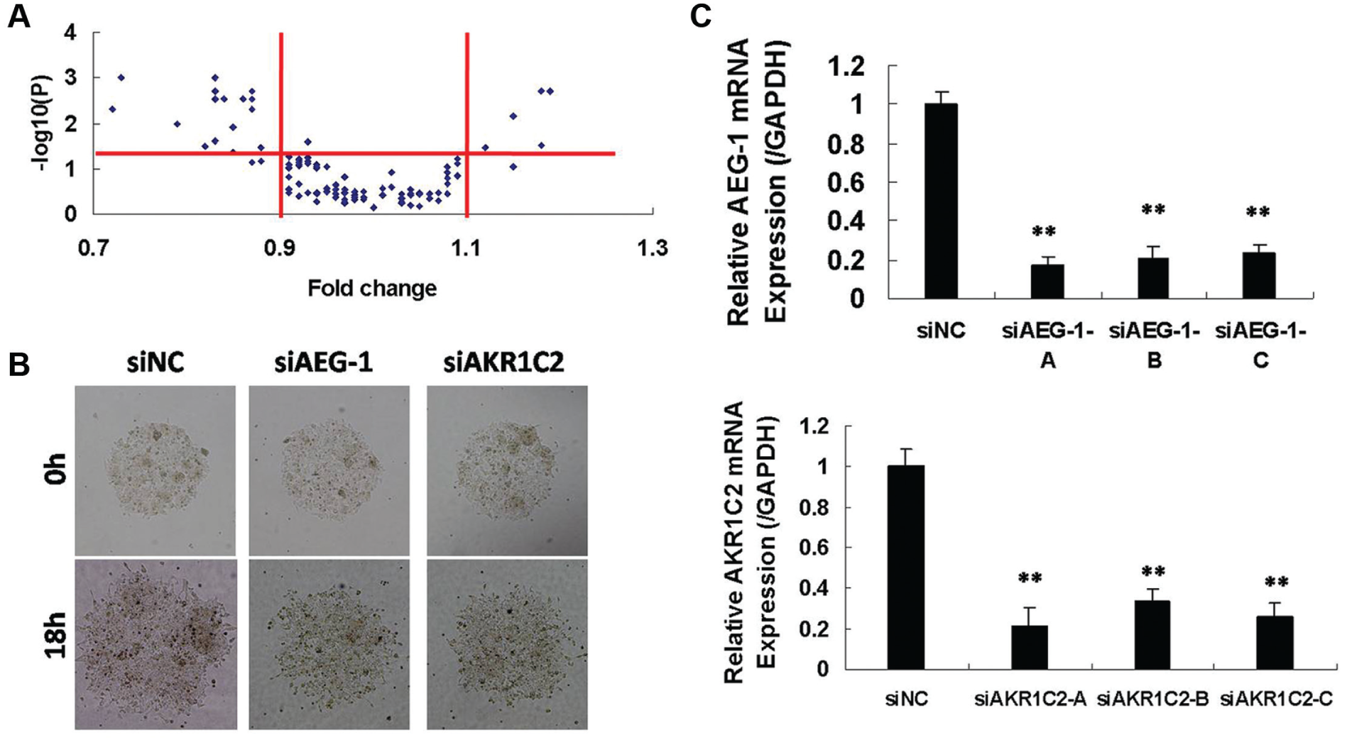
SAMcell functional screening in MHCC-97H cells to identify AEG-1 and AKR1C2 as positive regulators for liver cancer metastasis. (
The SAMcell method offers several advantages over traditional methods, such as transwell or wound-healing assays. SAMcell was first reported to be able to identify miR-23b as a powerful tumor suppression regulator.
7
The two hits from screening were verified in cell lines and clinical samples. In total, 6 migration-promoting hints and 18 migration-repressing hits were identified. To ensure the false-positive rate, we randomly selected 10 genes’ siRNA pools and investigated them by traditional transwell assay. None showed contradictory results with the former screening results. Among the migration-suppressing hits, there were a few well-reported metastasis regulators, such as VEGF, EGFR, and NRAS (
AEG-1 and AKR1C2 Promote Metastatic Behaviors in Liver Cancer Cells
To investigate the functions of AEG-1 and AKR1C2 in the metastasis of liver cells, we selected highly metastatic MHCC-97H cells and low metastatic potential Huh7 cells to perform functional investigations. As shown by transwell migration and invasion assay, the migratory and invasive capabilities in MHCC-97H were almost 3-fold greater than those of Huh7 (
In the highly metastatic MHCC-97H cell line, knockdown of AEG-1 or AKR1C2 suppressed cell migration (
Fig. 2A
), cell invasion (
Fig. 2B
), and cell proliferation (
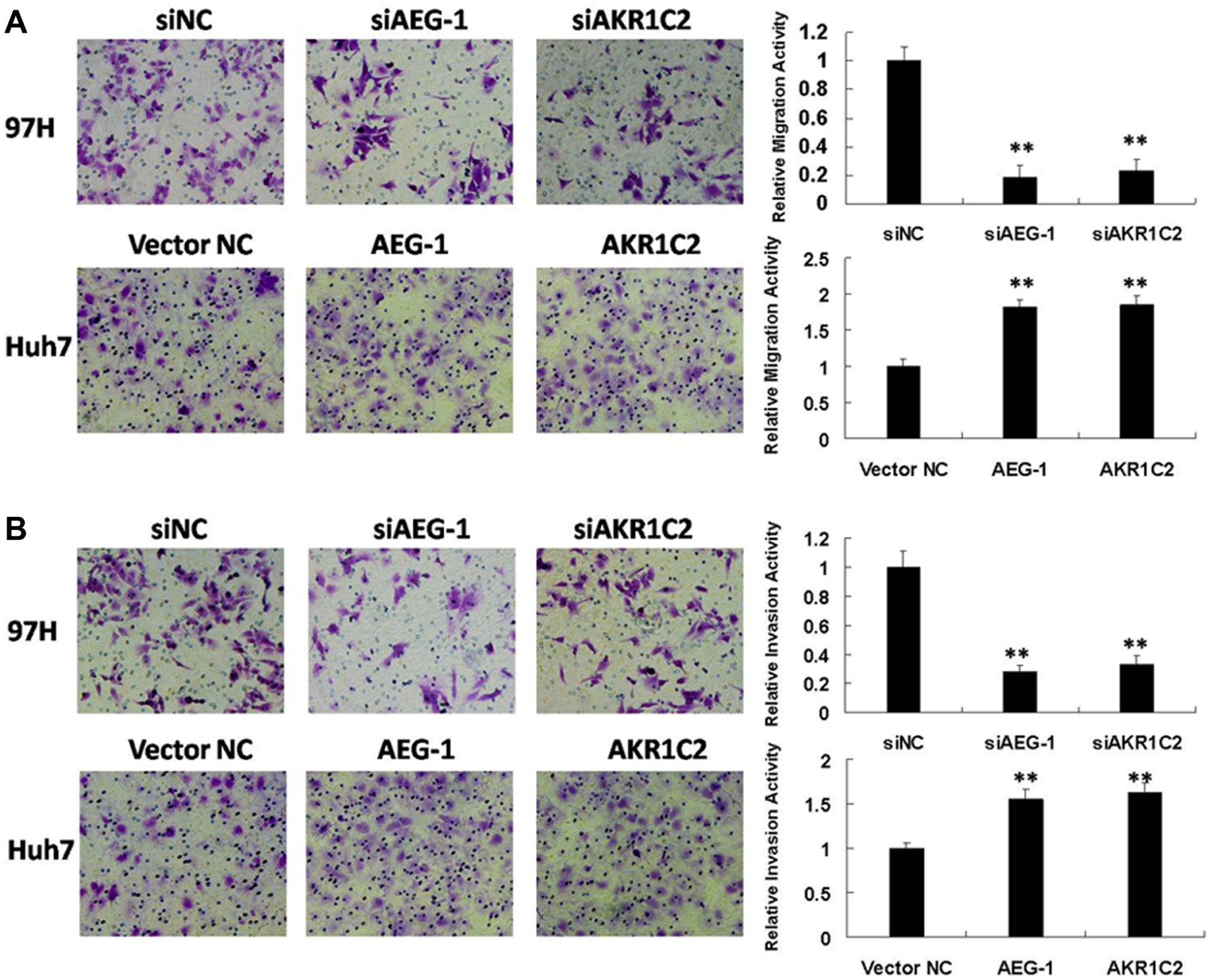
AKR1C2 and NF1 modulating liver cancer metastasis in vitro. (
AEG-1 and AKR1C2 Promote EMT Processes in Liver Cancer Cells
Manipulation of AEG-1 and AKR1C2 affected the process of epithelial to mesenchymal transition (EMT). In highly metastatic MHCC-97H cells, E-cadherin was expressed at a low level while vimentin was highly expressed. As shown in Figure 3A , E-cadherin was increased while vimentin was reduced after silencing AEG-1 or AKR1C2. That is, knockdown of AEG-1 or AKR1C2 could reverse the EMT process. In the low metastatic potential Huh7 cells, E-cadherin was reduced while vimentin was increased after overexpression of AEG-1 or AKR1C2 ( Fig. 3B ). That is, overexpression of AEG-1 or AKR1C2 could accelerate the EMT process. These results proved that AEG-1 and AKR1C2 are involved in the EMT process in liver cancer cells.
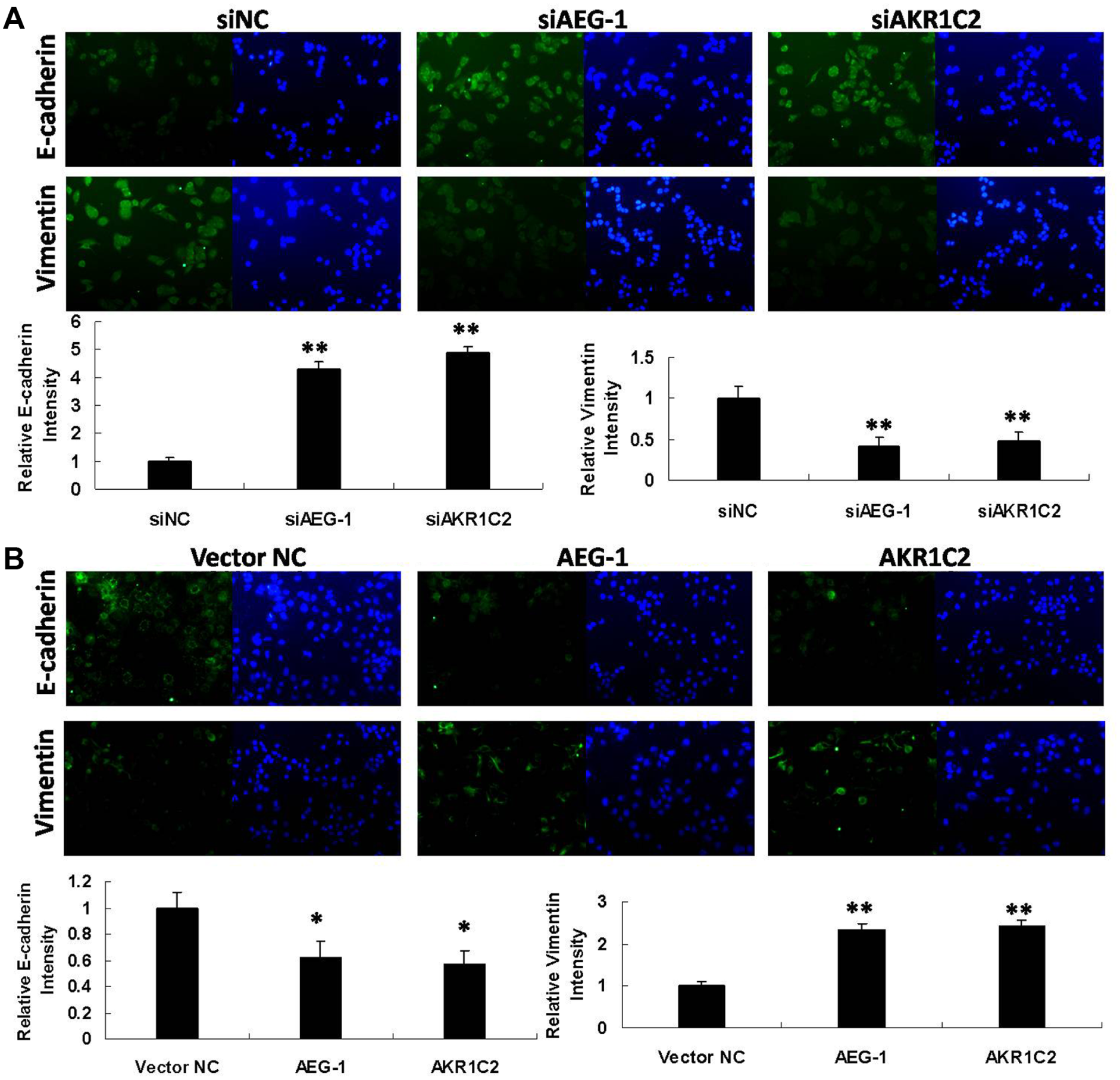
AEG-1 and AKR1C2 involved in epithelial to mesenchymal transition (EMT) in liver cancer cells. (
AEG-1 and AKR1C2 Promote Metastasis in Clinical Stages
Patient tissues from surgery were used to identify the gene expression patterns in liver cancer. These patients’ liver tumors had spread to other places; that is, metastasis had occurred. Immunohistochemistry was conducted on tumor or normal adjunct tissues. In total, we tested 20 pairs of samples from 20 different liver cancer patients. In all 20 cases, AEG-1 or AKR1C2 was expressed at high levels in tumor tissues compared with normal adjunct tissues ( Fig. 4 ). GPC-3, which is a well-known metastatic protein, was used as a positive control. These results proved that more progressive liver cancer tissue expressed a correlating higher level of AEG-1 or AKR1C2, in accordance with our functional screening and experimental validation results.
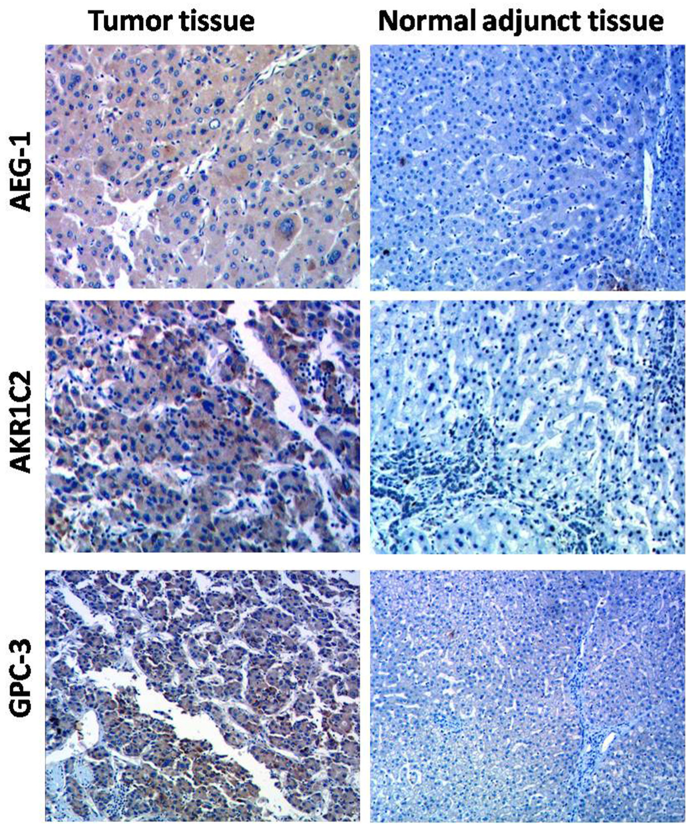
Immunohistochemistry detection of clinical human liver cancer samples. The antibodies are indicated. GPC-3 was used as a positive control.
Our results proved that AEG-1 and AKR1C2 functioned as key factors in metastasis progression and that inhibition of AEG-1 or AKR1C2 is of potential clinical use in the future. Human AEG-1 is a protein with 582 amino acids and a molecular mass of 64 kDa. It occurs downstream of Ha-Ras and c-Myc, and its overexpression leads to activation of downstream PI3k/Akt and NF-κB and the Wnt pathway. 10 Clinical tissue immunohistochemistry detection indicated that high expression levels of AEG-1 and AKR1C2 occurred in tumor tissues, compared with normal adjunct tissues. Chemical inhibitors might be of potential clinical use and could provide combination therapies with chemotherapy. In summary, we believe that these findings provide new insights into their physiological and therapeutic importance in liver cancer.
Footnotes
Acknowledgements
We thank Dr Hanshuo Zhang at the Peking University for critical reading of this manuscript and insightful discussions.
Declaration of Conflicting Interests
The authors declared no potential conflicts of interest with respect to the research, authorship, and/or publication of this article.
Funding
The authors disclosed receipt of the following financial support for the research, authorship, and/or publication of this article: This research is funded by National Natural Science Foundation of China (grant 81472328), Liver Disease AIDS Foundation of You An Hospital, Capital Medical University (BJYAH-2011-034), Army Youth Cultivation Project of Science and Technology (14QNP101), National Science and Technology Support Project (2012BAI15B08), and Key Project of National Communicable Disease (2012ZX10002015-002).
References
Supplementary Material
Please find the following supplemental material available below.
For Open Access articles published under a Creative Commons License, all supplemental material carries the same license as the article it is associated with.
For non-Open Access articles published, all supplemental material carries a non-exclusive license, and permission requests for re-use of supplemental material or any part of supplemental material shall be sent directly to the copyright owner as specified in the copyright notice associated with the article.