
Abstract
Drug discovery often requires the screening of compound libraries on tissue cultured cells. Some major targets in drug discovery belong to signal transduction pathways, and PerFix EXPOSE* allows easy flow cytometry phospho assays. We thus investigated the possibility to further simplify and automate this assay, to allow the direct screening of drugs targeting signaling pathways. We show here the sensitivity of this fully automated assay on human growth hormone (hGH)-driven JAK/STAT5-activated IM-9 cells, and we discuss the throughput of this system, which is compatible with medium-throughput drug screening. Because the kit works directly on whole blood samples, ex-vivo assays are also possible with this approach, which could allow for the screening of drugs under more physiological conditions.
Introduction
The JAK/STAT pathway is involved in many cellular signaling events, mainly in growth, immunity, and hematopoiesis. JAK2 acts in the signaling cascade initiated by hormones like growth hormone (GH) and erythropoietin; cytokines such as interleukins 3, 6, 12, 13, and 23; granulocyte colony-stimulating factor; and interferon-γ. Associated diseases include myeloproliferative neoplasm, polycythemia vera, essential thrombocythemia, primary myelofibrosis, Down’s syndrome–associated B-cell acute lymphoblastic leukemia, B-cell acute lymphocytic leukemia, primary mediastinal B-cell lymphoma, and Hodgkin’s lymphoma.1,2
Some major targets in drug discovery belong to signal transduction pathways, but given the relatively low number of JAK family members compared to the many receptors served, the development of specific regulators of cytokine activity at this stage is a tremendous challenge. To function in biochemical pathways and signaling cascades, participating protein complexes need to be transient. Fortunately, according to our previous results, the interaction between GH receptor and JAK2 appears to be much more transient than anticipated. 3 This feature is important and promising for the development of specific drugs that could block this interaction and be useful for specific pharmacological interventions.
The proof-of-target readout when screening these drugs is the phospho-epitope directly involved in the targeted pathway. Once cells are activated, detection of phosphorylation events can be done by Western blot techniques, or more recently by flow cytometry. The latter technique allows for the study of more complex systems (e.g., mixed cellular populations and multiple combined markers) and in more “physiological” situations (whole blood or bone marrow versus cell lines).
Unfortunately, phospho-epitope staining procedures for flow cytometry currently require numerous dedicated buffers and protocols, a total work time of 2 to 4 h, 0 °C or 37 °C incubations, and 4 to 8 washing steps, rendering the automation of such assays nearly impossible.
Some companies have developed solutions to automate these complex cell preparation procedures (e.g., the M5020 FlowCytoPrep Sample Prep System; MSP Corp., Shoreview, MN). Because samples are treated one by one in a cell chamber, this strongly limits the size of the experiments. There are, however, several high-throughput fully automated systems (e.g., BioCel; Agilent, Santa Clara, CA) that are well suited for plate-based assays but limited to adherent cells (enzyme-linked immunosorbent assay or cytology type of assays), because washing cells in suspension is still a challenge for these systems. By contrast, the new PerFix EXPOSE* kit has been developed to simplify and shorten the staining procedure to allow performance of the whole assay within 1 h, requiring only 2 wash steps and a brief 37 °C incubation. Here, we show that it is possible to further simplify this procedure by removing the wash steps and the 37 °C incubation steps, rendering the whole process fully automatable using a simple pipetting robot (as proposed by many vendors, such as Beckman Coulter, Pasadena, CA; Agilent; and Tecan, Männedorf, Switzerland).
In this report, we devised a simple, sensitive, and rapid protocol to screen drug libraries for their effect on the GH-driven JAK/STAT activation by flow cytometry. As a proof of concept, we optimized the p-STAT5 staining (one of the most challenging phospho-epitopes in flow cytometry) in a cell line (IM-9) stimulated with human GH (hGH). We evaluated the precision of the assay, validated it with two known inhibitors of the JAK/STAT pathway, and applied it to the screening of the Prestwick Library (1200 compounds; Prestwick Chemical, Illkirch, France). The throughput in this simple format is calculated at approximately 1 well/min in a 96-well format, and it could be easily increased as described in the Discussion. The protocol may further enable the monitoring of drug effects on multiple cytokine receptors at the same time, using the variety of dyes and detectors offered by all flow cytometers.
Materials and Methods
Materials
The PerFix EXPOSE (Phospho Epitopes Exposure Kit; Beckman Coulter) was used to fix and permeabilize cells. Samples were acquired on a FC500-MPL cytometer from Beckman Coulter (2 lasers: 5 colors, equipped with the Multi-Plate Loader that allows direct reading of 96-well plates), and data were analyzed with Kaluza* software (Beckman Coulter). Human lymphoblast cells IM-9 were derived from the blood of a patient with multiple myeloma and have been shown to be an Epstein–Barr virus (EBV)-transformed B lymphoblastoid cell line. Cells were maintained in RPMI 1640, 10% fetal calf serum, penicillin, streptomycin, containing 4.5 g/L glucose and 1 mM sodium pyruvate. hGH was purchased from R&D Systems (Minneapolis, MN) and aliquoted in phosphate buffered saline (PBS)–0.2% bovine serum albumin (BSA) at 1 µg/mL. Anti-Phospho STAT5-PE conjugate (Tyr694, clone C71E5) is from Beckman Coulter. The 1200 compounds tested in this study were obtained from Prestwick Chemical. These compounds were plated in 96-well plates, at 10 µM, in PBS containing 4% DMSO, 100 µL/well, and stored at −20°C until use. Inhibitory controls were purchased from Chemietek (Indianapolis, IN): BMS-911543 and INCB018424 (Ruxolitinib), resuspended in DMSO at 10 mM or 250 µM, then diluted in PBS at the indicated concentrations ( Fig. 4B ). Regular 96-well polystyrene U-shaped well plates were used (Greiner Bio-One, Courtaboeuf, France), allowing for a safe orbital shaker mixing.
Fluorescent-Based STAT5 Activation Assay According to the High-Throughput PerFix EXPOSE Procedure
Automation of the p-STAT5 staining assay was completed on a regular span-8 Biomek NX with fixed probes (Beckman Coulter) and is depicted in Figure 1 . An orbital shaker is included on the deck of the instrument to provide mixing when appropriate. Typically, 2×10⁵ IM-9 cells in 20 µL were distributed in the 96-well plate and incubated at room temperature (RT) with 5 µL of the compound (final concentration with cells, 2 µM and 0.8% DMSO) for 15 min. Then, 5 µL hGH in PBS–BSA (final hGH concentration, 50 ng/mL) was added, and cells were incubated for another 15 min. The reaction was terminated by the addition of 5 µL of fixative reagent (PerFix EXPOSE #1), followed by a 10 min incubation. Forty microliters fetal bovine serum (FBS) and 90 µL permeabilizing reagent (PerFix EXPOSE #2) were added to each well and incubated for an additional 10 min at RT. Ten microliters of the fixed cell suspension were transferred to a second plate containing 20 µL of staining reagent (PerFix EXPOSE #3) supplemented with anti-p-STAT5-PE (1 µg/mL final concentration). After 30 min, 200 µl of the wash reagent (PerFix EXPOSE #4) prepared from the original 20× concentrated solution was added to each well. Samples were then analyzed in a flow cytometer.

Automated PerFix EXPOSE procedure: experimental design as explained in the Methods section.
Results and Discussion
hGH-Activated IM-9 Cells
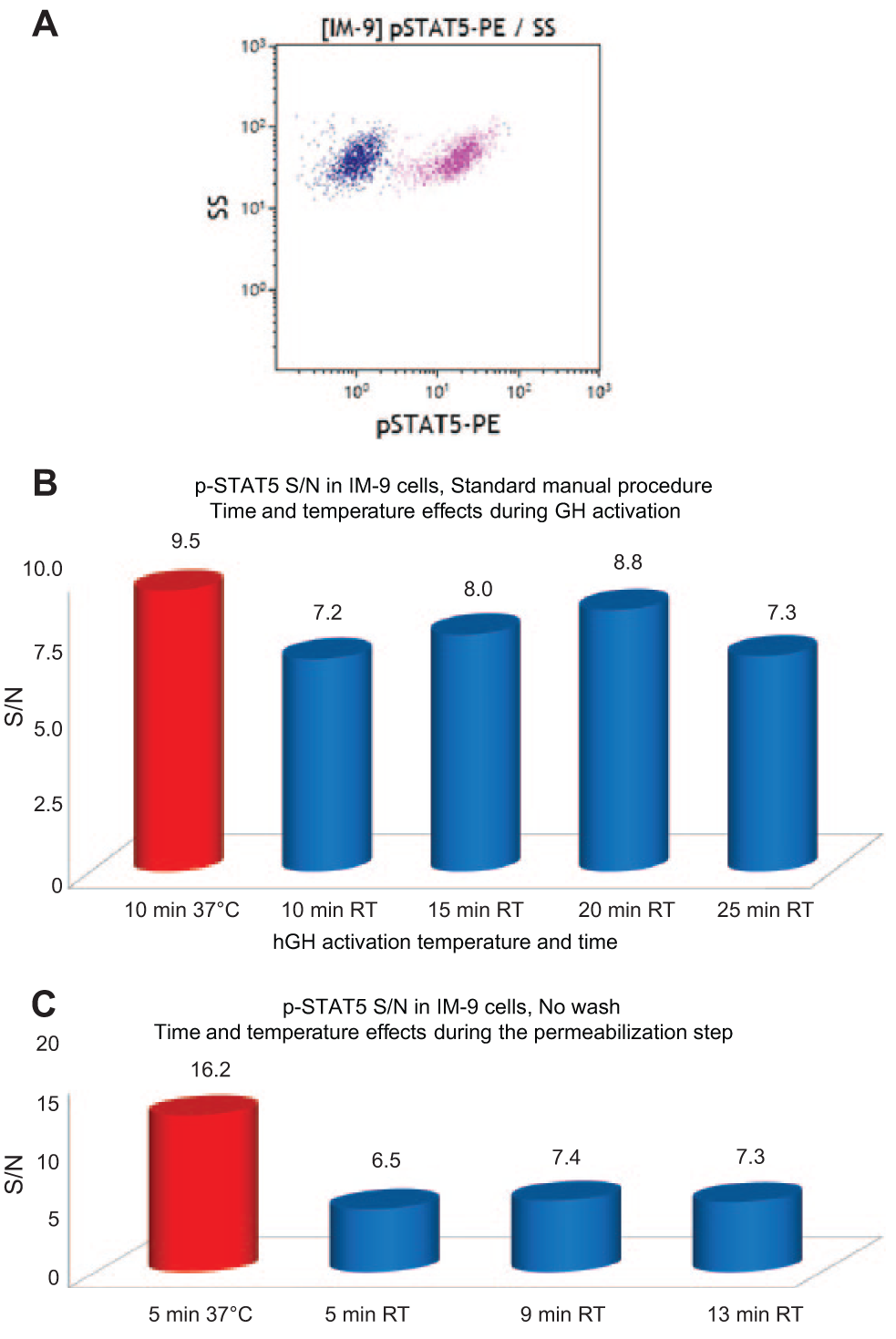
Human IM-9 lymphoblasts have detectable levels of endogenous GH receptor and have been used in many cytokine receptor studies.4,5 To demonstrate the feasibility of an assay by flow cytometry using IM-9 cells, we first measured the effect of hGH, using the standard PerFix EXPOSE procedure (as recommended in the “Instructions for Use”: in 5 mL tubes, with 2 wash steps and 2 incubation steps at 37 °C). Figure 2 illustrates the STAT5 activation on stimulation with hGH in IM-9 cells. The signal-to-noise ratio [S/N = mean fluorescent intensity (MFI) of positive (activated) cells / MFI of negative (nonactivated) cells] of 6 to 16 in these experiments enables unambiguous phospho-epitope detection by flow cytometry. The side scatter (SS) is a measure of the granularity or shape of the cells, and it is not affected by the activation. We conclude that IM-9 cells are suitable for hGH receptor–based high-throughput assay development.
(
hGH Activation at 25 °C versus 37 °C
To test the effect of the activation time and temperature, we incubated the IM-9 cells with hGH for 5 to 25 min at RT (controlled to 25 °C) or for 10 min at 37 °C ( Fig. 2B ). The cells were then processed following the standard procedure. The hGH signaling to STAT5 in these cells is only slightly decreased and delayed when the temperature is reduced to 25 °C. Incubating cells with hGH for 20 min at RT was optimal; this temperature and these conditions were applied in the following experiments.
Permeabilization–Lysis Step (R2 Reagent) at 25 °C versus 37 °C
To verify the feasibility of running the assay without permeabilization at 37 °C, we treated the IM-9 cells with hGH for 20 min at RT and permeabilized the cells with reagent R2 for 5 min at 37 °C or for 5, 9, and 13 min at RT (controlled to 25 °C) ( Fig. 2C ). The experiment depicted here was performed with a no-wash procedure in which other parameters (antibody titration, background blocking with serum, etc.) had already been optimized. This explains the improved S/N ratio observed for the positive control. We have previously determined that p-STAT5 staining is reduced using the PerFix EXPOSE assay if this step is run at lower temperatures (whereas other easily accessible phospho-epitopes are insensitive to this effect). We confirm here that the S/N ratio is decreased about twofold when the temperature is reduced to 25 °C. Nevertheless, the signal is still strong, and other factors can be further optimized. We thus applied the 9–10 min incubation time in the following experiments.
Final Optimization
The relative ratio of the R2 reagent (lysis and denaturation effect) versus FBS (protective effect) has been evaluated and optimized, along with the volume of this mix that is transferred into the staining reagent R3 (data not shown). The final format for this assay procedure is described in the Methods section and depicted in Figure 1 . It provides S/N ratios ranging from 15 to 25. We also verified that a final concentration of 0.8% DMSO had no significant impact on the activation ( Fig. 3 ). In further experiments (see Fig. 4 ), all activated points produced an MFI of 15–25, and the nonactivated controls produced an MFI of 1 to 1.5, thus confirming this S/N ratio of 15–25. This range of signal was observed among different experiments at different days, and it was therefore due to the sum of various variables: the sample variability (cell preparation and cell concentration), lab temperature, and flow cytometer. By contrast, the variability of the results within an experiment (same sample, a few plates prepared and analyzed) was very low.
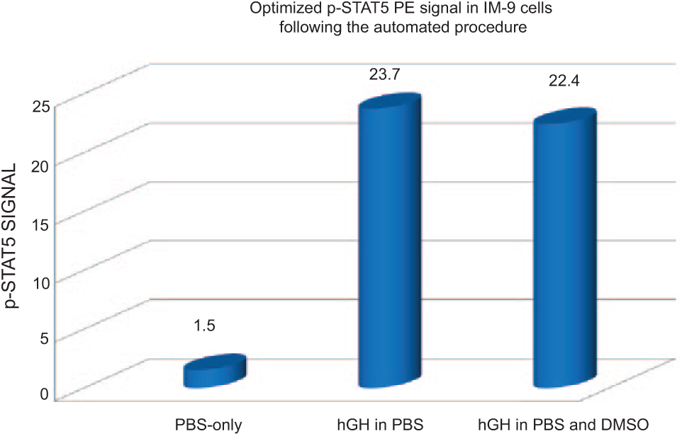
Optimization of pSTAT5-PE signal in an automated procedure. Representative results after the final optimization yielding a low background signal in nonactivated cells, with a high p-STAT5 signal in human growth hormone (hGH)-activated cells. DMSO in the phosphate buffered saline (PBS) buffer did not affect the activation (right column).
Validation. (
We estimated the variability of the assay by treating the same sample in multiple wells, both nonactivated (n = 24) and hGH activated (n = 24). The values of this experiment were, respectively: means and SD: 0.95 ± 0.14 and 24.2 ± 2.4, leading to a Z-factor of 0.67. This variability [roughly 10% coefficient of variation (CV)] was likely due to pipetting imprecisions, and we optimized these parameters that are common to all automated actions (pipetting speed, moving speed, position in the well, etc.). This resulted in a further reduction of the variability: means and SD: 1.12 ± 0.04 and 19.53 ± 0.98 (roughly 5% CV), leading to a Z-factor of 0.83. From that point, the pipetting procedure remained unchanged, and all further experiments demonstrated similar performances.
It is important to note here that such automation procedures require compromises among the pipetting precision of the robot, the limited volume in a well, the reagent volume ratios, and the quality of the staining. This explains why we processed the sample in one well, and then continued the procedure on a fraction of this in a daughter well ( Fig. 1 ). When doing so, it is necessary to start with a concentrated cell suspension to analyze enough cells at the end within a reasonable timeframe.
Validation
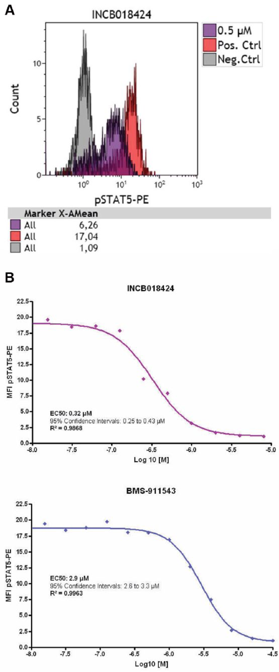
We measured the inhibitory effect of two commercial JAK inhibitors (INCB018424 ruxolitinib and BMS-911543).6,7 A representative flow cytometry histogram is presented in Figure 4A , and a full titration experiment is presented in Figure 4B . The titration was reproduced in 3 independent experiments, including 1 with a manual transfer of the plate to a 37 °C water bath, ensuring that both inhibition and activation occurred at 37 °C. This experiment provided similar results, indicating that the temperature change to 25 °C does not significantly affect the inhibitory effect of these compounds. Overall, we measured an IC50 for INCB018424 (ruxolitinib) of 0.5 +/− 0.29 µM, and an IC50 for BMS-911543 of 3.9 +/− 0.95 µM. Whereas we did not find a published value for BMS-911543, the IC50 for INCB018424 (ruxolitinib) is described to range from 0.1 to 0.5 µM depending on the cell type and assay, 6 which is in very good concordance with our measurements.
Prestwick Library Screening
The Prestwick compound library contains 1280 small molecules and 100% approved drugs (approved by the US Food and Drug Administration, European Medicines Agency, and other agencies). It presents the greatest possible degree of a drug-likeliness. The active compounds were selected for their high chemical and pharmacological diversity as well as for their known bioavailability and safety in humans. The library was designed to reduce the risk of generating low-quality hits, reduce the cost of the initial screening, and accelerate lead discovery. Two plates of the library have been tested as described, and no hit could be identified among the 192 tested molecules (final concentration, 2 µM). We then pooled 4 wells together from the remaining plates (thus, the final concentration decreased to 0.5 µM) to increase the throughput. Based on the precision of the assay (CV on positive MFI of 4–6%), we anticipated being able to detect an inhibitory compound at a concentration producing approximately 20% inhibition, indicating that a compound with an IC50 equal to or higher than 1 µM should be detected.
We found no hit among the other 1000 molecules tested this way. This was very clear because the method produced no false-positive points [we retested only a few points that were at the low end of the distribution (mean, −2 to −3 SD) to make sure we did not miss any hits, and they were all confirmed negative].
Thus, although the library contains kinase inhibitors such as gefitinib and erlotibin (epidermal growth factor receptor kinase inhibitors), 9 lapatinib ditosylate (an anti-Her2 tyrosine kinase inhibitor), 10 parthenolide (an IκB kinase inhibitor), 10 imatinib (a BCR-ABL kinase inhibitor), 11 and vatalavib (a tyrosine kinase inhibitor), 12 none of these interacted, under the current conditions, with the growth hormone receptor (GHR)-driven signaling pathway. This is not really a surprise, because none of them have been previously shown to interact directly with this pathway, and also because this pathway is rather straightforward (GHR → JAK → STAT), leaving room only for a GHR or JAK inhibitor.
Throughput Considerations
Sample preparation was executed on the deck of the system without user intervention. No other active device was required because the assay was performed at RT without any wash step. A 96-well plate was processed in approximately 85 min. Because the 8-needle pipetting pod was on rest most of the time (incubation times), throughput could easily be further enhanced by interleaving multiple plates (2 or 3), with no risk of changing the performance of the assay. Using a dual pod (span-8 and multichannel) system would further enhance the throughput because the multichannel head would facilitate the transfer of compounds and cells, thus shortening the overall assay time and allowing interleaving more plates (but this has to be validated). Pooling of compounds can also be used to increase throughput when the expected hits are rare. An important consideration when performing medium- to high-throughput cytometry will then be the selection of the cytometer due to data acquisition times. Systems have been developed to increase the speed of acquisition (e.g., the Intellicyt platforms); thus, cytometry now becomes a viable platform for medium-throughput applications.
Conclusions
Flow cytometry is a powerful technique for analyzing cellular events in all cell suspensions. Thanks to the simultaneous use of from 5 to more than 10 detectors, it allows the dissection of complex phenomena in heterogeneous populations of cells, requiring only hundreds of cells per analysis. Phosphorylation events are key players in the physiology of cells and diseases, but have proven to be among the most difficult to detect by flow cytometry. Here, we demonstrate that PerFix EXPOSE allows the full automation of flow cytometry phospho assays, and we proved the concept that this method can apply to libraries of compounds. The limiting factors are essentially the availability of well-concentrated cell suspensions and good markers. Because the kit works directly on whole blood samples, ex vivo assays are also possible with this approach, which could allow for the screening of drugs under more physiological conditions. Ultimately, one can envision a personalized screening approach in which abnormal cells would be treated ex vivo with an array of available drugs to find the most appropriate molecule and its optimal dose. Automation of the phospho assays was easily accomplished on a standard liquid handler with an orbital shaker. RT processing eliminated the need for costly incubators. Higher throughput can be achieved by off-deck storage devices, interleaving plates, and pooling of compounds.
Footnotes
Acknowledgements
*PerFix-EXPOSE, Kaluza, and Biomek are for research use only. They are not for use in diagnostic procedures. Kaluza is a trademark of Beckman Coulter, Inc. Beckman Coulter, the stylized logo, is a registered trademark of Beckman Coulter, Inc. and is registered in the USPTO.
Declaration of Conflicting Interests
The authors declared no potential conflicts of interest with respect to the research, authorship, and/or publication of this article.
Funding
The authors disclosed receipt of the following financial support for the research, authorship, and/or publication of this article: This research was supported in part by the Dutch Technology Foundation STW, which is the applied science division of NWO, and the Technology Program of the Ministry of Economic Affairs, grant 11155: Targeting the Jak2–growth hormone receptor interaction for treatment of cancer.