
Abstract
A novel, simple, and rapid method, named cell membrane affinity extraction (CMAE)–offline liquid chromatography time-of-flight mass spectrometry (LC-TOF-MS) was developed for screening and identifying antimicrobial peptides from Jatropha curcas meal protein isolate hydrolysates (JCMPIH) obtained by proteolytic enzyme (pepsin, trypsin, protamex, neutrase, flavourzyme, papain, alcalase, and acid protease) hydrolysis. A cationic antimicrobial peptide (CAILTHKR, JCpep8) was successfully isolated and identified by this method. Antimicrobial assay indicated that JCpep8 was active against the tested microorganisms (Escherichia coli ATCC 25922, Shigella dysenteriae ATCC 51302, Pseudomonas aeruginosa ATCC 27553, Staphylococcus aureus ATCC 25923, Bacillus subtilis ATCC 23631, Streptococcus pneumoniae ATCC 49619) with minimal inhibitory concentration values ranging from 29 to 68 µg/mL. JCpep8 induced significant morphological alterations of the tested microbe surfaces, as shown by transmission electron microscopy, indicating strong membrane disruption. The results showed that CMAE-offline LC-TOF-MS could be a promising method for discovering high-throughput screening antimicrobial peptides from JCMPIH.
Keywords
Introduction
Increasing resistance of all microbes is a worldwide problem. With the problem growing, the development of alternative therapies needs to be explored. 1 Antimicrobial peptides (AMPs) with broad-spectrum activity, which can physically destroy the bacterial membrane through several targets to kill the microbes, may be considered a promising candidate to overcome the problem. 2 Furthermore, protein hydrolysates are recognized as the best, cheap, accessible, and safe source of AMPs.3,4 However, the existing chromatographic techniques, which take too much time for discovering and high-throughput screening (HTS) of AMPs, hamper the industrial development of AMPs.5,6
Biochromatography7,8 as an emerging high-tech tool, which uses the enzymes, receptors, antibodies, transport proteins, and other biological macromolecules as the stationary phase, has been used for screening active ingredients in traditional Chinese medicine and exploring new mechanisms of drug action. In the field of biochromatography, reports about cell membrane chromatography (CMC), which can screen the component binding to the cell membrane and its receptor according to its chromatographic retention characteristic, are especially active.9–11 However, the column pressure, column temperature, mobile phase solvents, pH, flow rate, and other conditions, which often conflict with the biological activity of the cell membrane, have limited the application of CMC. 12
Based on the membrane-binding activity of AMPs, 13 CMC can be used to screen AMPs. However, until now, there have been few reports about HTS of AMPs by CMC.
To solve the above problems, in this article, an improved CMC (Escherichia coli cell membrane affinity extraction [CMAE]–offline liquid chromatography time-of-flight mass spectrometry [LC-TOF-MS]) was developed for screening and identifying AMPs from Jatropha curcas meal protein isolate hydrolysates (JCMPIH). Combined with an antimicrobial assay, our CMC bioaffinity chromatography method may provide an efficient HTS scheme to identify AMPs from complex samples.
Materials and Methods
Materials
The powder of J. curcas meal protein isolate (JCMPI) was obtained from the School of Food Science and Technology, Jiangnan University. Macroporous spherical silica (3–7 µm) was purchased from Sepax Technologies, Inc. (Suzhou, China). All the following reference strains were provided by Wuxi disease prevention and control center (Wuxi, China): gram-negative bacteria (Escherichia coli ATCC 25922, Shigella dysenteriae 51302, Pseudomonas aeruginosa ATCC 27553), gram-positive bacteria (Staphylococcus aureus 25923, Bacillus subtilis 23631, Streptococcus pneumoniae ATCC 49619), Penicillium expansum ATCC 1117, Aspergillus niger ATCC 16404, and Saccharomyces cerevisiae ATCC 40075. The strains were maintained as frozen stocks in appropriate broth plus 20% (v/v) glycerol. All the proteolytic enzymes (pepsin, trypsin, protamex, neutrase, flavourzyme, papain, alcalase, and acid protease) were obtained from Novozymes (Wuxi, China). Tris was purchased from Sigma-Aldrich (Shanghai, China). All other chemical solvents used were of analytical grade and purchased from Sinopharm chemical reagent factory (Shanghai, China).
Preparation of E. coli Cell Membrane (ECM) and ECM Stationary Phase
The experiment was performed according to Yang et al. 14 with some modifications. The mid-log phase of E. coli was centrifuged at 5000 g for 5 min, and then the precipitate was washed by the Tris-HCl buffer (50 mmol/L, pH 7.2) and centrifuged again. To completely remove culture medium and metabolites, the washing step was repeated 10 times. The collected E. coli cells were suspended in the buffer, then ultrasonicated using an ultrasonic cell disruptor (JY98IIID; Xinzhi Biotech Co., Ningbo, China) under the condition of ultrasonic power (500 W; ultrasonic treatment for 12 s with a 5-s interval) for 45 min in an ice bath at 4 °C and then centrifuged at 12 000 g for 20 min at 4 °C. The resulting precipitate was the E. coli cell membrane (ECM).
Activated silica (0.5 g) and the suspension solution of ECM were mixed slowly at 4 °C. The adsorption of the ECM on the activated silica surface was shaken for 5 h until equilibrium was reached. Then the filtrate in the reaction mixtures was removed by filtering (3 µm; Millipore, Billerica, MA), and the obtained precipitate (adsorbent) was resuspended in Tris-HCl buffer, sonicated for 30 min in an ice bath at 4 °C, and filtered (3 µm; Millipore) again. The precipitate was collected and named the ECM stationary phase (ECMSP).
Adsorption Isotherm of ECM
The experiment was performed according to Bilgili et al. 15 with some modifications. Activated silica (100 mg) was added to a series of reaction tubes containing a series of concentrations of ECM suspension in Tris-HCl buffer (50 mmol/L, pH 7.2). The reaction tubes were shaken in an ice bath at 4 °C for 5 h until the adsorption equilibrium was reached. Then the filtrate in the reaction mixtures was removed by filtering (3 µm; Millipore), and the obtained precipitate (adsorbent) was resuspended in Tris-HCl buffer, sonicated for 30 min in an ice bath at 4 °C, and filtered (3 µm; Millipore) again. The filtrate was collected again. The adsorbed amount of the ECM protein immobilized on the activated silica surface was determined by subtracting the amount of ECM protein in the filtrate from the original amount of ECM protein. The protein content of filtrate was determined by the method described by Bradford. 16 In addition, the maximum adsorption capacity of ECM protein on the silica surface and the Langmuir constant was calculated by the following equation:
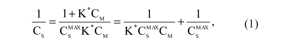
where Cs and Cm are, respectively, the concentration of ECM protein on the silica surface and in the solution; Csmax is the maximum adsorption capacity of ECM protein on the silica surface; and K*is the Langmuir constant.
JCMPIH Preparation
The experiment was performed according to Zhang et al. 17 with some modifications. The protein content of JCMPI, determined by the Kjeldahl method, was 97.26%. JCMPI was reconstituted in distilled water to obtain a solution containing 10% (w/v) of protein. The solution was digested with proteolytic enzymes (pepsin, trypsin, protamex, neutrase, flavourzyme, papain, alcalase, and acid protease) to different degrees of hydrolysis (DH, 7%, 9%, 11%, 13%, 15%, and 17%). The hydrolysis conditions of enzymes are shown in Table 1 . The hydrolysis was stopped by heating at 95 °C for 15 min. Hydrolysates were clarified by centrifugation (4500 g, 15 min), and then the supernatant was lyophilized and designated as JCMPIH. The values of DH were calculated using the following formula:
Optimum Condition of Proteolytic Enzymes a
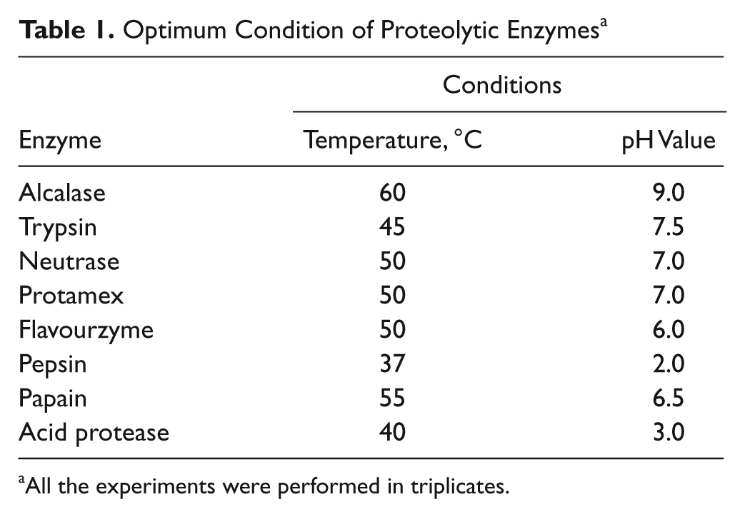
All the experiments were performed in triplicates.

where AN1 and AN2 are, respectively, the amino nitrogen content of the protein substrate before hydrolysis (mg/g protein) and after hydrolysis (mg/g protein), and Npb is the nitrogen content of the peptide bonds in the protein substrate (mg/g protein).
Measurement of Antibacterial Activity of JCMPIH
The experiment was performed by the liquid growth inhibition assay according to Bulet et al., 18 with some modifications. The mid-log phase of E. coli was adjusted to 105 colony-firming units (CFU)/mL. Aliquots (1 mL) of different JCMPIH (2 mg/mL) and 1 mL of the E. coli cell suspension were added to the reaction tubes and incubated for 12 h at 37 °C, and aliquots of phosphate-buffered saline (PBS; 50 mM, pH 7.4) tested under the same condition were used as a control. The optical density (OD600) was read on a Unico 2100 spectrophotometer (Unico, Shanghai, China). The antibacterial activity unit (U) was estimated by the following formulas:

where A and A0 are, respectively, the absorbance in the sample and in the control.
ECM Affinity Extraction/High-Performance Liquid Chromatography Fingerprint
ECMSP and the JCMPIH with the highest antibacterial activity were added to the reaction tubes and shaken to combine at 30 °C for 1 h. The filtrate in the reaction mixtures was collected by filter (3 µm; Millipore), and the obtained precipitate was washed with 50 mM PBS buffer until there was no residual JCMPIH detected by reverse-phase high-performance liquid chromatography (RP-HPLC) and filtered. Then all the filtrate was collected and named the ECM affinity extraction effluent (ECMAEE).
The fingerprints of the JCMPIH with the highest antibacterial activity and ECMAEE were obtained by RP-HPLC (Amethyst C18: 250 × 4.6 mm, 5 µm; HPLC system, Waters, Milford, MA). The two solvent reservoirs contained the following eluents: (A) 0.05% (v/v) trifluoroactic acid (TFA) and (B) 100% acetonitrile. The elution program consisted of a gradient system (5%−80% B in 40 min) with a flow rate of 0.5 mL/min. The specific binding peak obtained by comparing the two fingerprints was collected and analyzed by LC-TOF-MS.
Peptide Sequence Analysis
The experiment was performed according to Yergey et al. 19 A mass spectrometric experiment was performed with LC-TOF-MS (4700 Proteomics Analyzer; Applied Biosystems, Foster City, CA). All spectra were measured under the following conditions:
MS: reflector positive, CID (OFF), mass range (400–3200 Da), focus Mass (1200 Da), fixed laser intensity (6000), digitizer: bin size (1.0 ns)
MS/MS: 1 kV positive, CID (ON), precursor mass windows (relative 80 resolution [FWHM]), fixed laser intensity (7000), digitizer: bin size (0.5 ns)
α-Cyano-4-hydroxycinnamic acid (Aldrich, Steinheim, Germany) was used as a matrix. The concentration of peptide solution was 2 mg/mL. In total, 0.5 mL peptide solution plus 0.5 mL matrix was deposited on the sample slide and left to dry at room temperature. The resulting spectra were analyzed and compared.
Minimal Inhibitory Concentration
The minimal inhibitory concentration (MIC) was determined with a liquid growth antimicrobial assay. 20 The mid-log phase of culture was adjusted to 105 CFU/mL. Purified antimicrobial peptide stock solution was prepared with PBS (50 mM, pH 7.4). The purified antimicrobial solution was sterilized by filtration (0.22 µm; Millipore) and diluted twofold serially in sterile PBS (10 mM, pH 7.4). Purified antimicrobial peptide solution (50 µL) was incubated in sterilized 96-well plates with 100 µL media and 100 µL test microorganisms disposed as described above. Then, 50 µL PBS (50 mM, pH 7.4) tested under the same condition was used as control. MIC was considered the lowest purified antimicrobial peptide that showed no increase in the optical density (OD600) read on the microplate reader (Multiskan MK3; Thermo Scientific, Waltham, MA) after a 12-h incubation.
Transmission Electron Microscopy
The effect of a purified antimicrobial peptide on the ultrastructural morphology of the S. aureus ATCC 25923 cell was assessed by using transmission electron microscopy (TEM; H-7000, Hitachi, Tokyo, Japan). 21 After being treated with purified antimicrobial peptide at the MIC for 24 h at 37 °C, S. aureus ATCC 25923 cells were immediately washed three times with PBS and fixed with 2.5% (v/v) glutaraldehyde.
Hemolysis Assay
Hemolytic assay was performed using rabbit red blood cells (RRBc) in liquid medium as previously reported. 22 Briefly, the erythrocytes were washed six times by using PBS (2 mM, pH 7.4). Serial dilutions of the peptide were used, and after being incubated for 1 h at 37 °C, the mixture of the antimicrobial peptide/RRBc was centrifuged at 3000 g for 10 min and the absorbance in the supernatant measured at 595 nm. One hundred percent hemolysis was achieved by adding Triton X-100 to the control cells.
Statistical Analysis
All the experiments were performed in triplicates. The average value and standard deviation were calculated. The data were analyzed using SPSS 13.0 statistical software (SPSS, Inc., an IBM Company, Chicago, IL).
Results
Adsorption Isotherm of ECM
The maximum adsorption capacity of ECM on the surface of silica is the most important parameter, which can affect the chromatographic behavior of solutes, so it should be determined first. 23 The adsorption isotherm of ECM was determined according to the Langmuir equation. Based on the linear relationship of 1/Cs versus 1/Cm, K* and Csmax were calculated from the slope and intercept according to equation (1). The adsorption isotherm of ECM is shown in Figure 1 . The values of K* and Csmax were 56.31 mL/mg and 2.96 mg/g, respectively. It can be seen from the adsorption isotherm ( Fig. 1A ) of ECM that when the initial membrane protein concentration in the solution was greater than 0.056 mg/mL, the adsorption capacity was greater than 0.9 Csmax. At this moment, the adsorption isotherm changed smoothly, indicating that the adsorption capacity was approximately saturation, which showed that the interaction between ECM and silica was significant. Therefore, in the preparation of ECMSP, according to the adsorption isotherm of ECM, the initial concentration of the membrane suspension (Cm) was used to control the content of the ECM on ECMSP, which was helpful for guaranteeing the good reproducibility and stability of ECMSP at the same time. Similar results were reported by He et al. 23
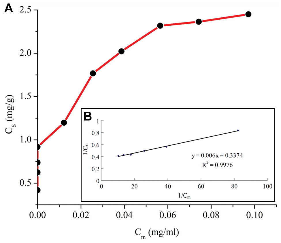
Adsorption characteristics of on activated silica. Adsorption isotherm curve (
Surface Characteristics of ECMSP
In an aqueous solution, the groups of Si-OH on the surface of silica display strong and irreversible adsorption of biological macromolecules (protein and lipid molecules in the cell membrane). For this reason, the cell membrane can be firmly adsorbed on the surface of silica. 24
To confirm this view, the surface of silica was investigated with the electron microscopy technique. As shown in Figure 2 , the scanning electron micrograph of silica carrier ( Fig. 2A ) and ECMSP ( Fig. 2B ) was magnified 5000 times. As can be seen from this picture, the pure silica carrier was significantly different from ECMSP, which showed that the ECM has been irreversibly adsorbed on the surface of the silica carrier and the surface of silica is completely covered by the ECM as a single entity to form the ECMSP. A similar phenomenon has also been found in other studies.9–11
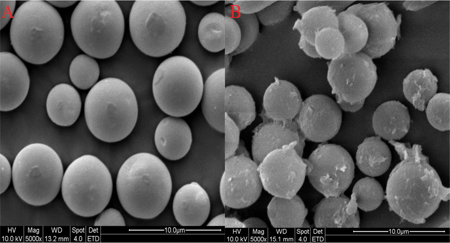
Scanning electron micrograph of (
Antibacterial Activity of JCMPIH
Many studies have reported that AMPs can be liberated by hydrolysis with proteolytic enzymes from the natural proteins.3,4 Therefore, we attempted to isolate AMPs by hydrolysis with proteolytic enzymes (pepsin, trypsin, protamex, neutrase, flavourzyme, papain, alcalase, and acid protease) from JCMPIH. As shown in Figure 3 , the antibacterial fragment was screened. The highest antibacterial activity, among the JCMPIH resulting from proteolytic enzymes, was observed in the Protames hydrolysates with hydrolysis of 13% (PSH13), which inhibited up to 67.76% of bacterial growth. The other JCMPIH actually increased bacterial growth. Similar results were reported by Mine et al. 25 Therefore, PSH13 was selected for further study.
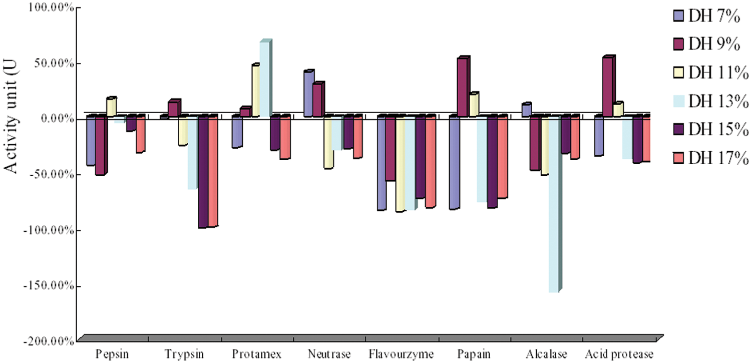
Antibacterial activity of Jatropha curcas meal protein isolate hydrolysates (JCMPIH). DH, degree of hydrolysis.
ECM Affinity Extraction/HPLC Fingerprint–Peptide Sequence Analysis
The stationary phase is prepared by fixing cell membrane at a carrier (silica) in the traditional cell membrane chromatography, which is according to the method of liquid chromatography to explore the interactions between drugs and the cell membrane or membrane receptors.9–11,26,27 Because of the protection of cell membrane activity in this method, the chromatographic conditions (the column pressure, column temperature, mobile phase flow rate, etc.) are often restricted.
To improve this method, the CMC was divided into two steps in our study. First, ECMSP and samples were added to the reaction tubes and shaken to combine under physiological conditions. Second, the effluent was collected for analysis by LC-TOF-MS, and compared with the standard HPLC fingerprint, which can easily and rapidly screen and identify specific binding components. This membrane solid-phase extraction technology, which can take the biological activity of cell membrane into account, can overcome the shortcomings of CMC derived from chromatographic conditions.
The reproducibility of the ECM affinity extraction (ECMAE)/HPLC fingerprint was also tested. The results showed that the relative standard deviation (RSD; %) of retention time (RT) of each peak was no more than 3% (n = 10). The precision between each experiment (n = 10) involved meeting the assay requirements. Validation was feasibility of the ECMAE/HPLC fingerprint method.
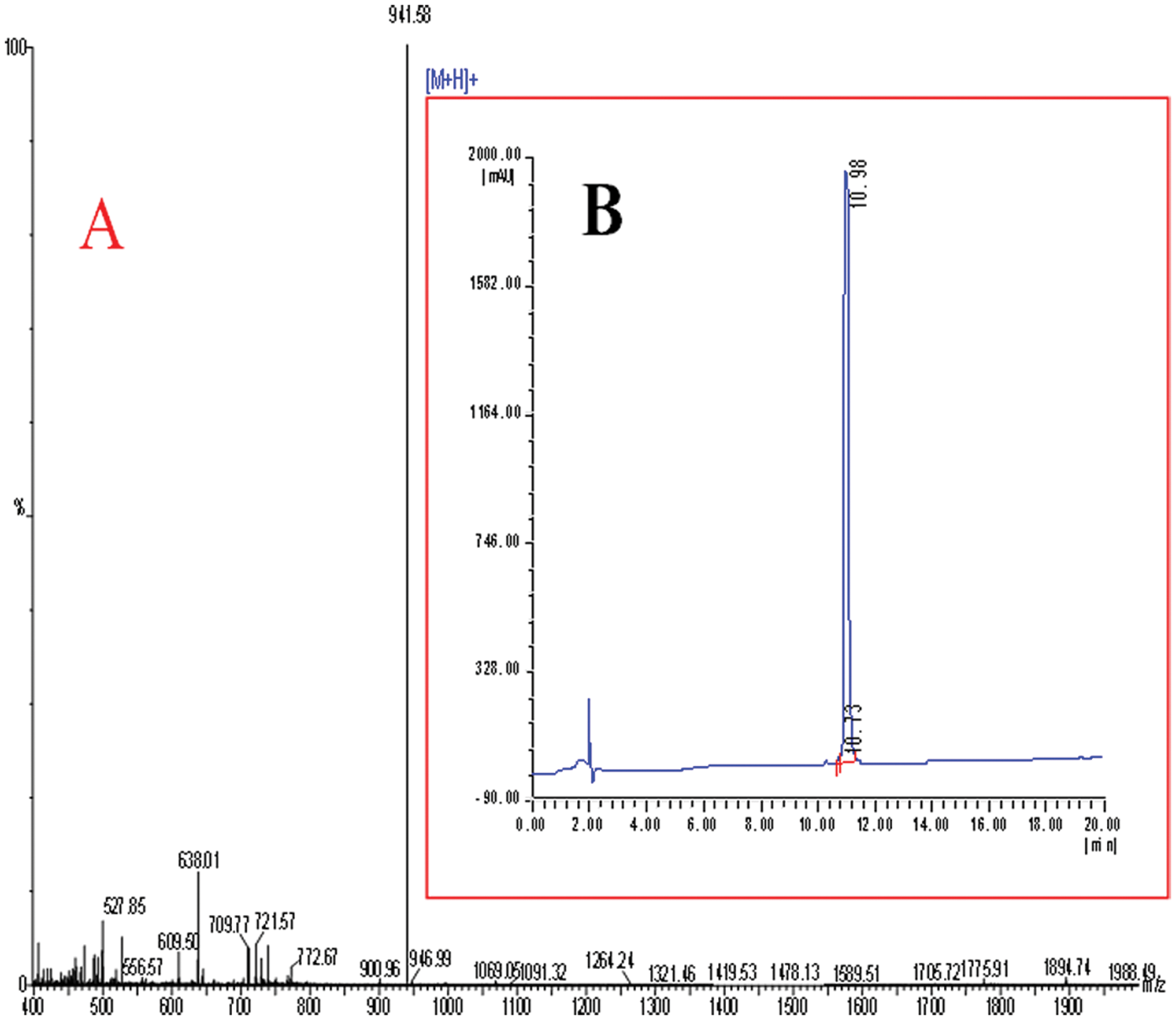
The HPLC fingerprints of PSH13 and ECMAEE are shown in Figure 4 , which shows that three specific binding peaks (peaks 1, 2, and 3) were manually collected and prepared as the same concentration. Then the antimicrobial activities were tested. We found that peak 2 had the highest antibacterial activity (95%), estimated by equation (3), compared with the other two peaks. The mass spectrometry of peak 2 is shown in Figure 5A . By MS/MS analysis, peak 2 was composed of eight amino acids with amino acid sequences of CAILTHKR (Cys-Ala-Ile-Leu-Thr-His- Lys-Arg). There are no matching AMPs between the National Center for Biotechnology Information (NCBI) database and the Antimicrobial Peptide Database (APD), suggesting that peak 2 is a novel peptide, designated as “JCpep8.”
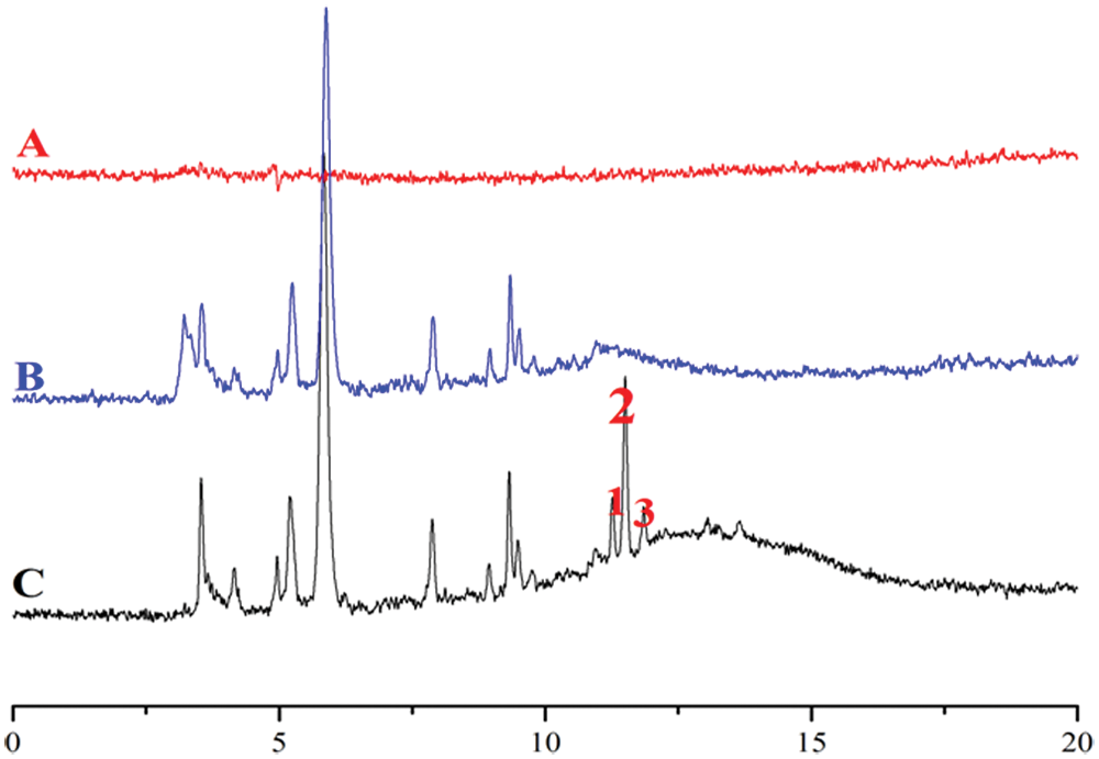
High-performance liquid chromatography (HPLC) fingerprints. (
(
JCpep8 is a cationic antimicrobial peptide with a total net charge of +3 and total hydrophobic ratio of 50%. As is known, cationic antimicrobial peptides are likely to first be attracted to the net negative charges in bacterial surfaces, 2 and the hydrophobicity is helpful for AMPs coating on the surface of the bacterial membrane with the hydrophobic face toward the lipid components and the polar residues binding to the phospholipid head groups. 13 Therefore, JCpep8 is consistent with the general characteristics of AMPs. For the subsequent experiments, JCpep8 was manually collected and freeze dried, the purity of which was 98.8% ( Fig. 5B ).
MIC of JCpep8
As shown in Table 2 , JCpep8 was active against both gram-negative (E. coli ATCC 25922, P. aeruginosa ATCC 27553, and S. dysenteria ATCC 51302) and gram-positive bacteria (S. aureus ATCC 25923, B. subtilis ATCC 23631, and S. pneumoniae ATCC 49619) with MIC values ranging from 29 to 68 µg/mL except fungi (P. expansum ATCC 1117 and A. niger ATCC 16404) and yeast (S. cerevisiae ATCC 40075). The JCpep8 had similar MIC against E. coli in comparison with antimicrobial peptides reported by Jang et al. 28 In addition, E. coli ATCC 25922 was the most sensitive to JCpep8. The results suggest that JCpep8 could control some bacteria efficiently.
Minimal Inhibitory Concentrations (MICs) of JCpep8
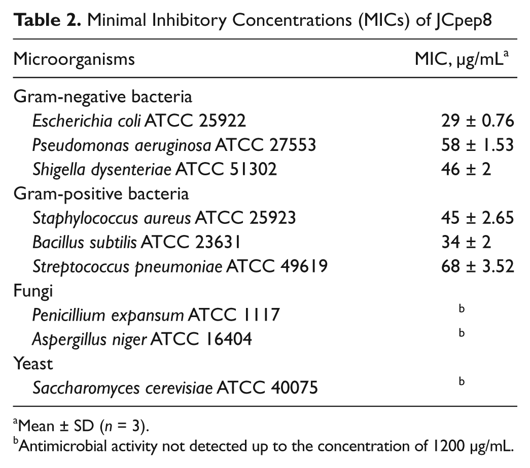
Mean ± SD (n = 3).
Antimicrobial activity not detected up to the concentration of 1200 µg/mL.
To elucidate the nature of the killing mechanisms of JCpep8, S. aureus ATCC 25923 treated with JCpep8 for 24 h were analyzed by TEM. As shown in Figure 6 , compared with the control, the treated cell had clear morphological changes. When treated up to 24 h, a ghost-like appearance and the lysed cell were observed.
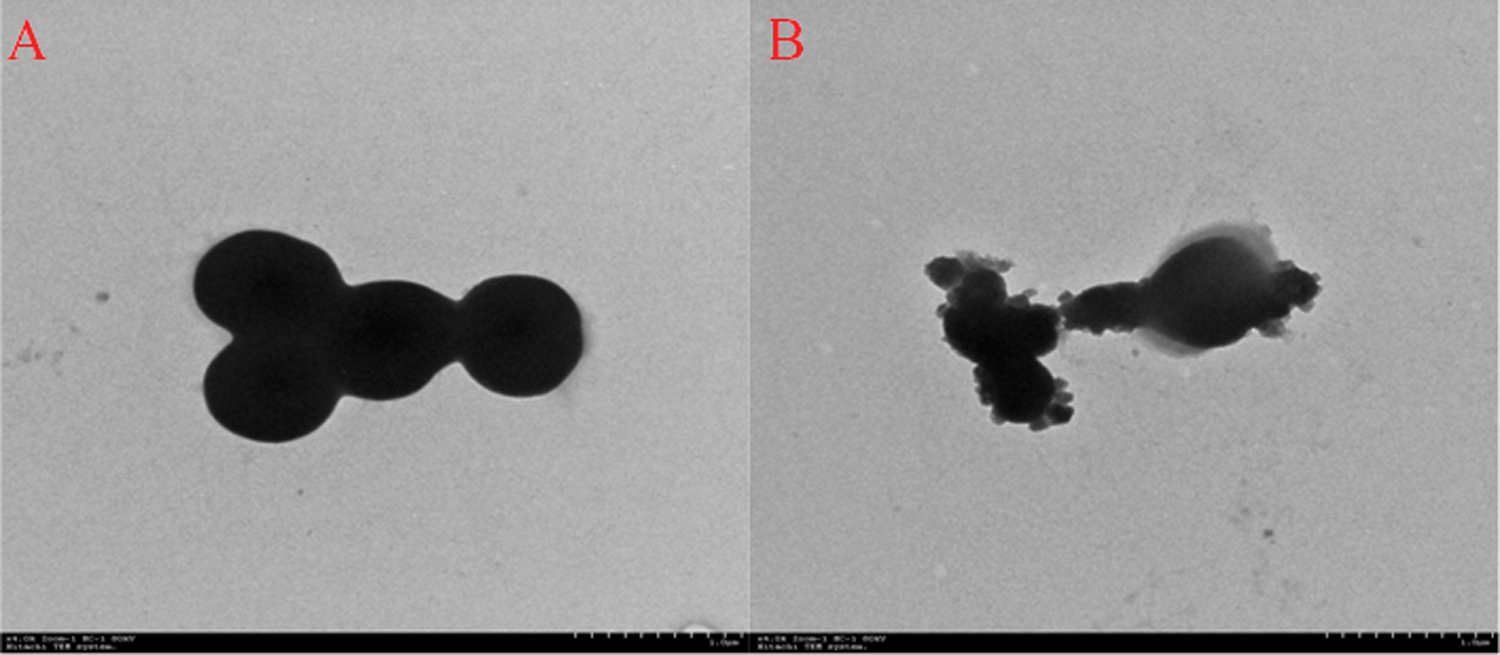
Morphological changes of Staphylococcus aureus ATCC 25923 upon incubation with JCpep8. (
Hemolytic Activity
Some AMPs exhibit hemolytic activities. 22 To assess the cytotoxicity of JCpep8 against mammalian cells, we measured the percentage of hemolysis against RRBc at various concentrations (200–1200 µg/mL) in this study. Based on a previous report, 22 a hemolytic rate more than 5% showed the emergence of hemolysis. After 1 h of incubation, there was no hemolysis at peptide concentrations lower than 600 µg/mL. However, a high level of hemolytic activity was observed at peptide concentrations higher than 800 µg/mL.
Discussion
CMC, a new bioaffinity chromatography, can be used to effectively study the interaction between receptor and drug by the chromatographic characteristics of drug on the stationary phase prepared by immobilizing cell membrane onto a silica carrier. It has been demonstrated that CMC has been successfully used to screen the active components in traditional Chinese medicine. Although this general chromatography method was published earlier, it remains largely underevaluated in the available literature. In addition, to protecting cell membrane activity of the biomembrane chromatography column, other studies restricted the development of CMC technology by the chromatographic conditions (the column pressure, column temperature, mobile phase flow rate, etc.).9–11,26,27
Therefore, in our study, the improved CMC (ECM affinity extraction–offline LC-TOF-MS) was used to screen AMPs in JCMPIH. In brief, first, the HPLC fingerprint of JCMPIH was developed. Second, JCMPIH was added into CMC, and interaction was performed at physiological conditions. Then, no combined components were removed by filtering, and the filtrate was collected. Third, comparing the HPLC profiles of filtrate and JCMPIH, we easily found the components that combined with E. coli cell membrane (potential antimicrobial agents). Finally, we confirmed their antimicrobial activity by using a liquid growth antimicrobial assay. This improved CMC method, which can avoid the limitations of the chromatographic conditions and eliminate the interference of many non-antimicrobial agents, had the characteristic of high efficiency and high selectivity.
The data from our study confirm earlier observations and demonstrate that the CMC approach can be employed to screen AMPs. However, it must be emphasized that there is significant potential for interference from sources of AMPs. Also, it should be clearly noted that AMPs from different sources produced different antibacterial mechanisms because of their inconsistent size, charge, and hydrophobicity. Despite these important concerns, we suspect that this method can be used to explore the application of biomembrane chromatography in screening of AMPs from different sources. More work about fully identifying peptides from some utility is in progress in our laboratory.
Footnotes
Acknowledgements
The authors thank Liya Niu for critical and insightful comments.
Declaration of Conflicting Interests
The authors declared no potential conflicts of interest with respect to the research, authorship, and/or publication of this article.
Funding
The authors disclosed receipt of the following financial support for the research, authorship, and/or publication of this article: This work was financially supported by the Program for Postgraduates Research Innovation in University of Jiangsu Province (CXZZ11_0493) and Doctor Candidate Foundation of Jiangnan University (JUDCF09023).