
Abstract
Cardiovascular side effects are critical in drug development and have frequently led to late-stage project terminations or even drug withdrawal from the market. Physiologically relevant and predictive assays for cardiotoxicity are hence strongly demanded by the pharmaceutical industry. To identify a potential impact of test compounds on ventricular repolarization, typically a variety of ion channels in diverse heterologously expressing cells have to be investigated. Similar to primary cells, in vitro–generated stem cell–derived cardiomyocytes simultaneously express cardiac ion channels. Thus, they more accurately represent the native situation compared with cell lines overexpressing only a single type of ion channel. The aim of this study was to determine if stem cell–derived cardiomyocytes are suited for use in an automated patch clamp system. The authors show recordings of cardiac ion currents as well as action potential recordings in readily available stem cell–derived cardiomyocytes. Besides monitoring inhibitory effects of reference compounds on typical cardiac ion currents, the authors revealed for the first time drug-induced modulation of cardiac action potentials in an automated patch clamp system. The combination of an in vitro cardiac cell model with higher throughput patch clamp screening technology allows for a cost-effective cardiotoxicity prediction in a physiologically relevant cell system.
Introduction
S
The patch clamp technique is the gold standard for real-time investigation of ion channels, 1 and the automation of the method increases the throughput and makes it accessible to a wider audience due to its ease of use compared with the conventional patch clamp method. 2
To overcome the hurdles in working with primary heart tissue from human donors or animals, the use of stem cell–derived cardiomyocytes may be a viable option for safety screening of larger-compound libraries in the earlier stages of the drug development process.
Here we describe the electrophysiological characterization of mouse embryonic stem cell (mESC)–derived cardiomyocytes on an automated patch clamp platform with voltage clamp and current clamp capability for assessing action potentials and their pharmacological modulation.
These cardiomyocytes are readily available and 100% pure, in contrast to preparations of primary cardiac myocytes, which are usually contaminated by fibroblasts. 3
Moreover, the selected mESC-derived cardiomyocytes revealed their physiological relevance by functional integration into infarcted mouse hearts and prolonging the span of life of transplanted mice in comparison with the control group. 4
We show that these mESC-derived cardiomyocytes functionally express all essential cardiac ion channels and that typical cardiac action potentials can be elicited.
The combination of an automated patch clamp instrument together with a standardized and pure cardiac myocyte model enables scientists in basic or applied cardiology and toxicology to perform a cost- and time-effective screening.
Materials And Methods
Immunostaining
A vial containing at least 1 million cryopreserved mESC-derived cardiomyocytes (product name Cor.At cardiomyocytes, catalog no. XCAC-1010; Lonza, Walkersville, MD) was thawed according to the distributors instructions. 5 In brief, the vial with the mESC-derived cardiomyocytes was taken out of the gas phase of liquid nitrogen immediately before thawing. After thawing for 2 min in a water bath prewarmed to 37 °C, the complete content of the vial was transferred into a sterile 50 mL tube containing 8 mL of prewarmed culture medium (Cor.At Complete Culture Medium, catalog no. XCAM-250N; Lonza) supplemented with 0.1% of the puromycin solution that is provided with the mESC-derived cardiomyocytes. The vials were rinsed twice with 1 mL fresh culture medium, and the solutions were added to the 50 mL tube. After centrifugation for 5 min at 200g in a centrifuge equipped with a swinging bucket rotor, the supernatant was aspirated and the cardiomyocytes were resuspended in 500 µL of culture medium. To determine the number of viable cells, 10 µL of the cell suspension was transferred into a 1.5 mL reaction tube with 50 µL trypan blue solution (catalog no. T8154; Sigma, St. Louis, MO) and 40 µL phosphate-buffered saline (PBS) without Mg2+/Ca2+. The cell suspension was incubated for 5 min at 37 °C in a water bath before 10 µL of the cell was transferred into a Neubauer hematocytometer and trypan negative cells were counted.
Prior to seeding of the mESC-derived cardiomyocytes, a 24-well standard microtiter cell culture plate (MTP) was coated with 100 µL 1:100 diluted fibronectin solution (1 mg/mL, catalog no. F1141; Sigma) for 3 h at 37 °C in a CO2 incubator.
The mESC-derived cardiomyocytes were seeded at a density of 105 viable cells/cm2 into a well of the fibronectin-coated 24-well MTP and cultured for 2 wk. The culture medium was exchanged every second day.
Frozen stocks of mESC-derived cardiomyocytes are at least 99.9% pure and may contain up to 0.1% residual noncardiac cells. The presence of puromycin in the culture medium for 2 additional days is sufficient to obtain 100% pure cardiomyocyte cultures. 4
Monolayers of mESC-derived cardiomyocytes were fixed with 250 µL 0.4% paraformaldehyde per well of the 24-well MTP. Labeling was done using primary antibodies against α-sarcomeric actinin (1:400; Sigma-Aldrich, Taufkirchen, Germany; catalog no. A7811) and connexin 43 (1:400; Biotrend, Cologne, Germany; catalog no. CX 43 B12-A). Primary antibodies were visualized by secondary antibodies conjugated to Cy2 and Cy3 (Dianova, Hamburg, Germany; catalog no. 115-225-003 and 115-165-003, respectively). Immunostainings were documented with an inverted microscope (Axiovert 200; Carl Zeiss, Jena, Germany).
Cell culture and dissociation
Differentiation of genetically modified mouse embryonic stem cells and the antibiotic selection of the mESC-derived cardiomyocytes had been described previously in detail. 4 Vials of at least 1 million or 5 million viable mESC-derived cardiomyocytes (Lonza; catalog no. XCAC-1010 or XCAC-1050, respectively) obtained directly from the manufacturer (Axiogenesis, Cologne, Germany) were thawed as described above according to the distributor’s technical manual. 5
For the analysis in the automated patch clamp devices, cells were seeded at a density of 105 viable cells per cm2 culture area in one T-25 cell culture flask with 5 mL Cor.At Complete Culture Medium when a vial with 1 million viable mESC-derived cardiomyocytes was used. Two T-75 cell culture flasks each with 10 mL Cor.At Complete Culture Medium were used when a vial containing 5 million mESC-derived cardiomyocytes was taken.
After a preculture period of 2 to 4 d, mESC-derived cardiomyocytes were washed twice with 5 mL or 10 mL 4 °C cold PBS with Ca2+/Mg2+ for a T-25 or T-75 flask, respectively, and incubated in the buffer at 4 °C for 15 min. Afterward, the mESC-derived cardiomyocytes were washed once with 5 mL (T-25) or 10 mL (T-75) PBS without Ca2+/Mg2+ and dissociated with 2 mL (T-25) or 5 mL (T-75) prewarmed 1× trypsin/EDTA solution for 4 to 5 min in a humidified incubator at 37 °C and 5% CO2. The cell suspension was then transferred to 6 mL (T-25) or 20 mL (T-75) Cor.At Complete Culture Medium in a 50 mL tube, centrifuged for 2 min at 100g. After discarding the supernatant, the cell pellet was resuspended to a final density of 1 million cardiomyocytes per milliliter in the external patch clamp solution.
Patch clamp solutions
Internal solution: 50 mM KCl, 10 mM NaCl, 60 mM KF, 20 mM EGTA, 10 mM HEPES/KOH, pH 7.2. External solution (except for recordings shown in
(
Electrophysiology
Whole-cell patch clamp recordings were conducted according to Nanion’s standard procedure for the Patchliner. 2 The action potential traces shown represent an average response of 4 recorded action potentials (APs). The APs were normalized to the time point of the beginning of the upstroke. Equipment: NPC-16 Patchliner Octo and Port-a-Patch (Nanion Technologies, Munich, Germany). Patch clamp amplifier (EPC-10 single or quadro; HEKA Elektronik GmbH, Lambrecht (Pfalz), Germany), PatchControl HT, and PatchControl software (Nanion Technologies). Software for data acquisition (Patchmaster, HEKA Elektronik GmbH) and analysis (IGOR-Pro; WaveMetrics Inc., Lake Oswego, OR). NPC-16 or NPC-1 chips (single-use, disposable; Nanion Technologies). Cell culture flasks (175 cm2; BD Falcon, cat. no. 353028).
Results And Discussion
During the differentiation of the mESC, a maximum of 5% of all cells develop into cardiomyocytes.
6
Using transgenic mESC with the puromycin resistance cassette under the control of the cardiac α-myosin heavy chain promoter, pure cardiomyocytes can be selected from the large noncardiac myocyte cell population by the application of puromycin.
4
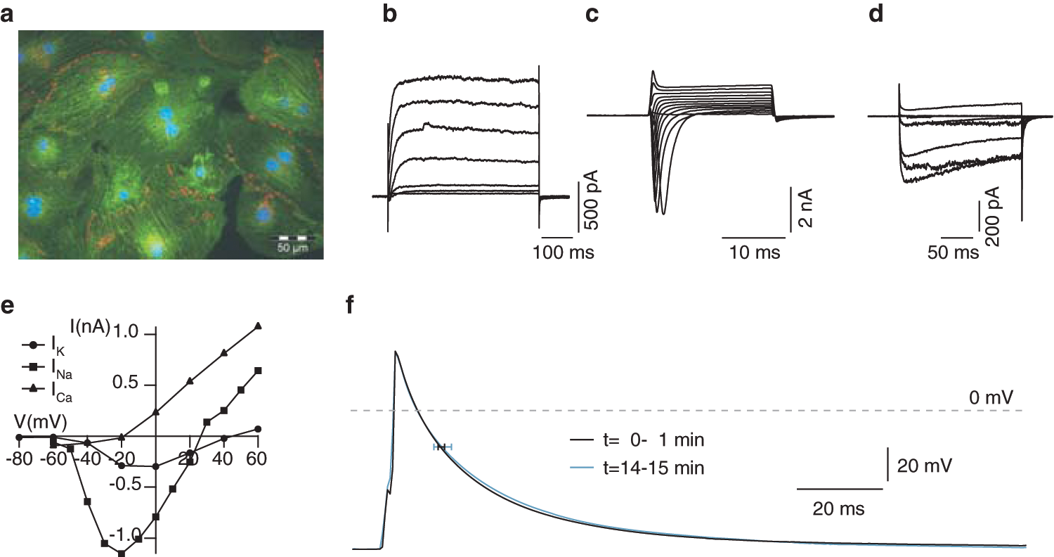
They show typical cardiac protein expression (cardiac α-actinin and connexin 43) and structure when cultured in monolayers
(
For long-term storage, the cells are deep frozen as single-cell suspensions and stored in liquid nitrogen or −150 °C deep freezers. When cultured overnight at an appropriate cell density, the thawed cardiomyocytes form spontaneously and synchronously contracting monolayers.
Single-cell suspensions of precultured cardiomyocytes were electrophysiologically characterized in the automated patch clamp (APC) systems (Port-a-Patch and Patchliner; Nanion Technologies GmbH). A unique feature of both APC systems is their capability to record action potentials in the current clamp mode and to perform recordings at physiological temperature in addition to the standard measurements of ion currents in the voltage clamp mode. We show that the mESC-derived cardiomyocytes have the electrophysiological properties of primary cardiomyocytes and also expected sensitivity to cardiotoxic compounds.
When recorded in the voltage clamp mode, the cells revealed essential cardiac ion currents including IK, INa, and ICa,L (
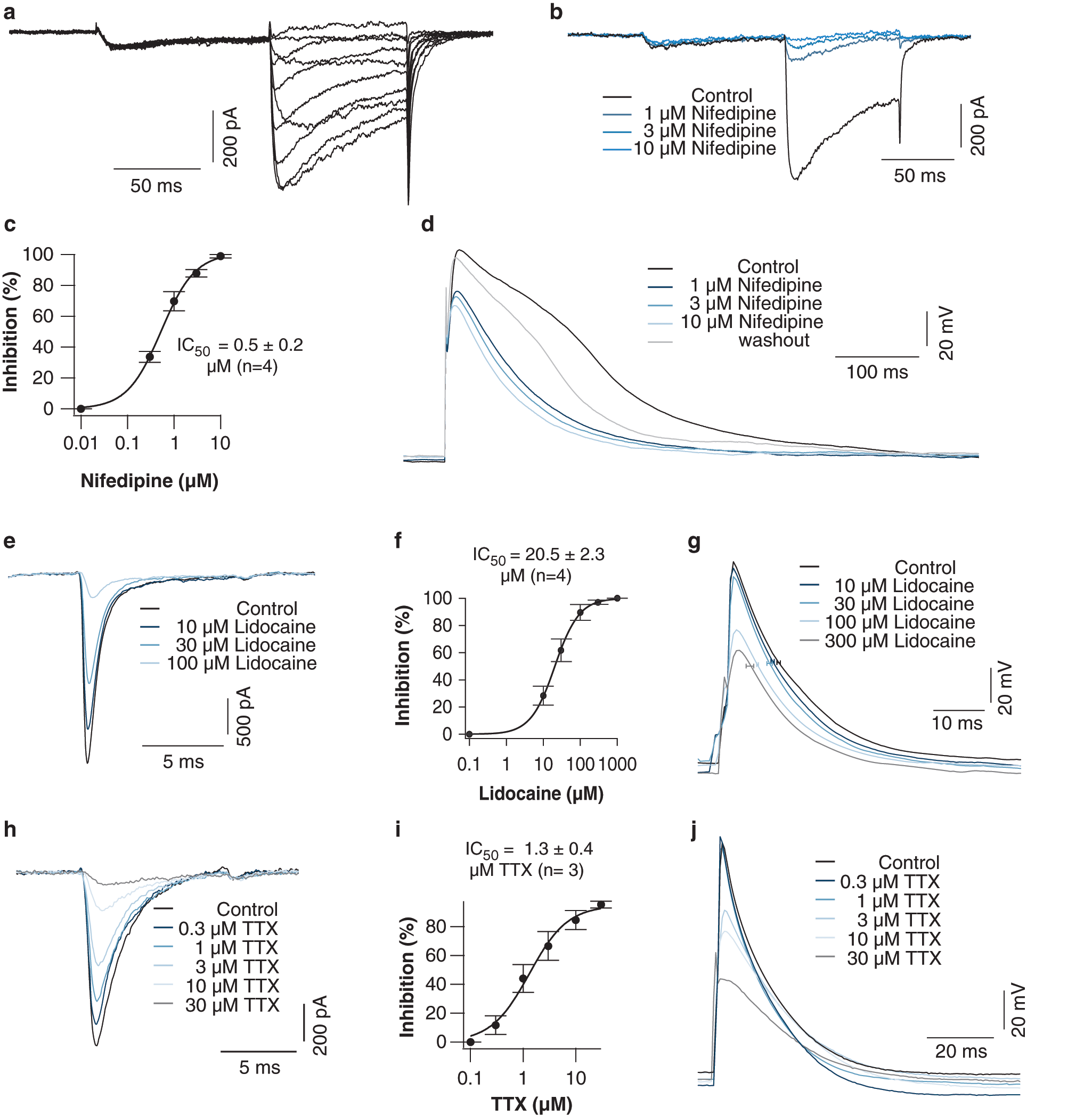
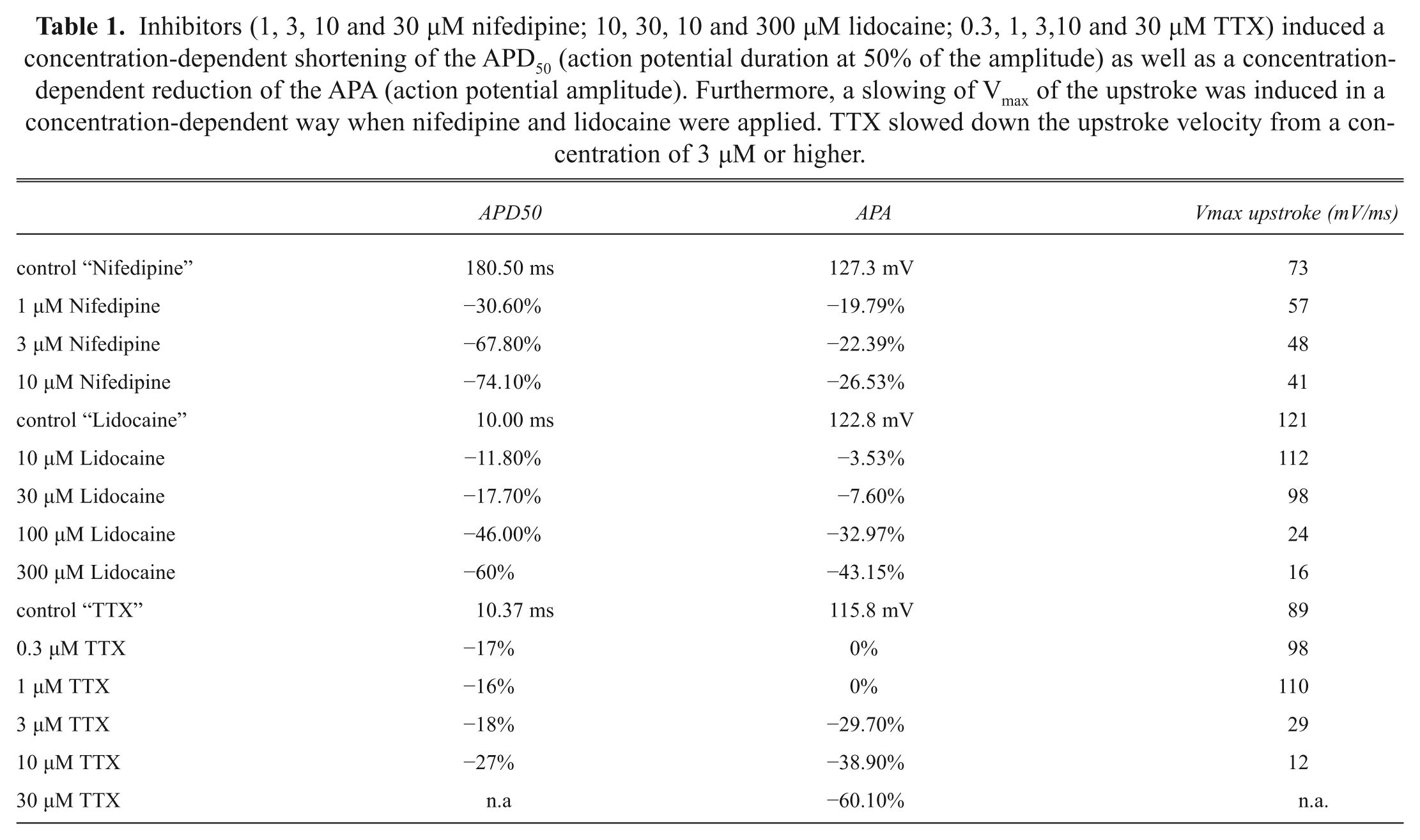
Inhibitors (1, 3, 10 and 30 µM nifedipine; 10, 30, 10 and 300 µM lidocaine; 0.3, 1, 3,10 and 30 µM TTX) induced a concentration-dependent shortening of the APD50 (action potential duration at 50% of the amplitude) as well as a concentration-dependent reduction of the APA (action potential amplitude). Furthermore, a slowing of Vmax of the upstroke was induced in a concentration-dependent way when nifedipine and lidocaine were applied. TTX slowed down the upstroke velocity from a concentration of 3 µM or higher.
Next, we looked at the antiarrhythmic agent lidocaine. Upon the application of increasing concentrations of the Na+ channel inhibitor, the current amplitude of the fast voltage-gated Na+ channel was inhibited in a concentration-dependent way (
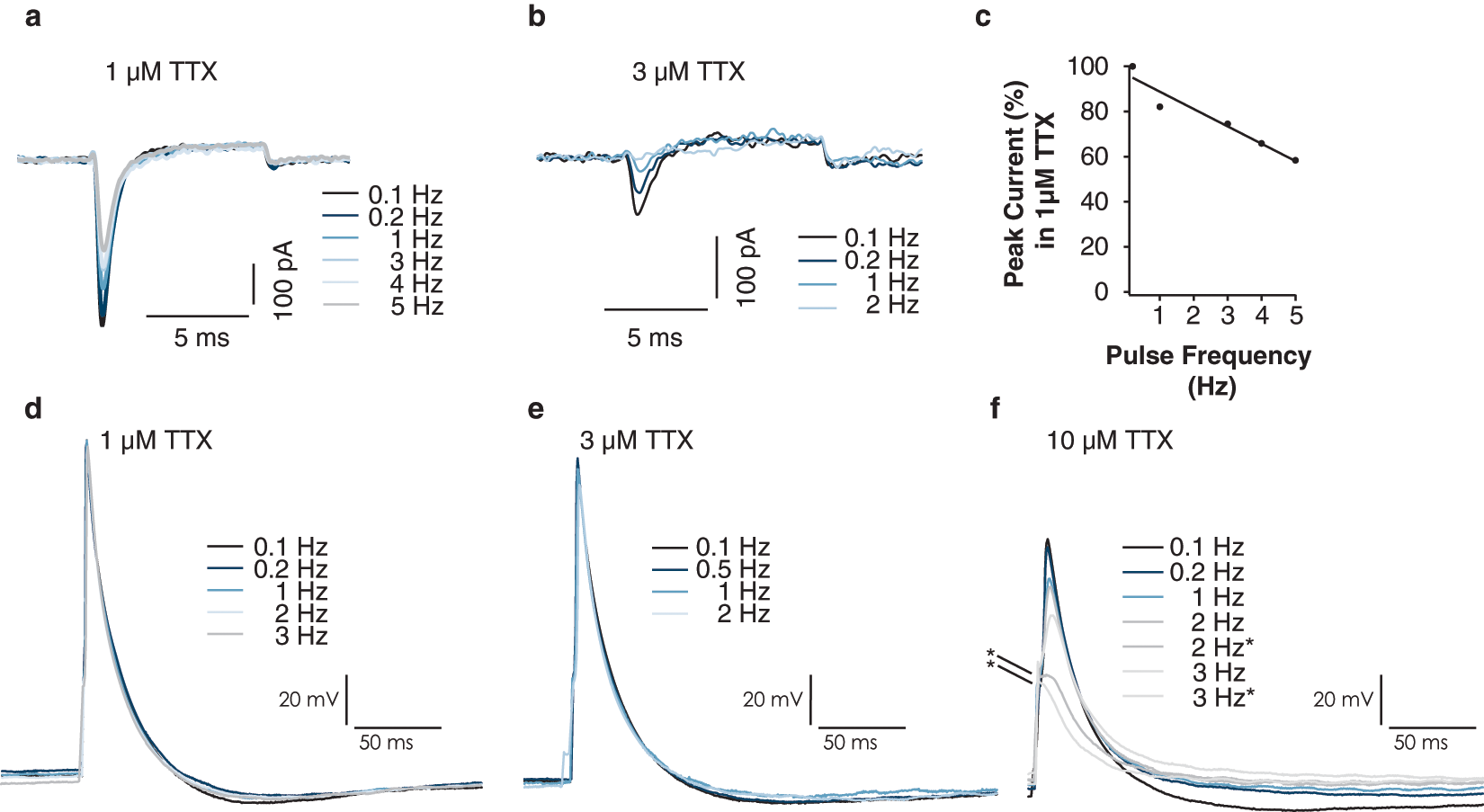
An important consideration in safety pharmacology testing is the blocking potency of compounds at frequencies of current inducing stimuli mimicking physiological heart rates. For this reason, we investigated the effect on efficacy of 1 and 3 µM TTX at different frequencies. When depolarizing the cells to 0 mV at frequencies from 0.1 Hz to 5 Hz (1 µM TTX) and 0.1 Hz to 2 Hz (3 µM TTX), we observed a gradual decrease of the current response, which represents a use-dependent inhibition of Na+ channels (
(
The mESC-derived cardiomyocytes revealed electrophysiological properties and the correct pharmacological modulation of these properties comparable to primary fetal and neonatal murine cardiac myocytes including functional IK current,7,8 which means they can serve as a model system for human cardiac safety pharmacology. Furthermore, this work demonstrates that the cells are suitable for the analysis on APC systems for the screening of safety pharmacological drug effects.
In the future, cardiomyocytes from pluripotent stem cells will gain more and more attention due to the predictive value for cardiac toxicity,14,15 the capability to reprogram somatic cells even from humans, 4 and their availability as ready-to-use high-purity cells.
This approach, mESC-derived cardiomyocytes analyzed on an APC with the flexibility of performing both voltage as well as current clamp recordings, offers the key advantage that drugs and their inhibitory effect on relevant cardiac ion channels can be analyzed in the voltage clamp mode, and their impact on the AP shape can be seen in the same experiment. mESC-derived cardiomyocytes have an advantage over heterologous expression systems because of their greater physiological relevance as multiple ion channels are expressed simultaneously in an environment more representative of the native situation. Although overexpressing ion channels in, for example, HEK cells can be useful for determining ion channel specificity of a particular compound, mESC-derived cardiomyocytes give an overview of all cardiac relevant ion channels. Furthermore, with mESC-derived cardiomyocytes being readily accessible compared with freshly isolated cardiomyocytes and at the same time the automated patch clamp assay being scalable, this opens the way for the pharmaceutical industry to perform high-throughput screening of new drug candidates with regard to drug safety and cardiotoxicity.
Footnotes
References
Supplementary Material
Please find the following supplemental material available below.
For Open Access articles published under a Creative Commons License, all supplemental material carries the same license as the article it is associated with.
For non-Open Access articles published, all supplemental material carries a non-exclusive license, and permission requests for re-use of supplemental material or any part of supplemental material shall be sent directly to the copyright owner as specified in the copyright notice associated with the article.