
Abstract
Members of the heat shock protein 70 (Hsp70) family of molecular chaperones are emerging as potential therapeutic targets. Their ATPase activity has classically been measured using colorimetric phosphate detection reagents, such as quinaldine red (QR). Although such assays are suitable for 96-well plate formats, they typically lose sensitivity when attempted in lower volume due to path length and meniscus effects. These limitations and Hsp70’s weak enzymatic activity have combined to create significant challenges in high-throughput screening. To overcome these difficulties, the authors have adopted an energy transfer strategy that was originally reported by Zuck et al. (Anal Biochem 2005;342:254-259). Briefly, white 384-well plates emit fluorescence when irradiated at 430 nm. In turn, this intrinsic fluorescence can be quenched by energy transfer with the QR-based chromophore. Using this more sensitive approach, the authors tested 55,400 compounds against DnaK, a prokaryotic member of the Hsp70 family. The assay performance was good (Z′ ~0.6, coefficient of variation ~8%), and at least one promising new inhibitor was identified. In secondary assays, this compound specifically blocked stimulation of DnaK by its co-chaperone, DnaJ. Thus, this simple and inexpensive adaptation of a colorimetric method might be suitable for screening against Hsp70 family members.
Introduction
M
Members of the Hsp70 family are composed of 2 domains: a substrate-binding domain (SBD) and a nucleotide-binding domain (NBD). 6 The SBD accommodates short stretches of peptide, and it seems to have a preference for the hydrophobic residues that might be exposed in misfolded or incompletely folded proteins. 7 Binding of the SBD to its substrates is regulated, in part, by the nucleotide state of the adjacent NBD: the adenosine triphosphate (ATP)–bound form has relatively weak affinity for substrates, whereas the adenosine diphosphate (ADP)–bound form binds tightly. 8 However, the intrinsic ATPase activity of these proteins is slow; for example, Escherichia coli DnaK has a Vmax of ~1 pmol ATP/µg enzyme/min. 9 This modest turnover allows regulation by co-chaperones, such as DnaJ. 10 DnaJ belongs to a family of co-chaperones that bind Hsp70s through a conserved J-domain. This protein-protein contact accelerates ATP turnover through an allosteric mechanism. 11
In envisioning strategies for inhibiting members of the Hsp70 family, 2 of their activities become apparent as potential targets. 5 One approach is to inhibit binding of the SBD to its substrates. This strategy is exploited by certain insect-derived, antibacterial peptides, 12 and a fluorescence polarization–based assay to identify similar compounds has been developed. 13 A separate strategy is to block nucleotide turnover. 14,15 This goal might be accomplished by either directly competing with nucleotide 16 or inhibiting the protein-protein interactions with the stimulatory co-chaperones. For example, we recently employed the well-known malachite green (MG) reagent to detect ATP hydrolysis by the DnaK-DnaJ combination. 17,18 Because the stimulatory activity of the J-domain dominates the phosphate signal, we anticipated that inhibitors found by this method would preferentially target the protein-protein contact. Indeed, we found new inhibitors in pilot screens that specifically block DnaK-DnaJ interactions, 19 and the resulting first-generation compounds have been used in a variety of disease models to reveal potential roles of Hsp70s. 20-22
To identify additional chemical scaffolds, we sought to screen larger compound collections in high-throughput ATPase assays. Unfortunately, our attempts to further miniaturize the absorbance platforms to low-volume 384-well microplates were frustrated by significantly decreased sensitivity. Guided by the work of Zuck and coworkers, 23 we explored whether sensitivity could be enhanced using energy transfer methodology. We were particularly interested in studying DnaK, as this chaperone has been implicated as an emerging antibacterial target. 4 Furthermore, DnaK’s weak ATPase activity makes it a particularly challenging test case. Briefly, we found that the fluorescence method improved sensitivity for phosphate and permitted screening of more than 55,000 compounds with good assay performance (Z′ ~0.6, coefficient of variation [CV] ~8%). These experiments yielded at least 1 new inhibitor of DnaK, which appears to block stimulation by DnaJ.
Materials and Methods
Reagents
Unless otherwise specified, all reagents were purchased from Sigma (St. Louis, MO). Escherichia coli DnaK was purified according to published procedures. 18 E. coli DnaJ was purified as previously described. 24
Small-molecule libraries
The MicroSource MS2000 library contains ~2000 bioactive compounds with a minimum of 95% purity. Briefly, the collection includes 958 known therapeutic drugs, 629 natural products and derivatives, 343 compounds with reported biological activities, and 70 compounds approved for agricultural use. The University of Michigan Center for Chemical Genomics (CCG) small-molecule library consists of 16,000 Maybridge HitFinder, 13,000 ChemBridge, 20,000 ChemDiv, 3000 National Cancer Institute (NCI), 450 National Institutes of Health (NIH) Clinical Collection (NCC) compounds, and ~20,000 natural product extracts. The activity of promising compounds was confirmed using repurchased samples from original vendors. Compound
High-throughput ATPase assay—absorbance method
The assay procedure was adopted from previous reports with modifications where indicated. 18 All components other than compounds were added by a Multidrop dispenser (Thermo Fisher Scientific, Inc., Waltham, MA). Stock solutions of 0.05% w/v quinaldine red (QR), 2% w/v polyvinyl alcohol, 6% w/v ammonium heptamolybdate tetrahydrate in 6 M HCl, and water were mixed in a 2:1:1:2 ratio to prepare the QR reagent. This reagent was prepared fresh prior to each experiment. For compound screening, a stock solution of DnaK and DnaJ was prepared in assay buffer (100 mM Tris-HCl, 20 mM KCl, and 6 mM MgCl2, 0.01% Triton X-100, pH 7.4) so that the final concentration of DnaK was 0.4 µM and DnaJ was 0.7 µM (unless noted). This solution (10 µL) was then added to each well of a 384-well clear plate (Thermo Fisher Scientific, Inc.). To this solution, 0.4 µL of either compound (1.5 mM) or DMSO was added to each well by Biomek HDR (Beckman, Fullerton, CA). Finally, 4 µL of a 7-mM ATP solution was added to begin the reaction. The plates were then incubated for 3 h at 37°C. After incubation, each well received 40 µL of the QR reagent, allowing 2 min of reaction time, and then quenched by addition of 32% w/v solution of sodium citrate (4 µL). The plates were then incubated for an additional 15 min at 37°C before measuring absorbance at 530 nm on a PHERAstar plate reader (BMG Labtech, Cary, NC).
High-throughput ATPase assay—fluorescence in white plates
The QR reagent was prepared exactly as indicated above. All components other than compounds were added by a Multidrop dispenser (Thermo Fisher Scientific, Inc.). The DnaK-DnaJ stock solution was prepared so that the final concentration of DnaK was 0.4 µM and DnaJ was 0.7 µM (unless noted). This solution (5 µL) was then added to each well of an opaque, white, low-volume, nonsterile, polystyrene 384-well plates (Greiner Bio-One, Monroe, NC). To this solution, 0.2 µL of either compound (1.5 mM) or DMSO was added to each well by Biomek HDR (Beckman). Finally, 2 µL of a 3.5 mM ATP solution was added to begin the reaction. The plates were then incubated for 3 h at 37°C. After incubation, each well received 15 µL of the QR reagent, allowing 2 min of reaction time, and then quenched by addition of 32% w/v solution of sodium citrate (2 µL). These plates were incubated for 15 min at 37°C and the fluorescence intensity measured (excitation 430 nm, emission 530 nm) on a PHERAstar plate reader. Standard curves were obtained using stock solutions of dibasic potassium phosphate.
Tryptophan fluorescence assay
The method for measuring binding to DnaK was carried out as previously described.
25
Briefly, DnaK (5 µM) in 1 mM ATP was incubated with the indicated concentration of compound
Enzyme-linked immunosorbent assay
The procedure for DnaK binding to luciferase was adapted from a previous report.
26
Briefly, firefly luciferase (0.2 mg/mL) was first incubated with 0.1 µM trypsin at 37°C for 1 h in HEPES buffer (40 mM HEPES, 8 mM MgCl2, 20 mM NaCl, 20 mM KCl, 0.3 mM EDTA, pH 7.2), and the reaction was quenched with 1 mM phenylmethanesulfonylfluoride (PMSF) and diluted to 5 µg/mL with phosphate-buffered saline (PBS) buffer (pH 7.4). An aliquot (50 µL) was then added to 96-well plates (Thermo Fisher brand; clear, nonsterile, flat bottom). After 1 h of incubation at 37°C, the wells were washed 3 times with 100 µL PBS-T (0.05% Tween-20). A solution of DnaK (at the indicated concentrations), compound
Results
Fluorescent energy transfer method has enhanced sensitivity for inorganic phosphate
The primary technical challenge in performing absorbance assays in high-density formats, such as 384- and 1536-well plates, is that the sensitivity is typically decreased ~2.5-fold by the restrictive well geometry.
27,28
In addition, absorbance assays demand flat, clear-bottomed microplates that consume more reagents than other well geometries (e.g., round, concave). However, absorbance assays are often inexpensive and robust, so there is interest in finding ways to adopt them to a higher density. One promising approach was reported by Zuck et al.,
23
who successfully converted a QR-based phosphate assay for use in a 384-well format (
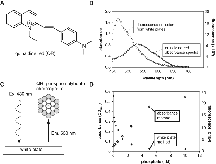
Model for converting an absorbance assay into a fluorescence quenching method. (
Before adapting this platform for use against DnaK, we were interested in discerning the lowest concentration of phosphate that could be robustly detected. Accordingly, we prepared phosphate standard solutions and directly compared the sensitivity of the absorbance- and fluorescence-based methods. For these experiments, we found that the linear detection range of the absorbance method was approximately 0.5 to 5 µM (
Fluorescence method has superior performance in pilot screens
To our knowledge, a high-throughput screening (HTS) application of this fluorescence method has not yet been reported. Therefore, we first performed a pilot screen in 384-well plates to gauge its performance. Briefly, we found that the combination of 0.4 µM DnaK and 0.7 µM DnaJ gave a signal that was linear for at least 2 to 3 h when incubated with 1 mM ATP (Suppl. Fig. S1). From these experiments, we developed a protocol that was adopted from the previous 96-well version (
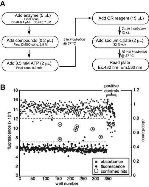
Comparison of screening performances between absorbance and fluorescence methods. (
Using these conditions, we screened the MS2000 collection of bioactive compounds. This library is known to contain 5 DnaK inhibitors; thus, it could be used to probe the assay performance.
21
Also, we directly compared the absorbance and energy transfer methods by splitting the reactions into either clear plates or low-volume white plates. Using this side-by-side approach, we confirmed our previous observations that the absorbance-based assay is a relatively poor HTS method, with a Z′ score of approximately 0.2. Moreover, this method identified only 1 of 5 known inhibitors and it gave 2 false positives (
High-throughput screen for inhibitors of DnaK’s ATPase activity
On the basis of the promising results from the pilot experiments, we screened an additional 55,400 small molecules and natural product crude extracts against the ATPase activity of DnaK-DnaJ (
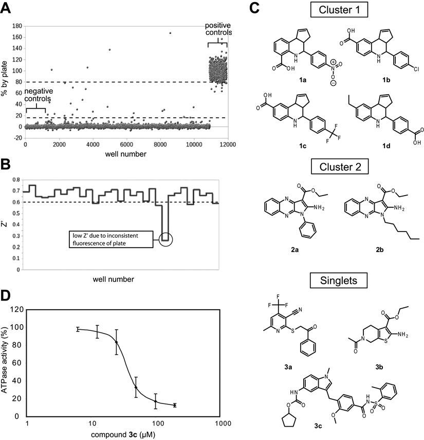
Screening of 55,400 samples. (
Samples were screened at a single concentration (between 10 and 40 µM) against DnaK-DnaJ, with the addition of 0.01% Triton X-100 to minimize false discovery of aggregators. From these screens, samples that exhibited effects of 35% or higher by plate were considered inhibitors. Samples with signals at least 3 times the standard deviation from the negative controls and samples on their given plate were also included. Of 55,400 samples tested, 598 fell into this category (hit rate: 1.1%). In our previous studies, activators of Hsp70s have also proven to be powerful chemical tools. 20,21 With the aim of potentially expanding the number of activators, the HTS results were reviewed for samples with signals of −2.5 standard deviations or lower. Using this modest criterion, 122 unique samples were identified as activators (hit rate: 0.22%).
To confirm their activities, the samples were subjected to confirmatory retesting in duplicate. Prior to this experiment, 10 compounds containing a heavy metal and 11 compounds with a molecular weight above 650 Da were excluded because they were expected to have unfavorable properties. In addition, the natural product extracts were excluded, and their analysis will be reported elsewhere. Thus, a total of 508 unique compounds (400 inhibitors and 108 activators) were assayed and 73 inhibitors confirmed. None of the activators confirmed upon retesting, although it should be noted that they exhibited weak activity in the primary screen, and the assay conditions were optimized to identify inhibitors. The Z′ factors for the confirmation assay ranged from 0.65 to 0.70.
The confirming structures were subsequently clustered to determine if series were present. Clustering at 65% + similarity with the fingerprints (Unity) and clustering algorithms (Optisim) by Benchware DataMiner produced 55 clusters. It has been shown that a compound with 85% or greater structural similarity to an active compound will have a ~30% probability of also being active.
30
Therefore, we analyzed our internal database of ~150,000 compounds and retrieved structures that have 85% or greater similarity to any of the confirmed inhibitors. On the basis of this analysis, we selected compounds that belong to 3 largest clusters for testing in dose dependence assays. These assays were performed using a 2-fold dilution series of 8 compound concentrations (1-125 µM). Of the 127 compounds tested, 70 showed dose-dependent inhibition curves. To minimize false positives, we then evaluated these 70 compounds for autofluorescence. Briefly, compounds were excluded if their intrinsic fluorescence (ex: 430 nm, em: 530 nm) at 5 µM was at least 10% of the positive control or if their dose dependence plateau was above the positive control (see examples in
To further evaluate the identified actives, we repurchased 4 examples that belong to the largest cluster (cluster I, containing 26 of the 36 remaining compounds):
Compounds belonging to the other clusters were studied in a similar manner. Briefly, we repurchased 2 compounds from cluster II (
Compound 3c binds DnaK, favors high-affinity binding to luciferase and blocks DnaK’s stimulation by DnaJ
To characterize compound
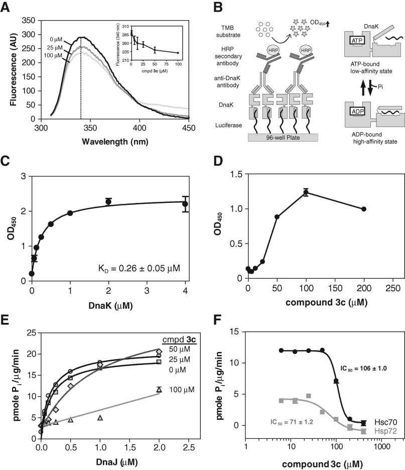
Compound
Because much of the signal from the ATPase assay is due to the stimulatory activity of the co-chaperone DnaJ, we hypothesized that compound
Finally, members of the Hsp70 family are highly conserved, with DnaK sharing nearly 50% sequence identity with human Hsp72 (HSPA1A) and bovine Hsc70 (HSPA8). To test whether compound
Discussion
Absorbance assays are widely used to monitor the activity of ATPases, GTPases, phosphatases, and kinases. They are widely used, in part, because they are robust, easy to use, and relatively inexpensive. However, these assays are typically performed in cuvettes or medium-density (i.e., 96-well) microplates. Although these formats are suitable for some studies, they are not preferred for high-throughput screens, especially in low volume. Adopting a method developed by Zuck et al., 23 we have successfully converted a QR-based absorbance assay to a 384-well format and screened a collection of >55,000 compounds. Although this general method was published in 2005, it remains largely underevaluated in the screening literature. Rather, other phosphate detection technologies, such as Phosphate Sensor (Invitrogen, Carlsbad, CA) and Transcreener™ ADP2 kit (Bellbrook Laboratories, Madison, WI), have been more widely used. 32 In our hands, this simple and inexpensive (approximately 1-2 cents/well) fluorescence method was robust and had good assay performance against a weak ATPase. However, it must be emphasized that there is significant potential for interference from autofluorescent or colored compounds, yielding false positives. Also, it should be clearly noted that occasional plates, from multiple manufacturers, produced very poor Z′ values due to their inconsistent fluorescence (see Suppl. Fig. S2). Despite these important concerns, we suspect that this procedure may be useful in certain settings, especially when cost is a primary concern.
Hsp70 family proteins have emerging potential as therapeutic targets; however, only a handful of relatively modest inhibitors are known.
5
Clearly, one challenge is the weak enzymatic activity of these chaperones, which hindered our own efforts to screen for new inhibitors in a low-volume, 384-well format. Using the energy transfer methodology, we embarked on a screening strategy against E. coli DnaK in combination with its co-chaperone, DnaJ. Screening of 55,400 molecules identified compound
Footnotes
Acknowledgements
The authors thank H. Larch and D. Walt for critical and insightful comments. This work was supported by grants from Thermo Fisher Scientific, the Alzheimer’s Association (IIRG-07-60067), the NIH (NS059690), and National Science Foundation (MCB-0844512) to JEG.