
Abstract
Although conventional high-throughput screens performed in vitro with purified protein kinases are powerful tools to discover new kinase inhibitors, they are far from ideal for determining efficacy in vivo. As a complementary approach, cell-based, target-driven secondary screens may help predict in vivo compound potency and specificity as well as evaluate bioavailability and toxicity. Here the authors report a simple protocol for treating K562 Bcr-Abl-expressing cells with small-molecule kinase inhibitors in 96-well filter-bottom plates followed by in-plate cell lysis. The lysates were assayed via a solid-phase kinase assay, allowing determination of apparent IC50 for known Bcr-Abl inhibitors as well as facilitating the screening of a small kinase inhibitor library. This approach may have further applications in generating lysates for analyzing kinase activity and inhibition in other nonadherent suspension cell lines.
Introduction
P
The Food and Drug Administration (FDA)–approved agents imatinib, nilotinib, and dasatinib have revolutionized the treatment of chronic myelogenous leukemia (CML) 2,3 by inhibiting Bcr-Abl, the fusion gene product of the Ph1 reciprocal 9:22 translocation with constitutive tyrosine kinase activity. 4,5 However, even more compounds are in clinical trials because mutations in the Bcr-Abl kinase cause resistance to these drugs. 6,7 With new compounds constantly being developed to overcome resistance in CML as well as to target oncogenic tyrosine kinases involved in other diseases, the demand for new PTK inhibitors will continue for the foreseeable future.
We have previously described a solid-phase kinase assay using peptides immobilized on 96-well hydrogel plates to detect the activity of recombinant c-Abl or Bcr-Abl present in lysates from the CML-derived cell line K562. 8 Here, aiming to measure targeted kinase activities in a more physiological context, we developed a protocol for treating K562 cells in 96-well filter plates with kinase inhibitors to obtain consistent amounts of lysate and detect Bcr-Abl activity with the solid-phase kinase assay. This simple, cell-based secondary assay may offer a rapid and inexpensive tool to select lead compounds from primary high-throughput screens while examining key drug properties such as cell permeability and toxicity, expediting the drug development process.
Materials and Methods
Materials
Imatinib mesylate was provided by Dr. Wendy Stock (University of Chicago, Chicago, IL). The 80-compound Screen-Well™ Kinase Inhibitor Library was purchased from BioMol (Enzo Life Sciences, Plymouth Meeting, PA). A collection of pyrido[2,3-d]pyrimidine-7-one class multitargeted kinase inhibitors was synthesized by the methods of Klutchko et al 9 and Boschelli et al. 10 All compounds were dissolved and further diluted in molecular biology grade DMSO (Sigma-Aldrich, St. Louis, MO).
Cell culture
K562 cells 11 (American Type Culture Collection, Manassas, VA) were grown at 37°C and 5% CO2 in RPMI-1640 media (Sigma-Aldrich) with 4 mM L-glutamine (Sigma-Aldrich) and 10% (v/v) fetal bovine serum (Gemini Bio-Products, West Sacramento, CA).
Bulk cell lysis and concentration analysis
K562 cells were lysed using PhosphoSafe Extraction Reagent (PER, Novagen, San Diego, CA) with 1× Protease Inhibitor Cocktail (PIC, Sigma-Aldrich). In brief, cells grown in T-175 flasks (Greiner Bio-One, Monroe, NC) were washed twice with 1 mL of cold phosphate-buffered saline (PBS; Fisher Scientific, Pittsburgh, PA) per 106 cells and incubated on ice for 20 min in 50 µL PER with 1× PIC per 106 cells. After brief agitation by vortex mixing, lysate was aliquoted into precooled 1.5-mL microcentrifuge tubes and centrifuged for 5 min at 16,000 g at 4°C. The protein concentration of the supernatant was analyzed with Coomassie Protein Assay Reagent (Pierce, Rockford, IL) by comparing absorbance at 595 nm to a bovine serum albumin standard.
Cell lysis in 96-well filter plate
The bottoms of 1-mL AcroPrep™ 96-well Filter Plates (Pall, East Hills, NY) with 3.0-µm glass fiber prefilters and 0.2-µm Bio-Inert filters were sealed with 3 layers of Parafilm (Pechiney Plastic Packaging, Menasha, WI). The plates were then filled with 106 cells per well suspended in 810 µL media. The cells were treated with 90 µL per well of either 10% DMSO or compound solution and shaken gently at 37°C for 1 or 2 h. Following drug treatment, the cells were lysed in the wells. First, the Parafilm was removed and the media were removed by centrifugation at 100 g for 5 min into a receiver plate. Then the cells were washed twice with 250 µL cold PBS per well by repeated centrifugation at 800 g at 4°C for 5 min. After a third 5-min centrifugation to remove residual PBS, 50 µL PER with 1× PIC was added per well, and plates were shaken on a Microplate Genie (Scientific Industries, Bohemia, NY) for 30 s and then incubated on ice for 20 min. The plates were shaken for another 30 s, and then cell lysates were collected in a chilled, sterile U-shaped 96-well plate (Greiner Bio-One) through centrifugation at 800 g at 4°C for 10 min. Another 25 µL PER with 1× PIC was added per well, the plate was briefly shaken, and flowthrough was collected by a 10-min centrifugation. The lysates were reserved on ice, and unused portions of lysate were flash frozen in liquid nitrogen and stored at −80°C. Protein concentrations were measured as above.
Peptide synthesis
Cys-Abltide (CEAIYAAPFAKKK) 12 and Cys-Abl ligand (CGGAPTYSPPPPPLL) 13 were synthesized on a Prelude Parallel Peptide Synthesizer (Protein Technologies, Tucson, AZ) using solid-phase Fmoc chemistry. The peptides were purified with an Agilent 1200 Series LC/MS (Santa Clara, CA) on an RP-C18 column and analyzed with an Applied Biosystems 4700 MALDI TOF/TOF MS (Foster City, CA). The peptides were dissolved in H2O, and concentrations were determined using Beer’s law at 280 nm.
Kinase reaction
The kinase assay was performed as previously described. 8 Briefly, Cys-Abltide and Cys-Abl ligand were attached to ez-rays™ plates (Matrix Technologies, Hudson, NH) and reacted with either 25 µg bulk lysate or 24 µL (~30 µg) lysate from the filter plate. Phosphorylated Cys-Abltide was probed with mouse antiphosphotyrosine recombinant clone 4G10 (Upstate, Charlottesville, VA) and horseradish peroxidase–conjugated antimouse IgG secondary antibody (GE Healthcare, Piscataway, NJ) and visualized using either SuperSignal West chemiluminescent substrate (Pierce, Waltham, MA) exposed to autoradiography film or Amplex Red substrate (Invitrogen, Carlsbad, CA) scanned by a Bio-Rad FX Pro Plus using the Alexa 532 filter (532 nm/588 nm). Quantitation of each well was performed with Quantity One software v4.6.2, with the diameter of the circular volume equivalent to the diameter of a well.
Assay evaluation
For assay evaluation, the cells were treated with 0 or 100 µM imatinib as positive and negative controls, respectively. The quantitated results were used to calculate the signal-to-background (S/B) and signal-to-noise (S/N) ratios, as well as the Z factor. 14 Z factor ranges from negative values to 1, and a value greater than 0.5 is generally accepted as indicating that an assay is sufficiently robust for high-throughput screening (HTS). The S/B and S/N ratios and Z′ factor were evaluated as follows 14 :

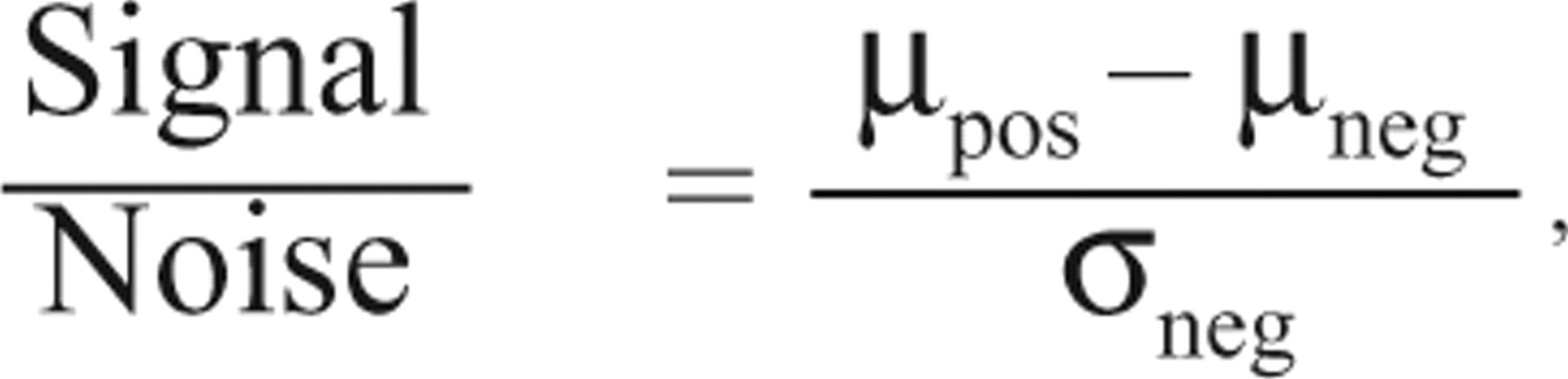

where µpos and σpos are the mean and standard deviation of the positive controls, and µneg and σneg are the mean and the standard deviation of the negative controls, respectively.
IC50 calculation
Prism v5.01 (GraphPad, La Jolla, CA) was used to calculate the IC50 values. Briefly, chemiluminescence and chemifluorescence were quantitated in QuantityOne, and the values were normalized to untreated samples. The drug concentrations were log transformed, and a sigmoidal dose-response (variable-slope) curve was fitted to the medians.
Results
96-well filter plate lysis validation
Previously, we developed a solid-phase kinase assay to measure the activity and inhibition of Bcr-Abl from K562 cell extracts using peptide substrates covalently attached to a 96-well hydrogel plate.
8
It provides a simple and robust platform to screen small molecules for inhibition of Bcr-Abl in vitro. To accelerate the examination of Bcr-Abl inhibition in cells, we developed a protocol to treat K562 cells in 96-well filter-bottom plates with various small-molecule inhibitors. The treated cells are then lysed in the filter-bottom plates and collected in a 96-well plate to yield cell extracts for the solid-phase kinase assay (
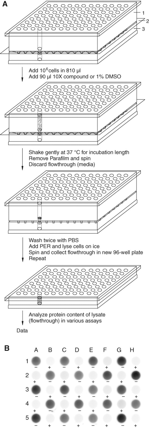
96-well filter plate lysis and lysate analysis. (
To measure whether lysis occurred reproducibly throughout the filter plate between positive and negative controls, cells in 48 wells were treated for 2 h with either 1% (v/v) DMSO (positive controls) or 100 µM imatinib (negative controls) and lysed. The protein concentrations were 1.25 ± 0.15 µg/µL and 1.24 ± 0.10 µg/µL for the positive and negative control groups, respectively. Lysate concentrations were consistent between treatment group and nontreatment group.
The lysates were then analyzed for Bcr-Abl activities in the solid-phase kinase assay.
8
In this kinase assay, a peptide substrate specific for Abl kinase (Cys-Abltide) and an Abl SH3 domain affinity peptide (Cys-Abl ligand) are tethered to a hydrogel surface through Michael addition chemistry. After performing the kinase reaction with the cell lysates, peptides are probed with an antiphosphotyrosine antibody and a horseradish peroxidase–conjugated secondary antibody and then visualized with either chemiluminescence or chemifluorescence. In the kinase assay, the lysates phosphorylated Cys-Abltide in each of 24 positive control wells and showed minimal background in all negative controls (
Several statistics were calculated for the kinase assay: the S/N ratio was 10.3, and the S/B ratio was 32.9. The Z factor considers both dynamic range and variability across multiple samples, with a value of 1 representing a theoretical perfect assay and values over 0.5 being suitable for high-throughput screens. In prior work using either purified c-Abl or Bcr-Abl in bulk K562 lysates, 8 we attained Z factors between 0.5 and 0.9 for the solid-phase kinase assay. In our present study, we were able to obtain a Z factor of 0.4, most likely due to well-to-well variations in the protein concentrations. Although unlikely to provide a robust tool for high-throughput screens in its present form, the assay appears to have sufficient sensitivity and specificity to compare compounds in a medium-throughput assay.
Apparent imatinib IC50
To evaluate and compare the in vitro and in vivo inhibition of Bcr-Abl, we performed 2 kinase assays in parallel to determine the apparent IC50 values of imatinib. Here, in vitro inhibition is defined as adding compounds to K562 lysates during the kinase assay. In vivo inhibition is defined as treating K562 cells with the same compound prior to lysis, then analyzing the lysates in the kinase assay. To compare Bcr-Abl inhibition in these contexts, a 2-fold dilution series of imatinib from 256 µM to 0.0156 µM was examined for kinase inhibition in vitro (
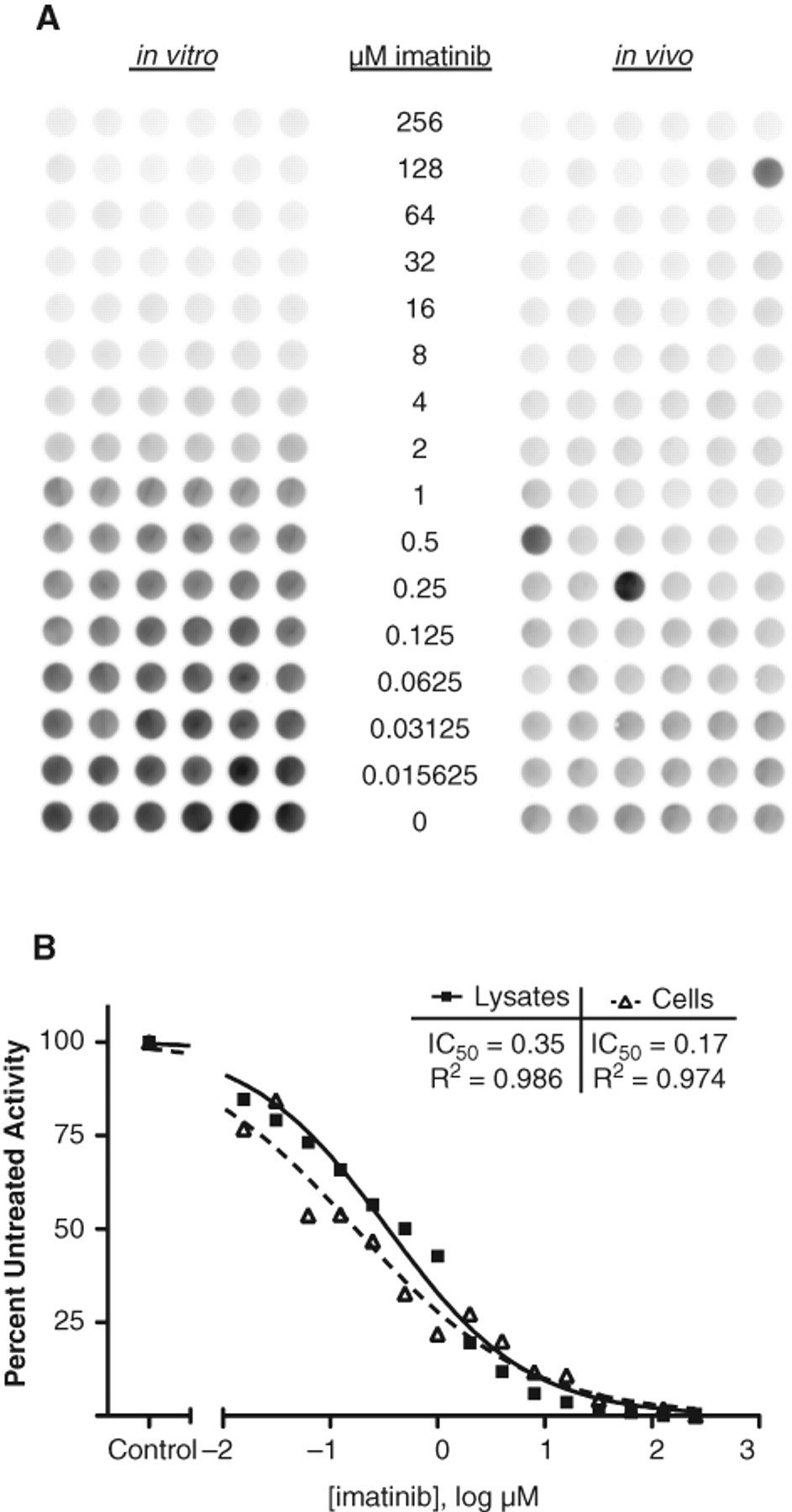
Imatinib dose responses in vitro and in vivo. (
Cell-based model screens
Pyridopyrimidine series
After validating the lysis protocol and studying the sensitivity and reproducibility of the kinase assay, we screened 16 pyridopyrimidine dual-specificity kinase inhibitors alongside imatinib, dasatinib, and 2 dasatinib-related compounds, using chemiluminescence for visualization. Compounds in this library were previously observed to display superior inhibition of Bcr-Abl compared to imatinib when applied to K562 lysates.
8
When K562 cells were treated with 0.04, 0.4, 4, or 40 nM of the compounds for 1 h, lysed, and then assayed for Bcr-Abl kinase activity, several significantly inhibited Bcr-Abl even at sub-nanomolar concentrations (
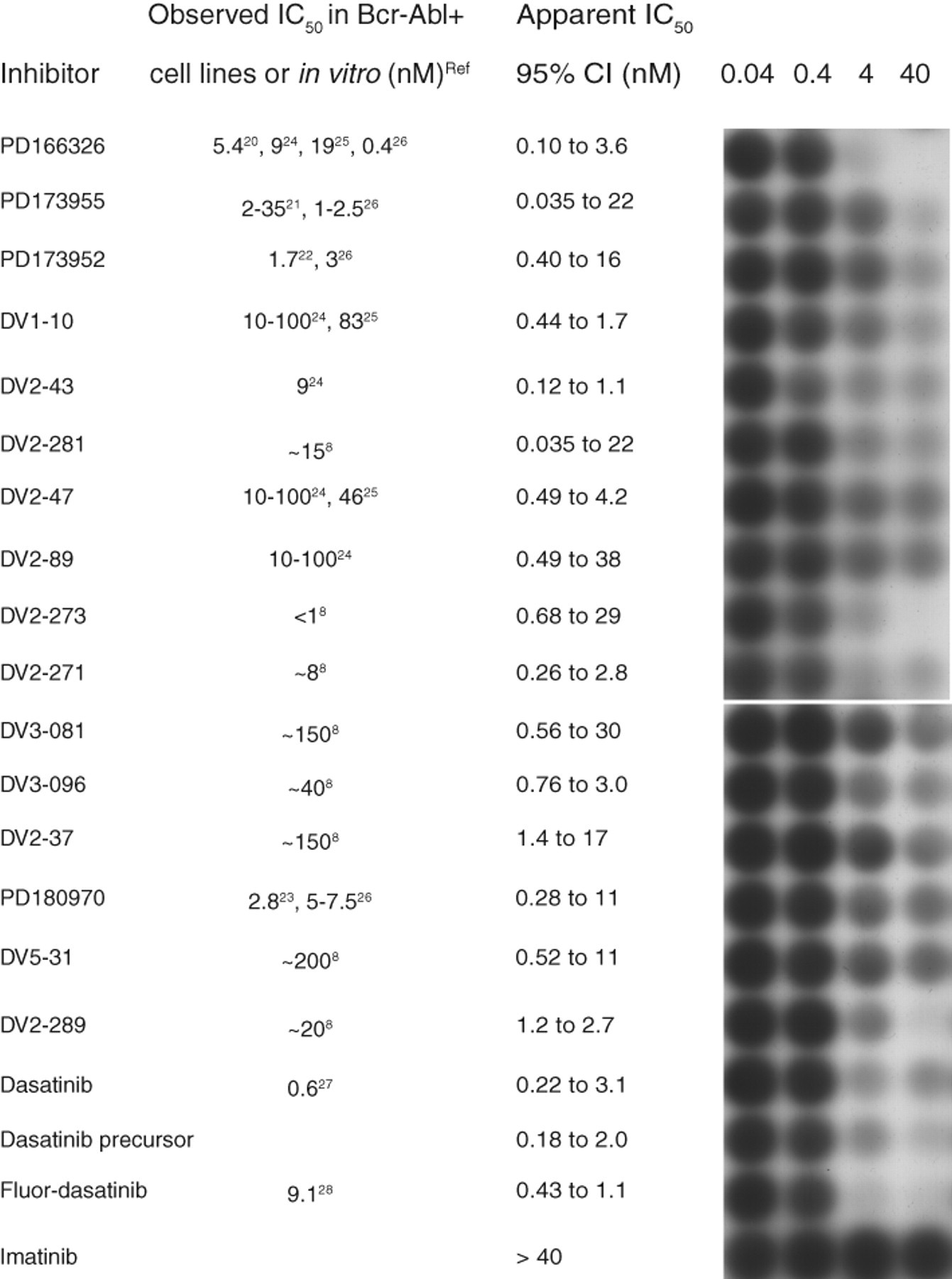
Model screen of potent kinase inhibitors. Compounds that strongly inhibited purified c-Abl in previous screens 8 were further characterized by in vivo assays. Briefly, cells were treated for 1 h prior to lysis. The collected lysates were added to the hydrogel plate with immobilized Cys-Abltide and Cys-Abl ligand. The resulting phosphorylated Cys-Abltide was probed with antiphosphotyrosine and visualized with chemiluminescence. Quantitation was performed using QuantityOne, and the IC50 curve was calculated using GraphPad Prism (v5.01).
BioMol ScreenWell™ Kinase Library
The BioMol ScreenWell™ Kinase Library is a collection of 80 compounds that show activity against diverse serine/threonine and tyrosine kinases.
8
To interrogate this library for indirect and/or synergistic effects on Bcr-Abl activity, we sensitized the in vivo and in vitro kinase assays by combining 5 µM imatinib along with 200 µM of each compound. Substrate phosphorylation was detected with chemiflurorescence (
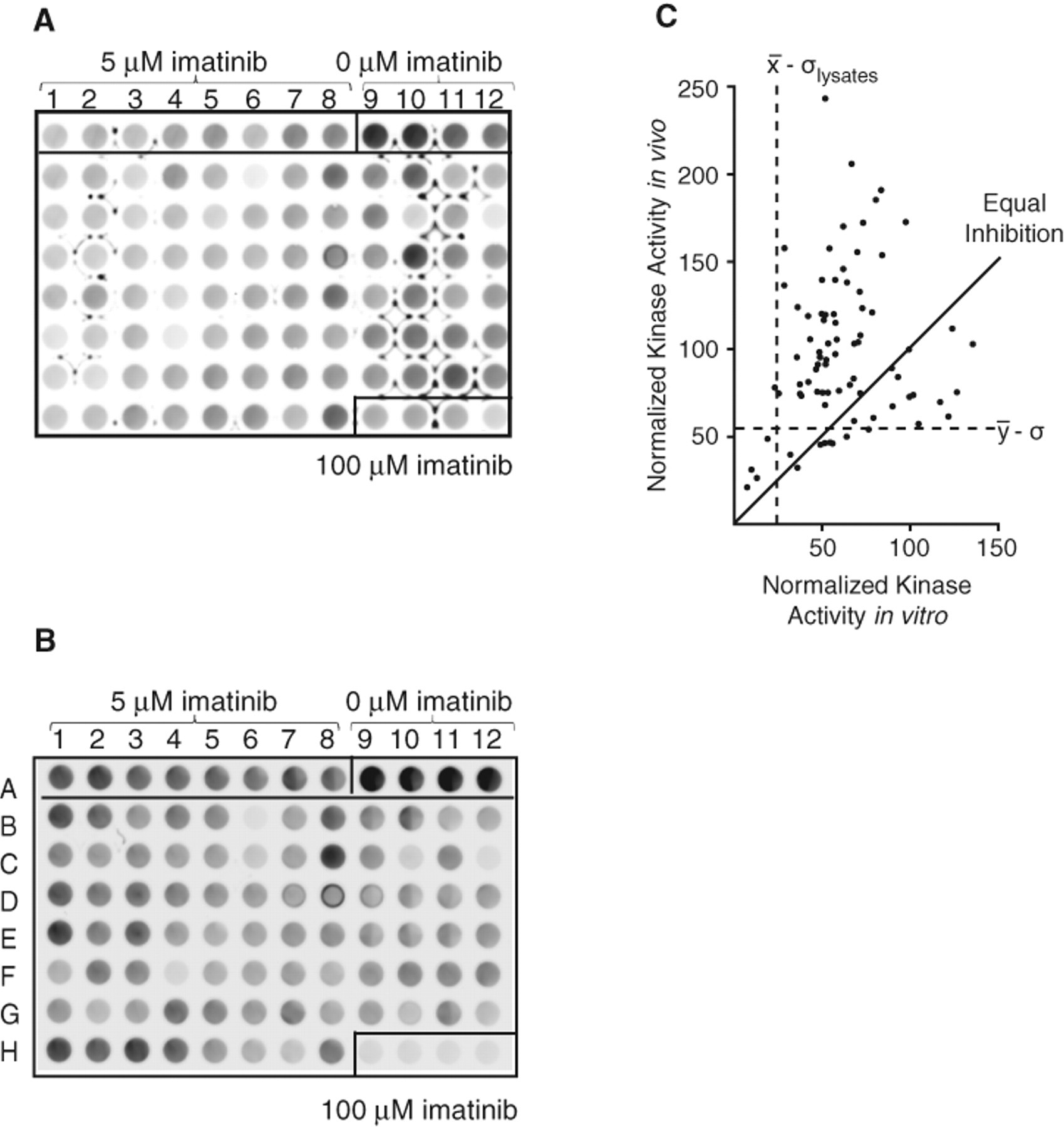
Model screen of BioMol kinase inhibitor library. (
In summary, we report here a medium-throughput screening protocol in which cells are treated with compounds in 96-well filter-bottom plates followed by in-plate lysis and analysis in a solid-phase kinase assay. Compared to the previous in vitro compound treatment, this protocol provides more information about the permeability and cellular efficacy of potential kinase inhibitors. Although this assay was developed to measure Bcr-Abl activity in K562 lysates, it is likely to be useful for other receptor and nonreceptor protein tyrosine kinases, 18 given the availability of specific kinase substrates, and could also be employed for additional nonadherent cells, such as the widely used Ba/F3 cells. 19 This protocol provides a medium-throughput scheme for generating lysates to accelerate sample preparation for cell-based assays requiring high-quality lysates and should be adaptable to robotic handling for increased throughput and consistency in the future.
Footnotes
Acknowledgements
We thank Won Jun Rhee and Stacey L. Kigar for synthesis and purification of the peptides, as well as Drs. Wendy Stock and Bayard Clarkson for generously providing reagents.
Funding for this work was provided by National Institutes of Health grants R33 CA10323 and R01 HG003864. SJK was a Fletcher Scholar of the Cancer Research Foundation and a Leukemia & Lymphoma Society Scholar.