
Abstract
Background
Coronary heart disease (CHD) has a relatively high mortality rate. This study aims to explore the expression level of miR-3150b-3p in CHD and its potential mechanisms of action.
Method
This study collected basic information from 120 hypertensive patients and 110 CHD patients. Serum levels of miR-3150b-3p were quantified using qRT-PCR. The diagnostic value of miR-3150b-3p was evaluated through the ROC curve, and Pearson analysis tested its correlation with CHD severity. In vitro, ox-LDL was used to induce HVSMCs. Cell viability was assessed using the CCK-8 assay, inflammatory factors were measured by ELISA, and oxidative stress levels were evaluated by detecting superoxide dismutase activity and malondialdehyde content. miR-3150b-3p-ARL5B interaction was verified by luciferase assay and RIP.
Results
Serum miR-3150b-3p is up-regulated in CHD patients, correlates with disease severity. ROC curve demonstrated that miR-3150b-3p has robust diagnostic value. In vitro experiments demonstrated that the miR-3150b-3p inhibitor significantly reduced ox-LDL-induced inflammatory factors, increased SOD activity, and decreased MDA content. In addition, miR-3150b-3p was verified to target ARL5B.
Conclusions
The level of miR-3150b-3p in the serum of CHD patients is significantly elevated and positively correlated with disease-related indicators of CHD. miR-3150b-3p can regulate cellular functions and targetARL5B, providing a new potential target for the early diagnosis and treatment of CHD.
Introduction
Coronary heart disease (CHD) is a cardiovascular condition caused by the narrowing of the coronary arteries, which can lead to angina or myocardial infarction.1,2 CHD has become a global issue and is one of the leading causes of death, especially in developed countries. 3 Hypertension is also a significant risk factor for cardiovascular diseases, exerting long-term effects on the cardiovascular system. 4 Currently, the diagnosis of CHD primarily relies on symptoms, electrocardiograms, and angiography, while treatment often involves medication, intervention, or surgery, 5 However, these methods still have limitations in early diagnosis, disease prognosis, and long-term efficacy. 6 The pathological basis of CHD is atherosclerosis, characterized by lipid deposition and smooth muscle cell proliferation. 7 The abnormal activity of these vascular smooth muscle cells (VSMCs) can lead to plaque rupture, resulting in acute coronary syndrome. 8 For instance, miR-18a-5p 9 and miR-30a-3p 10 may play a significant role in disease progression by regulating VSMCs and could serve as potential therapeutic targets.
Therefore, in-depth research into the mechanisms of VSMCs is of significant importance for understanding the pathogenesis of CHD and developing new therapeutic strategies.
MicroRNA is a small non-coding RNA that11–13 regulates cell proliferation, differentiation, apoptosis, and stress responses, 14 while CHD involves processes such as inflammation, lipid metabolism disorders, vascular endothelial dysfunction, and extracellular matrix remodeling. 15 Current research on miR-3150b-3p primarily focuses on cervical cancer 16 and lung squamous cell carcinoma. 17 However, cardiac metastasis from cervical cancer and lung squamous cell carcinoma may affect cardiac function, indirectly correlating with the risk of CHD.17–19 There are also literature reports indicating that RRAS2-hsa-miR-3150b-3p-ILF3-AS1 signaling axis plays a crucial role in atherosclerosis-related ischemic stroke. 20 miR-3150b-3p shows promise as a biomarker for cardiovascular risk assessment in patients with renal failure, but its mechanisms in disease and inflammation still require in-depth investigation. 21 Therefore, this study hypothesizes that miR-3150b-3p may play a significant role in CHD and could potentially serve as a diagnostic marker or therapeutic target. However, its specific functions and mechanisms require further in-depth investigation.
ARL5B is a crucial small GTPase involved in maintaining intracellular homeostasis, 22 and its dysfunction may disrupt Golgi apparatus function, increasing the risk of cardiovascular diseases. 23 The role of ARL5B in the blood-brain barrier suggests its potential involvement in the regulation of membrane trafficking and tight junctions. 24 Based on the above background, this study hypothesizes that miR-3150b-3p may influence cardiovascular cell function by targeting ARL5B, thereby playing a potential role in the pathogenesis of CHD, and further validation of this mechanism is conducted.
Given the current gaps and challenges in the research on miR-3250b-3p and ARL5B in CHD and hypertension. This article aims to delve into the clinical value of miR-3150b-3p in CHD and its regulatory role in VSMCs, with the goal of providing new theoretical foundations and potential targets for the diagnosis and treatment of CHD.
Materials and Methods
Research Subjects and Information Collection
This study enrolled 120 patients with hypertension (HTN, control group) and 110 patients with CHD who visited West China Hospital of Sichuan University from April 2024 to December 2024 and voluntarily signed informed consent forms. Inclusion criteria for the hypertension group: Untreated office blood pressure ≥140 mm Hg systolic or ≥90 mm Hg diastolic, confirmed on three separate days; Coronary angiography shows normal coronary arteries. Exclusion criteria for the hypertension group: Diagnosis of malignant tumors such as solid tumors or hematologic malignancies; Presence of hepatic or renal insufficiency; suffering from autoimmune diseases or infectious diseases; Patients with diabetes. Inclusion criteria for the CHD group: presence of typical angina symptoms, manifested as paroxysmal oppressive pain behind the sternum; The electrocardiogram shows ischemic changes, or myocardial ischemia episodes are recorded by ambulatory electrocardiogram monitoring; Coronary angiography or coronary CTA shows stenosis of ≥50% in the main branches of the coronary arteries. The exclusion criteria for CHD are consistent with those for hypertensive patients. All participating patients were first subjected to medical history collection and medical evaluation, excluding the aforementioned comorbidities, and had not taken any medications that might affect the expression of miR-3150b-3p or oxidative stress status within the three months prior to the commencement of the study.
Collect the basic information of the two study groups, including key indicators. Simultaneously, the Gensini score was calculated to assess the severity of coronary artery lesions.
Expression Detection of miR-3150b-3p
Take 5 mL of fasting venous blood, let it stand for 30 min, then centrifuge to obtain the upper serum, aliquot into RNase-free tubes, and store at −80°C. Total RNA was isolated using an RNA extraction kit (TIANGEN, Beijing, Biotech, China), and then the purified RNA was converted to cDNA with the HiScript II miRNA RT kit (Vazyme, Nanjing, China). Using miRNA-specific primers and probes, qRT-PCR detection was performed with three replicate wells for each sample. The primers used in reverse transcription and real-time quantitative PCR are as follows: The reverse transcription primer for miR-3150b-3p: CTCAACTGGTGTCGTGGAGTCGGCAATTCAGTTGAGAGAACCAA; U6 reverse transcription primer: 5′ AAAATATGGAACGCTTCACGAATTTG 3′; miR-3150b-3p (Forward Primer: 5′-TGAGGAGATCGUCGAGGTTGG-3′; Reverse Primer: 5′-TGGTGTCGTGGAGTCG-3′); U6 (Forward Primer: 5′-CTCGCTTCGGCAGCACA-3′; Reverse Primer: 5′-AACGCTTCACGAATTTGCGT-3′) ; ARL5B (Forward Primer: 5′-AGTGGGACTGGATAATGCAGGG-3′; Reverse Primer: 5′-ATCGCAGAGACTCCTGACCACC-3′); GAPDH (forward: 5′-GAAGGTGAAGGTCGGGAGTC-3′, reverse: 5′-GAAGATGGTGATGGGATTTC-3′). Among them, U6 and GAPDH were used as internal reference genes for miRNA and mRNA analysis, respectively, as their expression stability in serum has been validated by both literature and preliminary experiments,25,26 and quantification was performed using the 2−ΔΔCt method.
Cell Culture and ox-LDL Induction
Human vascular smooth muscle cells (HVSMCs) from ATCC (Manassas, VA) were cultured in RPMI-1640 with 10% FBS and 1% penicillin-streptomycin at 37 °C, 5% CO₂, with routine medium changes; logarithmic-phase cultures were used for experiments. Set up three independent cell culture experiments, with 5-8 replicate wells per group in each experiment. An atherosclerosis model was created in vitro with ox-LDL (50 μg/mL) 27 : cells received either ox-LDL-supplemented or control medium. Incubate in the same incubator for 24-48 h to induce atherosclerosis-related changes in the cells.
Cell Transfection and Functional Analysis
Divide ox-LDL-induced HVSMCs into two groups: the negative control group and the miR-3150b-3p inhibitor group. Lipofectamine™ 3000 (Invitrogen, USA) mediated transfection per manufacturer instructions. When the cells reach 70%-80% confluency, seed them into 96-well plates. Mix the miR-3150b-3p inhibitor or negative control with the liposome transfection reagent, add serum-free medium, and incubate at room temperature for 15-20 min to form complexes. Then add the mixture to the cell plate and gently shake to ensure thorough contact. After transfection for 6 h, replace with complete medium and culture for another 48 h before experiments. The transfection efficiency of this study was evaluated by qRT-PCR, achieving 85%, indicating successful transfection.
Cell Viability Detection
The effect of miR-3150b-3p on the viability of ox-LDL-induced HVSMCs was analyzed using CCK-8. Seed transfected cells into a 96-well plate. At 0, 24, 48, and 72 h, add 10 μL CCK-8, incubate 2-4 h, then measure OD at 450 nm. Each sample was set up with 5-8 replicates, and the experiment was repeated 3 times.
Inflammatory Factor Testing
After transfection, the cells were collected, the supernatant was harvested, and the secretion of inflammatory factors by HVSMCs induced by ox-LDL was detected via ELISA (R&D Systems). During the experiment, strictly follow the instructions of the kit, add standards and samples to the enzyme-linked plate, incubate, wash, and then add enzyme-labeled reagents for color development. Finally, the OD values at the corresponding wavelengths were assayed using an ELISA reader, and the concentrations of each inflammatory factor were calculated based on the standard curve.
Oxidative Stress Detection
The SOD and MDA assay kits from Beyotime Biotechnology (Jiangsu, China) were used to measure the activity and content. Collect cells to extract proteins, measure protein concentration, add reagents for incubation and color development, then perform the assay to calculate SOD activity and MDA content.
Target Gene Prediction and Validation
Predict the downstream target genes of miR-3150b-3p through the TargetScan database and analyze its binding sites with potential target genes. The interaction between miR-3150b-3p and its target gene in HVSMCs was evaluated using the luciferase reporter assay. The ARL5B 3′UTR fragment was cloned into the luciferase reporter gene vector to construct the WT plasmid, while the MT plasmid was constructed by mutating the miR-3150b-3p binding site. The miR-3150b-3p mimic or negative control was co-transfected with WT or MT plasmids into HVSMCs. 48 h post - transfection, luciferase activity was assayed.
The experiment was conducted with the Magna RIP™ Kit (Millipore) following the manufacturer's instructions. Collect the transfected HVSMCs, lyse them with RIP buffer, and incubate the lysate with magnetic beads and specific antibodies (anti-AGO2) for immunoprecipitation. Afterwards, the magnetic beads were treated with proteinase K to extract the bound RNA, and the levels of miR-3150b-3p and ARL5B were detected by RT-qPCR.
Statistical Analysis
Data were analyzed via SPSS 26.0 and GraphPad Prism. Values are presented as mean ± SD. The specific dataset analyzed in this study includes baseline characteristics (LDL-C, HDL-C, CK-MB, Gensini score, and cTnI), miR-3150b-3p expression levels, inflammatory factors, oxidative stress indicators, and target gene validation results. Two-group comparisons used independent t-tests, and multi-group comparisons used ANOVA. Correlation analysis used Pearson's test. Significance was set at P < .05. Prior to data analysis, the Shapiro-Wilk normality test was conducted to confirm that the data followed a Gaussian distribution (P > .05). For categorical variables such as gender, smoking history, alcohol consumption history, and diabetes history, the chi-square test was used for statistical comparison. This study emphasizes data integrity and authenticity, retaining all original data, including outliers. Because these outliers may reflect specific physiological states, individual differences, or particular stages of diseases, they contribute to a comprehensive representation of the characteristics of the study population. Deleting or adjusting outliers may introduce subjective bias, affecting the objectivity and generalizability of the results.
Results
Baseline Characteristics
According to the baseline data (Table 1), there were significant differences in LDL-C (3.42 ± 0.96 vs 2.88 ± 0.96) and HDL-C (1.26 ± 0.11 vs 1.34 ± 0.09) levels between the CHD group and the hypertension group, with P < .001. Indicators such as CK-MB, Gensini score, and cTnI were only detected in the CHD group, with values of 66.02 ± 4.65, 57.03 ± 3.78, and 1.75 ± 0.18, respectively. However, there were no statistically significant differences in age, sex ratio, BMI, smoking, drinking, TG, and TC levels between the two groups of patients (all P > .05).
Summary of Baseline Characteristics.

Note: HTN, hypertension; CHD, coronary heart disease; BMI, body mass index; TG, triglyceride; TC, total cholesterol; LDL-C, low-density lipoprotein cholesterol; HDL-C, high-density lipoprotein cholesterol; CK-MB, creatine kinase-MB; cTnI, cardiac troponin I. M/F, male/female; Y/N, yes/no. The date was presented as mean ± SD), or percentage (/).
Expression Characteristics and Correlation Analysis of miR-3150b-3p in CHD
Compared to the hypertension group (1.00 ± 0.12), miR-3150b-3p in CHD was markedly increased (Figure 1A, 1.18 ± 0.11, P < .0001). ROC curve analysis demonstrated that miR-3150b-3p exhibited good diagnostic efficacy for CHD, with an AUC of 0.880 (95% CI: 0.657-0.812, P < .0001). At the optimal diagnostic threshold of 1.075, the sensitivity reached 82.7%, and the specificity was 74.2% (Figure 1B). Furthermore, miR-3150b-3p showed significant positive correlations with LDL-C (r = 0.829, Figure 1C), CK-MB (r = 0.812, Figure 1E), Gensini score (r = 0.781, Figure 1F), and cTnI (r = 0.758, Figure 1G), P < .0001. miR-3150b-3p was negatively correlated with HDL-C (r = -0.840, Figure 1D).

The Expression of miR-3150b-3p in CHD and its Relationship with Clinical Indicators. (A) the Expression of miR-3150b-3p in the Hypertension and CHD Groups (****p < .0001); (B) Evaluation of the Diagnostic Efficacy of miR-3150b-3p; miR-3150b-3p Showed Significant Positive Correlations with LDL-C (C), HDL-C (D), CK-MB (E), Gensini Score (F), and CTnI (G),****P < .0001.
Multifactorial Logistic Regression Analysis of Risk Factors for CHD
The results of the multifactorial logistic regression analysis revealed that LDL-C (OR = 2.35, 95% CI = 1.15-4.78, P < .001), HDL-C (OR = 0.19, 95% CI = 2.58-10.82, P < .001), and miR-3150b-3p (OR = 11.80, 95% CI = 5.78-24.06, P < .001) were significant predictors of CHD occurrence (Table 2). The associations of age, gender, BMI, TG, and TC with CHD were relatively weak (P > .05).
Logistic Regression Analysis Predicts Risk Factors for CHD Occurrence.

Note: OR odds ratio; CI confdence interval; BMI, body mass index; TG, triglyceride; TC, total cholesterol; LDL-C, low-density lipoprotein cholesterol; HDL-C, high-density lipoprotein cholesterol.
Analysis of the Role of miR-3150b-3p in ox-LDL-Induced HVSMCs
The in vitro experiments in this study further elucidate the role of miR-3150b-3p in ox-LDL-induced HVSMCs. Compared to the control group, ox-LDL induction significantly upregulated the expression of miR-3150b-3p (P < .0001), and the use of miR-3150b-3p inhibitor effectively reversed this change (Figure 2A, P < .01). The cell viability assay results showed that ox-LDL treatment significantly reduced cell viability (P < .0001), while the addition of the miR-3150b-3p inhibitor significantly restored cell viability (Figure 2B, P < .01). Further detection of inflammatory factors revealed that, compared to the control group, the ox-LDL group exhibited significantly elevated levels of TNF-α (Figure 2C), IL-6 (Figure 2D), and IL-1β (Figure 2E) (P < .0001). The miR-3150b-3p inhibitor significantly reduced the levels of these inflammatory factors (P < .0001). In addition, oxidative stress indicators revealed that the SOD activity (Figure 2F) was decreased and the MDA content (Figure 2G) was increased in the ox-LDL group. However, the miR-3150b-3p inhibitor significantly increased SOD activity (P < .01) and decreased MDA content (P < .01), thereby ameliorating the oxidative stress state induced by ox-LDL.
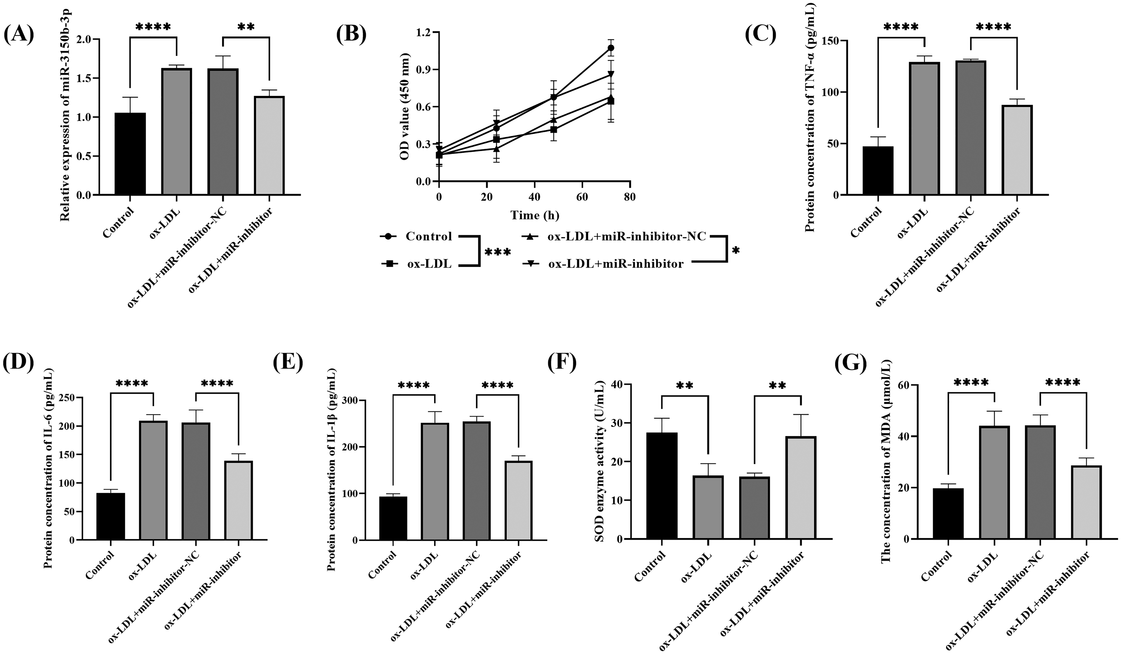
The Effect of miR-3150b-3p on ox-LDL-Induced HVSMCs. (A) ox-LDL Significantly Upregulated the Expression of miR-3150b-3p (P < .0001), and the miR-3150b-3p Inhibitor Reversed This Change (**P < .01); (B) ox-LDL Induction Reduced Cell Viability (****P < .0001), and the miR-3150b-3p Inhibitor Significantly Restored Cell Viability (**P < .01); ox-LDL Induction Significantly Increased the Levels of TNF-α (C), IL-6 (D), and IL-1β (E), While the miR-3150b-3p Inhibitor Markedly Reduced Their Levels (****P < .0001); ox-LDL Decreased SOD Activity (**P < .01, F) and Increased MDA Content (****P < .0001, G), and the miR-3150b-3p Inhibitor Reversed These Changes.
Validation of the Targeting Relationship Between miR-3150b-3p and ARL5B
TargetScan predicts that miR-3150b-3p binds to the 3′UTR of ARL5B with complete seed sequence complementarity (Figure 3A). The luciferase reporter gene assay validated the binding relationship between miR-3150b-3p and ARL5B. Compared to the NC-mimic group, the relative luciferase activity of WT-ARL5B in the miR-mimic group was significantly reduced (P < .0001), while MT-ARL5B showed no significant changes, indicating that miR can bind to and inhibit ARL5B expression (Figure 3B). In addition, compared to the IgG group, the level of ARL5B mRNA in the Ago2 antibody-enriched complex was significantly reduced (Figure 3C, P < .001).
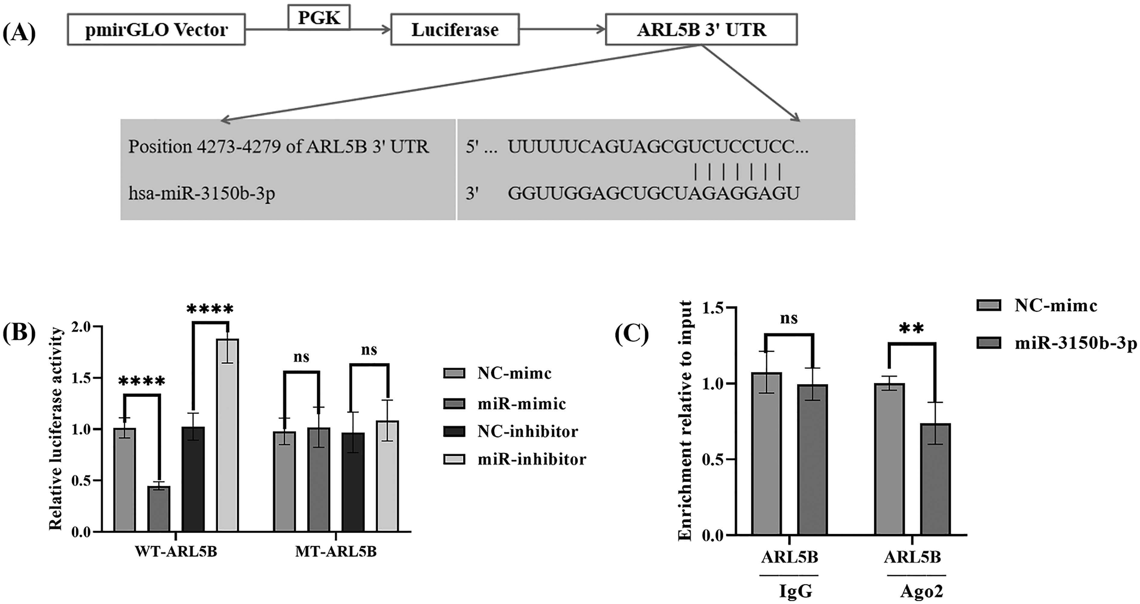
Validation of the Relationship Between miR-3150b-3p and ARL5B. (A) the Binding Site of miR-3150b-3p with the 3′UTR of ARL5B; (B) the miR-3150b-3p Mimic Significantly Reduced the Luciferase Activity of WT-ARL5B (****p < .0001), While no Significant Change was Observed in MT-ARL5B. (C) RIP Experiments Confirmed the Interaction Between miR-3150b-3p and ARL5B.
Discussion
CHD, as a common cardiovascular condition that poses a serious threat to human health, imposing a heavy burden on families and society. In-depth research into the pathogenesis of CHD, as well as the identification of effective diagnostic markers and therapeutic targets, is crucial for improving patient prognosis.
MicroRNAs (miRNAs) are non-coding RNAs with regulatory roles, 20 and their levels reflect cellular states, serving as potential biomarkers for disease diagnosis.28,29 miR-3150b-3p, as a novel miRNA, holds potential diagnostic and therapeutic value in various diseases. In hepatocellular carcinoma (HCC) linked to hepatitis B virus, miR-3150b-3p binds to ASCL1′s 3′UTR, silences it, alters von Willebrand factor expression, and modulates the malignant progression of tumors. 30 In HCC, miR-3150b-3p suppresses HCC cell proliferation, migration, and invasion by targeting circNEIL3 and LAMC1. 31 Similarly, In atherosclerotic plaque-related ischemic stroke (IS), miR-3150b-3p in the RRAS2-ILF3-AS1 regulatory axis may play a key role in IS progression. 20 A new study indicates that the expression of miR-3150b-3p is significantly upregulated in patients with CHD, 32 which is consistent with the findings of this study. This study also found that miR-3150b-3p was upregulated in the serum of patients with CHD, closely associated with the severity of the condition, and may be involved in the pathological process of CHD. Indicators such as LDL-C, HDL-C, CK-MB, Gensini score, and cTnI are related to the occurrence and progression of CHD, playing a crucial role in its diagnosis, risk assessment, and prognosis evaluation. 32 The findings of this study are consistent with these reported trends, showing a significant positive correlation between miR-3150b-3p and the aforementioned indicators (P < .0001). Compared with other miRNAs, the miR-3150b-3p in this study demonstrated significant advantages in the diagnosis of CHD, with an AUC of 0.880, which is higher than that of let-7c (0.654)、miR-155 (0.620)、miR-145 (0.670)、miR-92a (0.520) and miR-22-3p (0.642).33,34 These results collectively support the potential of miR-3150b-3p as a biomarker and therapeutic target in CHD.
HVSMCs are a crucial cell type in the vascular system, playing a significant role in maintaining vascular contraction, relaxation functions, and the integrity of the vascular wall.35–37 ox-LDL is a significant inducer of atherosclerosis, capable of triggering a series of pathological changes in HVSMCs.38,39 Ox-LDL significantly affects HVSMCs by promoting inflammation, enhancing cell adhesion, inducing apoptosis, and stimulating extracellular matrix synthesis. 40 For example, the ox-LDL-induced HVSMCs cell model significantly promotes cell proliferation and migration while inhibiting cell apoptosis, simulating the pathological environment of coronary atherosclerosis. 41 In this study, ox-LDL induced HVSMCs to reduce cell viability and exacerbate inflammatory responses and oxidative stress. Furthermore, in the ox-LDL-induced HVSMCs cell model, miR-3150b-3p expression was up-regulated, and its inhibition can effectively alleviate the pathophysiological changes induced by ox-LDL. Therefore, miR-3150b-3p may act as a driving factor for VSMC injury. The downregulation of miR-483-5p promotes the survival ability of HUVECs and inhibits the apoptosis of HUVECs. 42 This study further revealed that the inhibition of miR-3150b-3p effectively alleviates the pathophysiological changes induced by ox-LDL. This echoes the finding that elevated levels of miR-3150b-3p in clinical samples are associated with CHD indicators. This further confirms the bidirectional causal relationship between miR-3150b-3p and VSMC injury, as well as its potential as a driving factor. Therefore, the comprehensive diagnosis integrating in vitro study results with clinical context is conducive to the development of miR-3150b-3p as a potential biomarker and therapeutic target for CHD, providing a theoretical basis for future clinical applications.
ARL5B (ADP-ribosylation factor-like protein 5B) is a small GTPase that participates in various intracellular processes such as vesicular transport, signal transduction, and cytoskeletal reorganization.43,44 Studies have shown that ARL5B can act as a negative regulator of the MDA5 signaling pathway, inhibiting MDA5-mediated production of type I interferons and pro-inflammatory cytokines, thereby preventing excessive inflammation and the occurrence of autoimmune responses. 45 This study confirmed the miR-3150b-3p and ARL5B binding, indicating miR-3150b-3p may play a significant role through ARL5B, which warrants further in-depth investigation. However, there is currently no direct research exploring the therapeutic potential of ARL5B in vivo, particularly in the context of cardiovascular diseases. Therefore, future studies could further investigate the regulatory role of ARL5B in in vivo models to evaluate its therapeutic effects.
This study comprehensively explored the role of miR-3150b-3p in CHD through clinical and cellular research, but there are still some limitations. Firstly, the small sample size might lead to selection bias, compromising the generalizability of the findings. Secondly, although in vitro cell experiments provide insights into molecular mechanisms for research, cell models cannot fully replicate the complex physiological and pathological environments in vivo. Future studies should expand the sample size to enhance the generalizability of the results, while also developing in vivo animal models to more accurately simulate the human environment, in order to comprehensively explore the role of miR-3150b-3p in CHD.
Conclusion
In this study, miR-3150b-3p was significantly upregulated in the serum of CHD patients and positively correlated with the severity of the disease, demonstrating its potential as a diagnostic marker.In vitro experiments have demonstrated that the miR-3150b-3p inhibitor can reduce inflammatory factors and improve oxidative stress levels. Furthermore, the study confirmed that miR-3150b-3p binds to ARL5B, revealing its molecular mechanism. These findings provide new biomarker candidates for CHD diagnosis and lay the foundation for developing therapeutic strategies based on miR-3150b-3p.
Footnotes
Abbreviations
Funding
The authors received no financial support for the research, authorship, and/or publication of this article.
Declaration of Conflicting Interests
The authors declared no potential conflicts of interest with respect to the research, authorship, and/or publication of this article.