
Abstract
Background:
A large myocardial infarction (MI) initiates progressive cardiac remodeling that leads to systolic heart failure (HF). Long-term heart rate reduction (HRR) induced by the I f current inhibitor ivabradine (IVA) ameliorates left ventricular (LV) remodeling and improves systolic performance in young post-MI rats. However, the beneficial effects of chronic IVA treatment in middle-aged rats remain to be determined.
Methods:
A large MI was induced in 12-month-old rats by left coronary artery ligation. Rats were treated with IVA via osmotic pumps intraperitoneal in a dose of 10.5 mg/kg/d (MI + IVA) and compared with MI and sham-operated animals 12 weeks after MI.
Results:
Heart rate in MI + IVA rats was on average 29% lower than that of rats in the MI group. Left ventricular remodeling was comparable between post-MI groups, although MI + IVA rats did not show the compensatory thickening of the noninfarcted myocardium. Chronic HRR had no effect on transverse cardiac myocyte size and capillary growth, but it reduced the collagen content in noninfarcted myocardium. Left ventricular systolic performance remained similarly impaired in MI and MI + IVA rats. Moreover, abrupt IVA withdrawal led to worsening HF and reduction of coronary reserve.
Conclusion:
Our data reveal that chronic IVA-induced HRR does not provide sustainable benefits for LV systolic performance in middle-aged rats with post-MI HF.
Introduction
A large myocardial infarction (MI) triggers progressive structural alterations in the heart, leading to left ventricular (LV) remodeling, systolic dysfunction, and eventually heart failure (HF). 1 Over the last several decades, a growing understanding of pathophysiological mechanisms responsible for LV remodeling and the development of HF 2 has allowed the identification of key therapeutic targets that may help to improve the clinical prognosis of patients with MI-induced systolic HF.3,4 Among such targets, chronically elevated heart rate has long been recognized as a major risk factor for progression of LV remodeling and systolic dysfunction.5,6 Therefore, heart rate reducing drugs, such as β-adrenergic blockers, have been included in the practice guidelines for management of acute and chronic HF7,8 to impede maladaptive LV remodeling and improve mortality and morbidity in patients with systolic HF.
Recently, the Systolic Heart failure treatment with the I f inhibitor ivabradine Trial (SHIFT) study 9 revealed that the use of the novel heart rate-lowering drug ivabradine (IVA), a selective inhibitor of the pacemaker I f current, significantly reduced cardiovascular mortality and hospitalization in patients with systolic HF.10,11 Hence, since 2012, the European Society of Cardiology practice guideline has recommended IVA for patients with symptomatic HF either in combination with standard therapy or when β-blocker therapy is contraindicated or not tolerated. 8 Most importantly, the SHIFT echocardiographic (ECG) substudy demonstrated that IVA therapy, administered on top of guideline-based therapy, reversed LV remodeling and improved systolic performance in patients with systolic HF, 12 suggesting that “pure” heart rate reduction (HRR) with IVA can modify the progression of disease. Unfortunately, the presence of multiple comorbidities 13 and the influence of other medications in patients with HF, especially β-blockers, makes it difficult to identify a specific impact of IVA-induced HRR on progression of HF in the course of a clinical study. 11
On the other hand, the rat coronary ligation model of MI-induced HF remains the most widely used experimental animal model to establish the benefits of novel therapies for the treatment of systolic HF. 14 Indeed, during the last decade, several research groups,15 –20 including our own,21 –23 have explored the benefits of IVA-induced HRR in a rat model of post-MI HF. Most of these studies did find that IVA therapy alone is able to ameliorate LV remodeling and improve contractile performance of the post-MI hearts,15 –17,20 –22 suggesting a beneficial impact of “pure” HRR on HF progression in post-MI rats.
Unfortunately, all the above-mentioned investigations have essential shortcomings that may limit the applicability of their results to conditions in humans. For instance, although it is well-established that systolic HF is prevalent in older individuals, 13 studies with the extended IVA treatment regimens (12- or 13-week long) were done only on young or young-adult rats.15,16,18 –20 However, considering the fact that the murine myocardium demonstrates substantial aging-related differences in normal structure24 –27 and its response to MI,28,29 old and young rats, may differ markedly in the progression of HF as well as in treatment outcomes. At the same time, our own studies conducted on middle-aged rats were limited to a relatively short (4-week long) treatment regimen that spanned primarily the healing/scarring phase of post-MI LV remodeling.21,22 Since adverse enlargement of the left ventricle, and therefore, HF progression continues long after the post-MI healing has ended, 30 our earlier studies did not allow us to determine whether the beneficial impact detected in post-MI middle-aged rats after 4 weeks of IVA-induced HRR would be maintained following a longer treatment period. Another crucial drawback in a number of previous studies was either a complete lack of information regarding the initial size of infarction15,19,20 or the use of rats with only moderate-size MI.17,18 However, it is well-established fact that in rodents, especially rats, only a large transmural MI can trigger progressive LV remodeling 31 that leads to the development of severe systolic HF. 32
Accordingly, the current study was designed to determine whether long-term IVA therapy can sustainably maintain the beneficial impact of “pure” HRR on LV remodeling and systolic performance in middle-aged rats with HF caused by a large MI. Furthermore, we tested the hypothesis that abrupt withdrawal of IVA therapy leads to worsening HF, as it has been previously reported in humans for other heart rate-lowering drugs such as β-blockers.33,34
Methods and Materials
All animal procedures were performed in accordance with the Guide for the Care and Use of Laboratory Animals published by the US National Institute of Health (NIH Publications No. 85-23, revised 1996) and approved by the University of Iowa Animal Care and Use Committee.
Animals and Experimental Protocol
All experiments were conducted on middle-aged male Sprague-Dawley rats (Harlan, Indianapolis, Indiana). A large transmural MI was induced in 12-month-old animals by the ligation of the left coronary artery near its origin under ketamine (100 mg/kg intraperitoneal [ip])/xylazine (10 mg/kg ip) anesthesia, as previously detailed. 35 In sham-operated rats (sham; n = 10), the ligature was pulled under the coronary artery but was not tied. Twenty-four hours after surgery, an echocardiographic (ECG) examination was conducted to confirm a large ischemic/infarcted zone in the left ventricle and to evaluate the systolic performance of the heart (Figure 1A). The rats with a large MI, ranging from 35.8% to 58.6% of the LV circumference (average 45.7% ± 1.5%), were then randomly assigned into 2 experimental groups: (1) untreated rats (MI; n = 10) and (2) IVA-treated rats (MI + IVA; n = 10). Following ECG examination, a solution of IVA hydrochloride (Institut de Recherches Internationales Servier, Courbevoie, France) in 5% dextrose was administered ip via ALZET osmotic pumps (2ML4; DURECT Corporation, Cupertino, California) at a dose of 10.5 mg/kg/d (Figure 1A). Untreated MI and sham rats received the ip pumps containing 5% dextrose only. The rats were housed under climate-controlled conditions at a 12-hour light/dark cycle and provided with standard rat chow and water ad libitum. In each rat, the heart rate was measured with a 4-week interval just before the replacement of depleted osmotic pump with a newly filled pump (Figure 1A). In the MI + IVA group, only the rats which showed a continuing reduction in heart rate greater than 25% from the level detected in sham animals were retained in the study.

A study timeline (A) and the level of heart rate in conscious, unrestrained rats during a 12-week post-MI period (B). Note, heart rate in MI (n = 10) and MI + IVA (n = 7) rats is expressed as percentage of mean values in sham-operated animals (n = 10). Values are means ± standard error of the means. ***P < .001 versus sham. IVA indicates ivabradine; MI, myocardial infarction; EchoCG, echocardiography.
Twelve weeks after the initiation of treatment, when the rats were 15 months old, an ECG examination was conducted to assess the LV dimensions and systolic performance. The osmotic pumps were removed and, 48 hours later (to allow for complete IVA clearance), the ECG parameters were reassessed and myocardial blood perfusion was evaluated (Figure 1A). After completion of the perfusion study, the rats were euthanized and their hearts collected for tissue sampling.
All data from the noninfarcted LV free wall (FW) of post-MI rats were derived from tissue ∼1.5 to 2 mm distal from the infarct edge (Figure 2). The data from post-MI rats were included in the study only if the infarct size was ≥50% of the LVFW.

Hematoxylin and eosin (H&E)-stained cross-sections of the left ventricle from sham, MI, and MI + ivabradine (IVA) rats. In each image, 2 black lines separate the interventricular septum (S) from the left ventricular (LV) free wall (FW). In post-MI hearts, the arrowheads indicate the edges of the transmural scar. Scale bars are 0.6 mm. MI indicates myocardial infarction.
Heart Rate Monitoring
Heart rate was monitored in conscious unrestrained rats by using cutaneous clips and a BioAmp differential amplifier coupled to a PowerLab data acquisition system (ADInstruments Inc., Colorado Springs, Colorado), as detailed previously.22,35
Echocardiographic Examination
Rats were lightly anesthetized with ketamine (50 mg/kg, ip) and 2-dimensional short- and long-axis images of the left ventricles were obtained using an Acuson Sequoia echocardiograph (Mountain View, California) equipped with an 8.0-MHz sector-array transducer, as previously detailed.22,35 Planimetric measurements were used to estimate the size of the ischemic/infarcted area. The LV mass, end-diastolic, and end-systolic volumes were calculated using the area-length method. Heart rate was determined by pulse-wave Doppler interrogation of mitral inflow. From these measurements, LV volume-to-mass ratio, stroke volume, cardiac output, and ejection fraction were calculated.
Regional Myocardial Perfusion Analysis
Baseline and maximal coronary blood flow were determined separately in the LVFW and septal myocardium using the neutron-activated stable-isotope labeled microsphere technique (BioPhysics Assay Laboratory Inc, Worcester, Massachusetts), as previously detailed.35,36 Briefly, the rats were anesthetized with a mixture of ketamine (100 mg/kg, ip) and xylazine (10 mg/kg, ip), placed under a Harvard rodent ventilator, and polyethylene catheters (PE-50) were inserted into the jugular vein, both femoral arteries, and into the left ventricle via the right common carotid artery. For each flow analysis, approximately 1 million microspheres were infused into the left ventricle, while a reference blood sample was withdrawn from the left femoral artery at 0.2 mL/min for 2 minutes by a programmable syringe pump. Mean arterial pressure (ie, perfusion pressure) was recorded via the right femoral artery using a fluid-filled pressure transducer attached to a bridge amplifier coupled with a PowerLab data acquisition system (ADInstruments Inc., Colorado Springs, Colorado).
In each rat, the microspheres labeled with 3 different isotopes were used to determine myocardial blood flow once under the baseline condition and twice after maximal coronary vasodilation. Maximal endothelium-independent coronary vasodilation was induced twice by infusion of dipyridamole via the left jugular vein in a dose of 6 mg/kg/min for 6 minutes each time. The highest attained value was utilized as maximal flow. At the end of the experiment, tissue and reference blood samples were prepared according to the manufacturer’s protocol and sent to the BioPhysics Assay Laboratory Inc for microsphere counting. From these measurements, regional myocardial blood flow was determined as (Cm/Cr) × Qr (mL/min), where Cm is the number of microspheres per gram of myocardium, Cr is the number of microspheres in the reference blood sample, and Qr is the withdrawal rate of the reference blood sample. Myocardial perfusion was then expressed as baseline and maximal coronary conductance (flow/perfusion pressure) per 100 g of tissue, and coronary perfusion reserve was calculated as maximal conductance divided by baseline conductance.
Ventricular Weight Measurement, Infarct Size Estimation, and Tissue Sampling
In each rat, the heart was arrested in diastole with 2% lidocaine-HCl, excised, and perfuse fixed on a Langendorff apparatus with 4% paraformaldehyde (PFA) in phosphate-buffered saline (PBS) for 20 minutes under the constant pressure (100 mm Hg). Then the hearts were transferred into a fresh 4% PFA/PBS solution and stored for 24 hours at +4°C. The atria and great vessels were removed; and the right ventricular FW and the left ventricle (LVFW plus septum) were briefly blotted dry with filter paper and separately weighed. The left ventricle was then cut transversely into 5 parallel slices with a multiblade guillotine.
In hearts with MI, all LV slices were digitized, and infarct size was estimated using Image-Pro Analyzer 7.0 software (Media Cybernetics, Inc, Rockville, Maryland), as detailed previously. 35 Briefly, in each digitized slice, the lengths of the entire FW and its portion occupied by the scar (both obtained at the midwall level) were measured, and the extent of the scarred area was estimated as the ratio between the length of the scar and the length of the entire FW. The mean of these ratios was calculated for each heart, and the infarct size was expressed as a percentage of the LVFW.
From each heart, 2 midventricular slices (at the level of the papillary muscles) were processed and embedded into paraffin for morphological examination. These slices are the most representative of the scale of LV remodeling and the extent of the transmural scar. 37 In the remaining LV slices, the transmural myocardium from the noninfarcted LVFW and septum was separately excised, weighed, and used as the tissue samples for microsphere counting in myocardial perfusion analysis (Figure 2).
Histology, Immunohistochemistry, Light, and Fluorescence Microscopy
Transverse 8.0-µm-thick serial sections were cut from paraffin-embedded LV slices and mounted onto microscope slides. From each heart, the histological sections were stained with hematoxylin and eosin (H&E), picrosirius red, toluidine blue, Masson trichrome, and Verhoeff elastic tissue stains. 38
Additional serial sections were immunostained with a rabbit antilaminin antibody (Sigma, St Louis, Missouri) in combination with an Alexa Fluor 594-conjugated Bandeiraea Simplicifolia-I (BSI) isolectin B4 (Molecular Probes, Inc, Eugene, Oregon) in order to outline the cardiac myocytes and to detect capillaries.22,35,36 An Alexa Fluor 488-conjugated goat anti-rabbit antibody (Molecular Probes, Inc) was used for visualization of an antilaminin antibody. All sections were coverslipped with ProLong Gold mounting medium containing 4′,6-diamidino-2-phenylindole, DAPI, (Molecular Probes, Inc) to counterstain the cell nuclei.
Some sections were immunostained with a monoclonal antibody against α-smooth muscle (SM) actin (Sigma) to identify vascular SM cells and myofibroblasts. The primary antibody was visualized using a peroxidase-conjugated antimouse secondary antibody (ImmPress Peroxidase Reagent kit; Vector Labs, Burlingame, California) followed by incubation with DAB substrate (ImmPACT DAB Peroxidase Substrate kit; Vector Labs). Finally, the sections were counterstained with hematoxylin 7211 (Richard-Allan Scientific, Kalamazoo, Michigan). The stained sections were examined under an Olympus BX53 microscope (Olympus America, Inc., Center Valley, Pennsylvania), and light and fluorescence images were captured into a computer using an Olympus DP72 digital camera (Olympus America, Inc., Center Valley, Pennsylvania).
Morphometry and Quantitative Image Analysis
Quantitative morphometric examination was conducted on digitized images using Image-Pro Analyzer 7.0 software (Media Cybernetics, Inc). The H&E and Masson trichrome-stained sections were used to obtain the following parameters of the left ventricle: LV cross-sectional area (CSA) and the mean diameter, LV cavity CSA and the mean cavity diameter, the average thickness of the FW and septum, the average thickness, and CSA of the scar. Using these measurements, the following global LV indices were computed: (1) LV cavity diameter to septum thickness ratio, 39 (2) the remodeling index − [LV cavity CSA of MI heart/average LV cavity CSA of sham hearts] × [LVFW thickness of MI heart/average LVFW thickness of sham hearts], 40 (3) the scar thinning ratio − the ratio between average thickness of the scar and the average thickness of the septum, and (4) the infarct expansion index − [LV cavity area/LV area] × [septal wall thickness/scar thickness].41,42
Picrosirius red-stained sections were used to determine the myocardial content of fibrillar collagen. The interstitial collagen content was estimated separately in the LVFW and septum as the volume fraction of the area occupied by fibrillar collagen and cardiac myocytes.
Toluidine blue-stained sections were used to estimate the density of mast cells separately in the LVFW and septum. Briefly, the areas of the noninfarcted FW and septum were determined under the low-power magnification (ob. ×2) and then the total number of mast cells within each area was counted under the high-power magnification (ob. ×40). Finally, mast cell density was expressed as the cell number per square millimeter of myocardium.
Vessels labeled with BSI-B4 lectin (less than 5 µm in diameter) were used to estimate capillary numerical density, as previously detailed.22,35 Cross-sectional area and numerical density of laminin-outlined transversely cut cardiac myocytes were determined in the same regions used for capillary analysis, and a capillary to myocyte ratio was calculated based on the numerical densities calculated for capillaries and cardiac myocytes, as previously detailed.22,35 All parameters were calculated separately in the following 2 LV regions: the FW epimyocardium and the septal endomyocardium. Only the areas in which the cardiac myocyte profiles showed the transverse plane and the presence of centrally located nuclei were used for evaluation. Overall, ∼340 to 940 cardiac myocytes per region (average 532.7 ± 22.6 profiles) were counted in each heart.
Using high-resolution images of transmural scars, which were digitally assembled with the use of Adobe Photoshop CS5 software (Adobe Systems, San Jose, California), the fractional volumes of the following scar components were determined as detailed previously 38 : collagen fibers in the interstitial space (interstitial collagen) and in the wall of residual blood vessels (intravascular collagen), elastic fibers, surviving and mummified dead cardiac myocytes, and α-SM actin positive cells in the wall of blood vessels (vascular SM cells) and in the interstitial space (nonvascular cells, presumably myofibroblasts).
Statistical Analysis
Data are expressed as the mean ± standard error of the mean. Statistical analysis was performed using GraphPad InStat 3.05 software (GraphPad Software, Inc, La Jolla, California). A 1-way analysis of variance followed by the Bonferroni post hoc test was used for multigroup comparison. An unpaired t test was used to assess intergroup differences. P < .05 was selected to denote a significant differences.
Results
Long-Term HRR With IVA
Continuing treatment with IVA consistently reduced heart rate in post-MI middle-aged rats by ∼28%, ∼27%, and ∼35% compared to sham-operated animals (P < .001) after 4, 8, and 12 weeks of drug delivery, respectively (Figure 1B). On average, heart rate in the MI + IVA group was ∼29% lower than that of rats in the untreated MI group for the duration of 12-week treatment period (Table 1).
Heart Rate in Untreated and Ivabradine-Treated Post-MI Rats During the Course of 12 Week Experimental Period.a
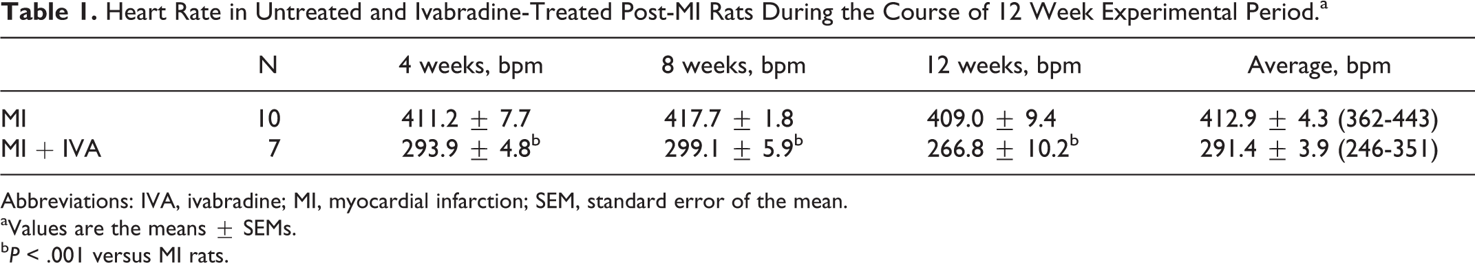
Abbreviations: IVA, ivabradine; MI, myocardial infarction; SEM, standard error of the mean.
aValues are the means ± SEMs.
b P < .001 versus MI rats.
Global and Regional LV Remodeling
In both post-MI groups, the left ventricle demonstrated a large transmural scar of the anterolateral wall and a markedly dilated LV cavity compared to sham-operated rats (Figure 2 and Table 2). Long-term HRR did not modify the size of MI and the major structural parameters of the left ventricle, including LV chamber and cavity transverse dimensions, septal thickness, and the indices reflecting the extent of post-MI chamber remodeling (Table 2). In addition, MI and MI + IVA rats revealed relatively comparable LV weights, suggestive of a similar degree of compensatory myocardial hypertrophy between 2 post-MI groups. Importantly, although the LV weight-to-body weight ratio appeared significantly higher in IVA-treated rats than in untreated MI and sham animals, it was likely a result of smaller body weight in the former. Furthermore, in contrast to untreated post-MI rats, continuing HRR prevented compensatory thickening of the remaining noninfarcted FW myocardium in MI + IVA rats (Figure 2) that in conjunction with a markedly expanded LV cavity could be indicative of higher LV wall stress in IVA-treated animals (Table 2).
Structural Parameters of the Left Ventricle in Sham, Untreated, and Ivabradine-Treated Post-MI Rats.a

Abbreviations: n, number of rats; LVFW, left ventricular free wall; BW, body weight; VW, ventricular weight; LVW, left ventricular weight; RVW, right ventricular weight; CSA, cross-sectional area; MI, myocardial infarction; LV, left ventricular; IVA, ivabradine; SEM, standard error of the mean.
aValues are the means ± SEMs. Remodeling index = (LV cavity CSA of MI heart/average LV cavity CSA of sham hearts) × (LVFW thickness of MI heart/average LVFW thickness of sham hearts). Expansion index = (LV cavity area/LV area) × (septal wall thickness/scar thickness); scar thinning ratio = scar thickness/septal wall thickness.
b P < .01 versus shams.
c P < .01 versus MI rats.
d P < .05 versus shams.
e P < .05 versus MI rats.
Cardiac Myocyte Size and Coronary Capillary Density
At the tissue level, 12 weeks of HRR did not substantially alter the transverse size and regional density of cardiac myocytes, myocardial capillary density, or the capillary to myocyte ratio compared to untreated post-MI rats (Table 3). However, IVA-treated animals revealed a significantly lower density of coronary capillaries and markedly reduced capillary to myocyte ratio within the surviving FW epimyocardium compared to age-matched shams (Table 3). These findings are indicative of either limited adaptive angiogenic response or adverse spatial rearrangement of tissue components within the ventricular wall of MI + IVA rats.
Cardiac Myocyte Diameter and Cross-Sectional Area, Capillary and Cardiac Myocyte Numerical Densities, and Capillary to Myocyte Ratio in the Left Ventricle of Sham, Untreated, and Ivabradine-Treated Post-MI Rats.a

Abbreviations: n, number of rats; CD, capillary density; MD, myocyte density; C/M, capillary to myocyte ratio; MI, myocardial infarction; IVA, ivabradine; SEM, standard error of the mean.
aValues are the means ± SEMs.
b P < .05 versus shams.
Interstitial Collagen Content and Mast Cell Density
Middle-aged rats from the MI + IVA group demonstrated a significantly lower content of fibrillar collagen within the interstitium of the noninfarcted FW and the septum compared to both untreated post-MI rats and age-matched sham-operated animals (Figure 3A). Moreover, the reduced collagen content in IVA-treated rats was associated with a relatively lower density of mast cells, especially in the interstitium of the noninfarcted FW (Figure 3B), suggesting a possible functional link between a small number of mast cells and the lower collagen content in post-MI rats with long-term HRR.

The content of interstitial collagen (A) and the density of mast cells (B) in the left ventricular (LV) free wall and septum of sham (n = 6), MI (n = 8), and MI + IVA (n = 7) rats. Values are means ± standard error of the means. *P < .05 and **P < .01 versus shams; §§ P < .01 versus MI rats. IVA indicates ivabradine; MI, myocardial infarction.
Structural Components of LV Scar
Twelve weeks of IVA treatment did not modify any of the major structural components within the transmural scar compared to untreated post-MI middle-aged rats, including the overall content of fibrillar collagen (Table 4), indicating that collagen turnover within the interstitium of noninfarcted LV myocardium and the scar are most likely controlled by the different mechanisms.
Structural Components of the LV Scar in Untreated and Ivabradine-Treated Post-MI Rats.a

Abbreviations: n, number of rats; CMs, cardiac myocytes; CSA, cross-sectional area; SMCs, smooth muscle cells; MI, myocardial infarction; LV, left ventricular; IVA, ivabradine; SEM, standard error of the mean.
aValues are the means ± SEMs.
b P < .05 versus MI rats.
Effect of Chronic IVA Treatment on LV Systolic Function
At the end of the experiment, the middle-aged rats from both post-MI groups had significantly larger end-diastolic and end-systolic volumes as well as the end-diastolic volume to LV mass ratio (a functional index of LV chamber remodeling) compared to sham-operated animals (Table 5). Moreover, IVA-treated and untreated post-MI rats demonstrated a comparable drop in ejection fraction, ∼54% and ∼58%, respectively, compared to age-matched shams (P < .001), suggesting severe HF in both post-MI groups. In addition, in MI + IVA rats, a significant reduction in heart rate in combination with unchanged stroke volume caused a marked ∼21% decline in cardiac output (P < .05) compared to sham-operated animals (Table 5). Furthermore, according to the percentage change in the values of LV volumes and ejection fraction that occurred between days 1 and 84 post-MI, the scale of LV structural and functional alterations appeared to be comparable between IVA-treated and untreated middle-aged rats (Figure 4).

The percentage change in ejection fraction, end-diastolic and end-systolic volumes, and stroke volume between 24 hours and 12 weeks after MI recorded in the same rats from MI (n = 10) and MI + IVA (n = 7) groups. Note in MI + IVA rats, all parameters were reevaluated 48 hours after abrupt termination of ivabradine treatment (without IVA, without ivabradine; see Material and Methods for details). Values are means ± standard error of the means. §§ P < .01 versus MI rats; † P < .01 versus MI + IVA rats in the presence of ivabradine. IVA indicates ivabradine; MI, myocardial infarction.
Global Dimensions and Functional Parameters of the Left Ventricle in Sham, Untreated, and Ivabradine-Treated Post-MI Rats.a

Abbreviations: EDV, end-diastolic volume; ESV, end-systolic volume; BW, body weight; MI, myocardial infarction; LV, left ventricular; IVA, ivabradine; SEM, standard error of the mean; n, number of rats.
aValues are the means ± SEMs. Note in MI + IVA rats, all parameters were reevaluated 48 hours after abrupt termination of ivabradine treatment (without IVA; see Material and Methods for details).
b P < .01 versus shams.
c P < .001 versus MI rats.
d P < .05 versus shams.
e P < .01 versus MI rats.
f P < .05 versus MI + IVA rats.
g P < .001 versus shams.
h P < .05 versus MI rats.
i P < .01 versus MI + IVA rats.
Effect of Abrupt IVA Withdrawal on LV Systolic Function and Myocardial Perfusion
To determine whether long-term IVA-induced HRR could cause the sustainable alterations in LV functional properties that might be masked by the presence of IVA or its metabolites, IVA delivery was abruptly terminated at the end of a 12-week experimental period for 48 hours to allow the complete IVA clearance from MI + IVA rats before reanalyzing their LV systolic performance and assessing the levels of regional myocardial perfusion.
Two-day long IVA withdrawal led to a significant ∼25% rise of heart rate in rats of the MI + IVA group, compared to the prewithdrawal level in the same animals, although it did not reach the level detected in rats from either untreated MI or sham groups (Table 5). The abrupt interruption of the HRR in animals from the MI + IVA group did not alter end-diastolic volume, end-diastolic volume-to-mass ratio, and stroke volume, but it markedly increased end-systolic volume by ∼33% (P < .05) and, as a consequence, significantly reduced LV ejection fraction by ∼28% compared to rats from the untreated MI group. It is important to note that a substantial decline in LV systolic performance found in these animals was even greater when compared to the level detected in the same rats when IVA was still present in their body, as revealed a ∼32% drop (P < .01) in ejection fraction (Table 5). Further analysis demonstrated that the absence of IVA-induced HRR (the MI + IVA group without IVA) had significantly exaggerated the extent of the dimensional and functional changes occurred in the left ventricle of MI + IVA rats over the 12 experimental weeks compared to untreated post-MI animals (Figure 4). Together, these findings suggest that abrupt IVA withdrawal following the extended period of treatment had a harmful impact on systolic HF in post-MI middle-aged rats.
On the other hand, the acute deterioration of LV systolic performance detected in MI + IVA rats after abrupt IVA withdrawal was not associated with significant alterations in regional myocardial perfusion as compared with the rats from the untreated MI group, although mean arterial pressure was markedly lower in MI + IVA rats at the baseline (Table 6), probably because of the weakened LV systolic capacity. In this context, coronary reserve in the noninfarcted FW myocardium and in the septum 48 hours after withdrawal of IVA was similar to coronary reserve in the untreated MI group (Figure 5). It also appears that significantly lower coronary reserve in MI + IVA rats (P < .05), as compared to shams, was not a consequence of impaired maximal perfusion, but rather due to higher baseline blood flow in the former, probably caused by increased oxygen consumption in FW cardiac myocytes in the absence of a protective effect of chronic HRR.

Coronary conductance and coronary perfusion reserve in left ventricular (LV) free wall and septum of sham (n = 6), MI (n = 8), and MI + IVA (n = 7) rats. Note in MI + IVA rats, all measurements were conducted 48 hours after abrupt termination of ivabradine treatment (without IVA, without ivabradine; see Material and Methods for details). Values are means ± standard error of the means. *P < .05 versus sham group. IVA indicates ivabradine; MI, myocardial infarction.
Coronary Blood Flow and Coronary Vascular Resistance in the Left Ventricle of Sham, Untreated, and Ivabradine-Treated Post-MI Rats.a

Abbreviations: n, number of rats; MAP, mean arterial pressure; CVR, coronary vascular resistance; MCVR, minimal coronary vascular resistance; MI, myocardial infarction; IVA, ivabradine; SEM, standard error of the mean.
aValues are the mean ± SEM. Note in MI + IVA rats, myocardial blood flow and coronary vascular resistance were assessed 48 hours after abrupt termination of ivabradine treatment (without IVA; see Material and Methods for details).
b P < .01 versus shams.
c P < .01 versus shams.
d P < .05 versus MI rats.
e P < .05 versus shams.
Discussion
This study on middle-aged rats with MI-induced HF, which have been treated with the heart rate-lowering drug IVA for 12 post-MI weeks, reveals several important findings. First, chronic HRR significantly decreases interstitial collagen content in the surviving LV myocardium, while preserving the structural integrity of the transmural scar. Such structural alteration in MI + IVA rats favors a better ventricular compliance and is manifested in a greater stroke volume and less attenuated ejection fraction. Second, long-term IVA-induced HRR prevents compensatory thickening of the noninfarcted FW myocardium that in association with a markedly dilated LV chamber probably results in an elevated ventricular wall tension. Third, chronic HRR with IVA is unable to prevent a decline in LV systolic performance mainly because of its inability to attenuate an adverse increase in end-systolic volume. Finally, abrupt termination of IVA-induced HRR markedly exacerbates LV systolic impairment, as indicated a significant drop in ejection fraction, and reduces coronary reserve due to elevated baseline perfusion.
Attenuation of pathologic LV remodeling, particularly after large MI, has been long considered to be a goal of a clinical practice guideline in preventing the development and progression of systolic HF.3,43 Among the key recommendations, the administration of heart rate-lowering drugs, for example, β-adrenoceptor antagonists or β-blockers, appeared to be one of the most beneficial therapeutic options for the long-term cardiovascular outcome in patients with an MI complicated by LV systolic dysfunction 44 and HF. 45 Nevertheless, since β-blockers can be contraindicated or not tolerated in some patients with HF because of the additional nonheart rate-related effects, 46 there has been a search for novel drugs with a “pure” heart rate-lowering effect during last 20 years. 47 One such therapeutic agents, IVA (a selective I f channel blocker), has recently been proven to be effective in patients with HF9,12 and is currently recommended by the European Society of Cardiology for use in adults with chronic HF. 8
Despite the use of IVA in modern-day clinical practice, the overall impact of “pure” HRR on MI-induced LV remodeling and, hence, progression of systolic HF is not yet fully understood. 11 To a great extent, this is caused by the following 2 reasons. First, in all clinical studies, IVA was always administered on top of the standard HF therapy9,12 that made differentiation between the specific effects of IVA-induced HRR and those of other drugs, especially β-blockers, extremely difficult. Second, because the majority of animal studies investigating the impact of “pure” HRR on progression of post-MI HF have been conducted on young or young-adult (2.5- and 3-month-old) rats,15 –20 their findings may not accurately reflect the outcome in humans, considering that systolic HF is predominant in older adults and the elderly patients. 13 In addition, a number of these studies utilized animals with a moderate size MI17,18 or failed to consider the size of infarcted region,15,19,20 although it is well-established that in a rat MI model only animals with the large infarct sizes (≥45% of the LV circumference) would develop progressive structural LV remodeling leading to noticeable systolic dysfunction and HF. 31 Therefore, we believe that our current observations in conjunction with the results from the previous reports performed on middle-aged rats with a large MI (greater than 50% of the LVFW)21 –23 can provide a more coherent account of possible LV alterations which may occur in aged post-MI human heart under the influence of “pure” HRR induced with IVA.
For instance, in contrast to the findings from the long-term studies conducted previously on younger post-MI rats by others,15,16,20 our current results did not reveal the sustainable improvement in systolic LV performance in middle-aged rats following a 12-week period of “pure” HRR with IVA. Moreover, although our own earlier investigations showed that a shorter (4 weeks) period of IVA treatment could ameliorate LV systolic dysfunction in post-MI middle-age rats,21,22 our current data indicate that by 12 weeks after infarction such treatment was not able to maintain the most of the favorable effects. Furthermore, it is important to indicate that the difference in LV contractile performance observed between IVA-treated middle-aged animals in our current study and younger post-MI rats from the previous long-term investigations seems to be independent of the extent of myocardial hypertrophy,15,16,18,20 especially considering the analogous absence of the compensatory ventricular wall thickening compared with untreated post-MI rats. 18 Moreover, the level of myocardial fibrosis remained significantly lower in all post-MI rats with chronic HRR.15 –17 Therefore, taking into consideration these findings as well as the long-known fact that aging itself could markedly alter the intrinsic contractile and metabolic properties of the ventricular cardiac myocytes,48 –51 we hypothesize that the age-related differences in myocyte energetics and/or contractile properties rather than the scale of myocardial hypertrophy and interstitial fibrosis might to some extent be responsible for the inability of the surviving myocardium of older post-MI rats to continually preserve the improved LV systolic capability even in the presence of persisting HRR.
On the other hand, it is important to draw attention to the fact that although the present study as well as our previous investigations21,22 demonstrated that the “pure” HRR was not able to stimulate a significant angiogenic response in surviving LV myocardium of the post-MI middle-aged rats, the substantial capillary growth has been repeatedly reported in their younger counterparts.15,16,18,52 Besides, it has also been shown that the continuing HRR could not produce the additional growth of coronary arterioles in the remaining myocardium of the post-MI middle-aged rats21,22 whereas such growth was evident in younger animals. 52 This phenomenon may plausibly be explained by the fact that the HRR in middle-aged post-MI rats, in contrast to younger animals, 52 was not able to produce the long-lasting activation in any major angiogenic growth factor/receptor systems existing in LV myocardium.21,22 Thus, we imply that the absence of more elaborate angiogenesis and arteriogenesis in the post-MI myocardium of the middle-aged rats can be another key factor that might impede the beneficial effects of the long-term IVA-induced HRR in comparison with younger animals.
It should also be considered that the inefficient expansion of coronary microvessels found in the residual FW myocardium of post-MI middle-aged rats may explain at least partially the worsening systolic HF detected in the present study after the abrupt withdrawal of IVA treatment. In fact, the beneficial implication of extensive angiogenesis within the ventricular walls of young post-MI rats with the chronic IVA-induced HRR has been initially revealed by Mulder et al 16 who found a sustainable improvement in LV systolic function even after the abrupt 3-day long cessation of the IVA treatment. Later, analogous observation has been done in our own study on middle-aged post-MI rats in which permanent HRR was acutely terminated for 2 days following 4 weeks of continuing IVA treatment. 22 However, because in the current study the marked deterioration in systolic function associated with the acute termination of long-term HRR had even exceeded of that in untreated post-MI rats, it is reasonable to suggest that other factors can also be involved.
Indeed, according to our current data, the post-MI rats from the IVA-treated group had a significantly thinner, than untreated rats, LVFW, whereas both groups of post-MI rats showed a comparable LV cavity expansion. In addition, it seems that IVA-treated rats had the substantially lower number of cardiac myocytes across the ventricular wall since the residual myocytes in the surviving FW myocardium had almost similar transverse dimensions in both post-MI groups. Most of the time, such adverse structural alteration of the LV chamber in post-MI heart has been proven to lead to the elevated regional wall tension,53,54 and hence, to impaired energy metabolism 55 and altered oxygen consumption 56 in remaining cardiac myocytes. Therefore, it is feasible to predict that the acute termination of chronic HRR, resulting in significant shortening of the myocardial perfusion time, in combination with the higher wall stress would cause a marked increase in oxygen demand by the residual cardiac myocytes that might be reflected in the compromised coronary reserve. In accordance with such notion, our current study failed to demonstrate a sustainable recovery of coronary reserve that was one of the key highlights for the beneficial impact of IVA-induced HRR in our earlier 4-week investigation on the post-MI middle-aged rats. 21 Most importantly to indicate that the deficiency in coronary reserve observed in this study was not related to the impairment in maximal endothelium-independent perfusion (a functional index of the arteriolar growth); instead it was primarily caused by the higher level of baseline coronary conductance, as a plausible evidence of the increased oxygen consumption in the remaining overloaded cardiac myocytes. Thus, we believe that the abrupt termination of the IVA-induced HRR in the middle-aged rats with substantial post-MI LV remodeling produced a highly unfavorable balance between a markedly elevated cardiac myocyte work load and a drastically diminished metabolic/oxygen supply leading to the acute deterioration of LV systolic function, and hence, significantly worsening HF.
Taken together, our data provided the first evidence documenting that long-term “pure” HRR with IVA does not provide sustainable benefits for LV systolic performance in middle-aged rats with severe post-MI HF. Moreover, we strongly believe that the abrupt termination of IVA-induced HRR in aged failing heart following an extended period of treatment can lead to acute worsening systolic HF.
Study Limitations
The current study examined the effect of IVA-induced HRR on LV structural remodeling, systolic performance, and myocardial perfusion only at 1 time point during a post-MI period. Therefore, it is difficult to corroborate whether persisting HRR would continue to modify these parameters at later post-MI stages. Furthermore, because only 1 level of HRR was utilized, the effects of other HRR regiments on the assessed parameters remained unknown. Additionally, in dramatic contrast to humans, the middle-aged rats were able to regularly survive a large MI that raises a concern regarding a resemblance between the pattern of LV remodeling in middle-aged rats and humans. Finally, our study focused exclusively on the effects of IVA-induced HRR, suggesting that our data cannot directly correspond to a condition in patients with HF in which these effects would always be intermixed with the impacts from other guideline-based medications.
Footnotes
Acknowledgments
The authors thank Ms Alice O’Connor for the excellent assistance with histological techniques.
Author Contributions
E. I. Dedkov and R. J. Tomanek contributed to conception and design, acquisition, analysis, and interpretation, drafted the article, critically revised the article, gave final approval, and agree to be accountable for all aspects of work ensuring integrity and accuracy. Y. Bogatyryov, D. S. McCooey, and R. M. Weiss contributed to acquisition, critically revised the article, gave final approval, and agree to be accountable for all aspects of work ensuring integrity and accuracy. L. P. Christensen contributed to acquisition and analysis, critically revised the article, gave final approval, and agrees to be accountable for all aspects of work ensuring integrity and accuracy.
Declaration of Conflicting Interests
The author(s) declared no potential conflicts of interest with respect to the research, authorship, and/or publication of this article.
Funding
The author(s) disclosed receipt of the following financial support for the research, authorship, and/or publication of this article: This work was supported by National Institute of Health grants RO1-HL62587 (to Dr Tomanek) and S10-RR026293 (to Dr Weiss), and in part by research funds from Institut de Recherches Internationales Servier (PHA-16257-067-USA; to Dr Tomanek) and the New York Institute of Technology College of Osteopathic Medicine (to Dr Dedkov).