
Abstract
Background
The past decade has shown a sharp incline in the human papillomavirus (HPV) infection associated oropharyngeal carcinoma cases, especially in men younger than 60 years old. Tonsils are one of the key sites, within the oropharyngeal region, which shows malignant changes due to HPV infection, and there is very limited literature to understand the specific dynamics in the tonsillar areas.
Objective
This critical review was undertaken to explore and unravel the bio-molecular interactions and the role of specific proteins associated with HPV infection induced tumorigenesis for the tonsils.
Design
A systematic search of the literature was performed utilising keywords and MeSH terms related to HPV and tonsillar carcinoma in PubMed, Scopus, Embase, and Web of Science without restrictions on dates until July 2023. All studies that reported on molecular biomarkers or genes/genetic proteins in the context of HPV associated tonsillar carcinoma were included in the study.
Results
Preliminary searches revealed a total of 2734 studies of which 23 satisfied the final inclusion criteria and were included. More than 25 proteins and biomarkers were identified, and their role in the malignant process was extracted and compiled. This review also presents a short excerpt on each of the molecules identified to provide a better understanding of the pathogenesis.
Conclusion
Given the rapidly increasing number of cases, there is an urgent need for more focused research on virally induced tonsillar cancers, to develop a better understanding, and for clarity of management and treatment.
Plain Language Summary
Human Papillomavirus is a common, sexually transmitted infection which effects more than 80% of people at least once in their lifetimes. In most cases it is cleared from the body spontaneously with little or no impact, but some types can persist and induce a cancerous change in the affected organs (cervix, penis, anal, oropharynx, tonsil). To elaborate on any specific site impacted by cancer, the understanding of molecular level events is critical, and information linking different events in the tonsils specifically is sparse, This review is an attempt to primarily collate all the HPV and tonsillar cancer data together, and generate an understanding of how cancer occurs, and different strategies to manage it.
Introduction
Human papillomavirus (HPV) viruses are a heterogeneous group with circular, double-stranded, non-enveloped DNA and more than 200 genotypes have been isolated. These viruses have an affinity to infect and proliferate in the epithelial and mucous cells. 1 HPV infection is a sexually transmitted infection, 2 with women having an 85% chance and men having and 91% chance of acquiring it, at least once in their entire lifetime. 3 Given the odds, it is not surprising that the global prevalence of this infection has grown considerably over the past few decades. 4 Global burden of cancer indicates that, HPV infections alone contribute towards more than 80% of anal, more than 30% of oropharyngeal, around 80% of vaginal, and 50% of penile cancers worldwide. 5
Thirty HPV genotypes are currently clustered as alpha-papillomavirus (‘mucosal HPV types’), characteristic of infecting oro-genital mucosal tracts.1 1 Although HPV viruses characteristically wash out of the system after a transient phase of usually subclinical asymptomatic infection, certain virus subtypes show persistent behaviours and can initiate malignant changes within the mucosal cells. 6 This subgroup is termed the “high-risk” subtypes, with currently 14 subtypes recognised under this category. 1
Tonsils have been identified as the most common site to be infected within the oropharynx. 7 Traditionally, the incidence of tonsillar cancers was attributed to smoking and alcohol habits, but more recently the cases have been associated with a high-risk HPV persistent infection. 8 Alarmingly, HPV infection-associated tonsillar cancer cases increased by 30% in a period of 4 years (2005 to 2009), whereas the tonsillar cancer cases not associated with HPV infections did not show similar trends. 9 Thus, indicating a risk of increasing number of high-risk HPV infections, inducing malignant changes. In 2012, the American Centre for Disease Control and Prevention (CDC) also declared oropharyngeal cancer as the most frequently diagnosed cancer linked with persistent high-risk HPV infections, and the prevalence has now well exceeded the prevalence for cervical cancer. 6 Although, the rising numbers are worrisome, the good news is that HPV infection-associated tonsillar cancers have a 25% higher 5-year overall survival rate, thus indicating a more favourable prognosis. 10 The detection of HPV DNA after chemotherapy or radiation therapy has shown to be predictor for risk of recurrence and survival outcomes. 11 This has clear clinical implications, which helps in determining risks, but data regarding assessment and evaluation of HPV-DNA in tonsillar cancers is sparse, and more research is recommended, exploring this connection.
It has been observed that not all high-risk HPV infections are persistent, and induce cancerous changes, but there are certain molecular interactions which ascertain persistence and progression of cancerous changes. The molecular pathogenesis of HPV infection in cervical cancer has been well researched and documented thus, providing an insight into its probable mechanism in the tonsils as well. The current knowledge based on research is limited and there is lack of dedicated evidence towards tonsils as an HPV affected site. Due to this gap, there is limited understanding of any unique circumstances around designing treatment strategies for hpv-associated tonsillar carcinoma. This review is an attempt to colate the different molecular events, in order to encourage the translation of this knowledge into clinical strategies. Viruses tend to induce cellular interactions and incite certain biomolecules to act in an uncontrolled manner, initiating cellular altercations and promoting tumorigenic environments. The cancer inducing viral - host interactions at a cellular level provide a clear understanding into the progression of the disease as well as encourage the development of targeted molecular therapy. This review aims to provide a comprehensive understanding of the bio-molecular interactions and to elucidate the normal and abnormal functions of proteins involved in HPV infections associated with tonsillar cancers.
Materials and methods
In order to ensure a comprehensive and complete representation of the current data related to HPV infection-associated tonsillar cancer, a systematic search strategy was employed. After extracting the relevant information, short excerpts on each biomarker and molecule identified, were presented. The possible role in tumorigenesis was highlighted and discussed. This review was inspired by the findings of a previously published systematic review, which evaluated the clinical behaviour of HPV infection-associated tonsillar cancers, as compared to tonsillar cancers not associated with HPV infections. 12 The findings of the study have been reported following the PRISMA guidelines 13 ; however, as the findings collated in the results are narrative in nature, a meta-analysis was not planned for this study. The study has been registered with PROSPERO (CRD42022306602).
Given that no human or animal experiments or data were involved in the curation of this study, no ethical/review board approval was sought.
Data Sources and Searches
A structured reproducible literature search (Appendix) was performed using keywords and MeSH terms related to human papillomavirus and tonsillar carcinoma. The search was performed in July 2023 in the PubMed (including the operators ‘OR’ and ‘AND’, in combination with subject terms (‘MeSH Terms’) and free text terms (‘Text Word’)) and modified to the Scopus, Embase and Web of Science databases. The academic search engines utilised for completing the search for this study were exhaustive, ensuring capture of all relevant data. Bibliographies of the relevant studies were then manually searched to identify additional relevant studies not captured by the search engines.
Study Selection
A predefined inclusion criterion was determined for this review (studies specifically evaluating HPV-associated tonsillar carcinoma). No restrictions on study design (cross-sectional, cohort and case-control, retrospective, case reports, case studies, and interventional studies) were imposed, excluding studies presenting secondary evidence, like systematic reviews. Titles and abstracts of all the identified studies were retrieved and analysed against the inclusion and exclusion criteria. All of the studies deemed fit to be included in the full-text review stage were then retrieved and evaluated. Excluded studies included duplicates, studies not including HPV infection-associated tonsillar cancer, or had no full text available.
Data Extraction and Quality Assessment
Data extraction from the final list of relevant studies was performed and collated to a Microsoft Excel document. Quality assessment of the shortlisted papers was done using the Joanna Briggs Institute (JBI) appraisal tools,1 14 which is essentially a series on nine questions assessed by two independent reviewers under ‘Yes’, ‘No’ or ‘Unclear’ categories. More information on the quality assessment of included papers has been published elsewhere. 12 The extracted and recorded data included: characteristics of the studies like country and year of publication, study design, biomarker studied, inference of study exploring the potential role of the particular protein in HPV-associated tonsillar carcinoma and the main findings were further explored.
Results
Study Characteristics
A total of 2734 studies were identified and transferred to the reference managing software, EndNote (X9.3.3). After removal of duplicates, titles and abstracts of 1508 studies were screened. Adhering to the inclusion and exclusion criteria, a total of 38 studies proceeded to the full-text review stage. After reviewing the full texts of the retrieved studies, 23 studies15–37 were deemed eligible to be included in the final review (Figure 1). PRISMA 2020 flow diagram for this systematic review which included searches of databases, registers and other sources.

aUSA – United States of America, IHC - Immunohistochemistry, ISH – in-situ hybridization, PCR – Polymerase chain reaction, MMA – Microsatellite marker analysis, WB – Western blotting, SBH – Southern Blotting Hybridization, TMA – Tissue microarray, FISH – Fluorescence in situ hybridization, pRb- Retinoblastoma tumour suppressor protein, miR - microRNA, FGFR3 - Fibroblast growth factor receptor 3, PIK3CA - Phosphatidylinositol-4,5-Bisphosphate 3-Kinase Catalytic Subunit Alpha, PTEN - Phosphatase and tensin homolog, AGR – Anterior Gradient, CD - Cluster Differentiation, MMP- Matrix metalloproteinase, 38 EFGR – Epithelial Factor Growth Receptor, HLA – Human Leucocyte Antigen, SLPI - Synaptotagmin like protein, LMP7 - Low mass protein-7.
Most of the studies used the polymerase chain reaction (PCR) technique,15–20,22–25,27,30–32,36 and the other popular choices were immunohistochemistry (IHC),15,19–22,25,26,28,29,32–35,37 in-situ hybridization (ISH),15,19,20,22 tissue micro-assay analysis (TMA),20,23 microsatellite marker analysis (MSA), 15 western and southern blotting techniques.15,36
Role of Different Proteins/Genes and Factors in Tonsillar Tumorigenesis Specifically Associated With HPV Infections.

Discussion and Review
More than 25 biomarkers were identified through this review (Table 2), and the following section presents a short excerpt on each. The majority of the papers included in the review are from Europe, and a few were from America, Japan and Korea. Given that the prevalence of HPV-associated cancers is higher in lower socioeconomic countries, this paper also highlights the need of more representative data and studies to develop a deeper clearer understanding of the same.
The role of each biomarker in prognosis, tumour formation and metastasis, in cancers of other sites as well as specifically, HPV infection-associated tonsillar cancers is discussed. This overview of the different biomarkers identified in HPV-associated tonsillar cancers can help guide molecular pathways specific to the tonsillar cancers (Figure 2). This will also help in enhancing an understanding of the molecular level events in theses lesions, which could impact potential screening, diagnosis and treatment strategies. Although clinical implications cannot be directly suggested at this stage, but it definitely has the potential to impact clinical management and deliver translatable outcomes for patients diagnosed with HPV associated tonsillar cancer. Expression of proteins/genes and factors in tonsillar tumorigenesis specifically associated with HPV infections.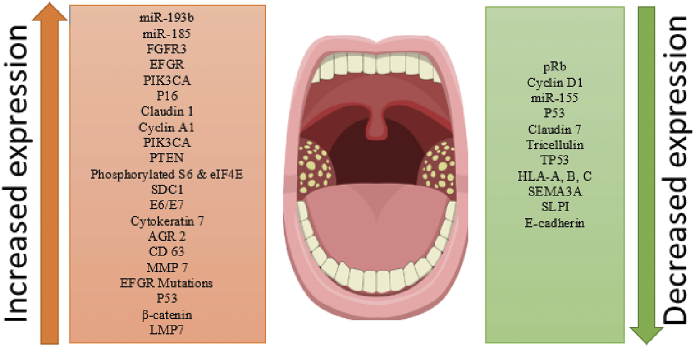
Retinoblastoma Tumour Suppressor Protein (pRb)
First identified in 1971 39 as a key gene (located on chromosome 13) involved in hereditary retinoblastoma, 40 pRb plays a significant role in regulating pathways associated with cell cycle, 41 cell growth, 42 inflammation, 43 differentiation, 43 replication, renewal, turnover and death.40,43–48 It has been speculated that in addition to cellular regulation, pRb is also involved in processes impacting tumour growth and extra-cellular interactions like genomic instability and cellular de-differentiation. 43 The distinct role of pRb as a tumour suppressor protein can be explained by its recognised role in key processes involved in carcinogenesis like proliferating in unsuitable environments and the tendency to metastasize. Another important aspect of pRb associated with tumour initiation and growth, cytokine regulation and research links pRb inactivation with increased secretion of pro-inflammatory cytokines like IL-6, 43 thus establishing an inflammatory tumour microenvironment. The role of pRb has been reported in most of the cancers like prostrate,49,50 breast,51,52 small-lung carcinoma,53,54 cervix55,56 etc.
Cyclins (D1 and A1)
Cyclins are critical proteins, first discovered in 1982, which facilitate the cell cycle and the processes involved in the cell cycle, by activating enzymes.57,58 The cyclin family consists of cyclins (A, B, D, E, F) and cyclin-dependent kinases (CDK1, 2, 4, 6), all of which have distinct functions in the cell cycle and dysregulation of these proteins have been associated with tumorigenic processes.59,60
Fibroblast Growth Factor Receptor 3 (FGFR3)
Located on chromosome 4, 61 the Fibroblast Growth Factor Receptor gene is responsible for coding this protein. FGFR3 is intricately related to tyrosine kinases. Tyrosine kinases are the specific enzymes within cells that orchestrate the transfer of a phosphate group from an ATP (Adenosine triphosphate) molecule to a protein with tyrosine residues, thus thanatophoric dysplasia, achondroplasia, and hypochondroplasia
p53
p53 is a tumour suppressor gene that was first identified in 1979 on chromosome 17. 62 It is critically involved in cell cycle regulation with either DNA reparation or apoptosis induction. Replication of damaged DNA involves p53 to be expressed and produced in the nucleus of the cell. Here, it undergoes chemical modifications 63 – phosphorylation and acetylation – allowing it to enter its functionally active form to transactivate target genes. The genes targeted are contingent on the extent of DNA damage, as the cell could undergo apoptosis or repair the DNA with the cell cycle being arrested at the G1/S checkpoint. 64 In the event of severe DNA damage, p53 activates pro-apoptotic genes of the Bcl-2 family, 65 including Puma and Noxa. Alternatively, in the latter case with p53-mediated cell cycle arrest, p21 undergoes transactivation to repair DNA before the cell returns to the normal cell cycle. 66 Dysfunctional p53 contributes approximately 38% – 50% of human cancers 67 with the majority of dysfunctionality being resultant of genomic mutations that prevent appropriate protein conformation. 68 Somatic p53 mutations are associated with various aggressive carcinomas 69 as they transactivate oncogenic and growth-promoting genes. 70
PIK3CA - Phosphatidylinositol-4,5-Bisphosphate 3-Kinase Catalytic Subunit Alpha
Phosphatidylinositol-4,5-Bisphosphate 3-Kinase Catalytic Subunit Alpha (PIK3CA) is a gene encoding p110 alpha (p110α) which is a catalytic subunit of phosphatidylinositol 3-kinase (PI3K). 71 PI3K is a critical enzyme mediating the progression of the cell cycle and growth by activating various downstream factors, including PDK1. 72 Cancer-specific mutations on PIK3CA were discovered in 2004 73 on chromosome 3, 71 with alterations increasing PI3K signalling, which in turn promotes growth factor independent development. 71 In patients testing HPV-positive and having head and neck squamous cell carcinomas, the genetic mutation was seen in 56%, whilst 34% 74 of HPV-negative patients, had the carcinoma and the mutation.
PTEN
Phosphatase and tensin homolog (PTEN) is a tumour suppressor enzyme that dimerizes 75 with another PTEN to allow it to bind to the cell membrane, where it dephosphorylates other enzymes, including PI3K. PTEN further aims to promote DNA repair and assist with chromosome stability. 76 Discovered in 1997 77 on chromosome 10, 78 loss of the PTEN protein is more frequently associated with cancer, than PTEN gene mutations which are seen in approximately 13.5% of all human cancers. 79 With the loss of the negative regulator for PI3K, the PI3K pathway 80 is activated with cellular proliferation and survival resulting. Mutated PTEN has a reduced ability to repair damaged DNA and maintain chromosomal stability, hence, a poorer prognosis of cancer is seen from higher penetrance. This is also an outcome of PTEN deficiency as DNA replication and mitotic spindle formation are hindered. 81 Genetic alterations in PTEN are seen in greater frequencies amongst HPV-positive patients with oral squamous cell carcinomas, in comparison to patients testing HPV-negative.82,83 Whilst somatic PTEN mutations are seen in various cancers, there is greater emphasis on endometrial cancers and glioblastomas. 84
Phosphorylated S6 and Phosphorylated eIF4E
Phosphorylated S6 (p-S6) and Phosphorylated Eukaryotic Translation Factor Initiation Factor 4E (p-eIF4E) are involved in specific mRNA translation during protein synthesis, 85 with phosphorylation of these individual proteins being heavily controlled. Phosphorylation is mitigated by downstream effects of the mammalian target of the rapamycin (mTOR) pathway with dysregulation being apparent in various cancers. 86 p-S6 and p-eIF4E were discovered in 1974 87 and 1976, 88 respectively, with elevated levels of either protein being indicative of uncontrolled cell growth 87 from increased protein synthesis. Enhanced p-eIF4E levels further suppress apoptosis with anti-apoptotic and pro-proliferative mRNAs 89 being translated.
Claudin (−1, −7)
Located across 12 chromosomes by over 17 genes, 90 the claudin family consists of over 24 membrane protein members, each playing a key role in tight junctions. With tight junctions mitigating epithelial cell polarity, loss of tight junctions is closely associated with metastatic potential. 91 An imbalance in claudin (−1, −7) is seen in increased tumorigenicity of breast tissue. Down-regulation of claudin-1 expression in breast epithelial cells has been seen to have neoplastic effects 92 with aggressive characteristics. This is similarly seen with claudin-7, as increased cellular discohesion is consistent with high-grade lesions. 93 Hence, tumour behavior has been seen to be heavily contingent on claudin (−1, −7) expression, with histological grade, invasiveness, and metastatic potential being determined. Tight junctions are further disrupted by HPV, 94 however, the association between HPV and the claudin members is limited since HPV is seen to instead degrade 95 MAGI-1.
Tricellulin
Analogous to its name, tricellulin is a protein found at junctions of three cells in the body, also called MARVELD2. 96 Intercellular junctional complexes include tight junctions, adherens junctions, and gap junctions, they intercede adhesion between connecting endothelial and epithelial cells. 97 Due to an epithelial barrier nature, it has been speculated that altered tricellulin expression is associated with advancing cancers, cellular invasion, and metastasis. 98 Research indicates the impact of altered tricellulin on NF-Κβ pathways and the epithelia-mesenchymal transformation pathways.98–100 Studies have reported an altered expression of tricellulin and tight junction proteins in colorectal cancers, 98 pancreatic cancers, 101 hepatocellular carcinomas, 102 and endometrial cancer. 103 Kondoh et al, 22 investigated the role of claudin-1,7 and tricellulin tight junction proteins in tonsillar cancers, and although there was some loss of expression for tricellulin junctional proteins in tonsillar squamous cell carcinomas, there was no loss of expression in the cases associated with HPV infections. This study states that HPV infections would not have an impact on the tight junctional proteins, and suggested alternative mechanisms of tumour invasion and metastasis. 22
Syndecan 1
Belonging to the four-member syndecan proteoglycan family, this protein is an integral membrane protein with a central core protein with multiple glycosaminoglycans (both chondroitin sulfate and heparin sulfate) side chains. 104 Due to the involvement of syndecan-1 in cellular processes like proliferation, migration and other cell-cell matrix interactions, it positions itself as an important protein during tumour growth and invasion. 105 The expression of Syndecan-1 in each tumour type is contextual and its relevance depends on the cell type and its significance. Szatmari T et al, 105 presents an explorative review of the prognostic role of syndecan-1 with relation to the cellular localisation in different organs. Lee SH et al, 23 studied the prognostic significance of syndecan-1 in tonsillar cancers and found that in cases with SDC1 positivity, the outcome was unfavourable.
Cytokeratin (CK) 7
Epithelial cells are structurally comprised of cell organs like mitochondria, ribosomes, Golgi bodies and, cytoskeleton proteins which constitute the structure and function of the cytoskeleton of the cell. 106 There is an approximately 20 different cytoskeletal proteins, the expression of which is retained by the tumour cells. Cytoskeletal markers are routinely used to diagnose tumours of unknown origin or poorly differentiated cells.107,108 Expression of CK-7 is strong in epithelial-derived tumours like colorectal cancers,109–112 ovarian cancers113,114 and cervical cancers. 115 Research with specifically HPV-associated tumours has revealed that CK-7 has a role in viral replication, thus promoting high-risk HPV-associated tumorigenesis. 116
Anterior Gradient (AGR) 2
Belonging to the protein disulfide isomerase (PD 1) family, the anterior gradient 2 (AGR2) is an estrogen-responsive developmentally regulated gene. 117 First recognised in breast cancer cells as an estrogen receptor target, 118 but now also observed as overexpressed in other tumours like ovarian, 119 pancreatic, 120 and colorectal cancers. 121
Cluster Differentiation (CD) 63
Encoded by the CD-63 gene, this protein is associated with intracellular vesicles inducing functions on the cell surface like cellular signalling for growth, proliferation and motility.122,123 Platelet activation is one of its recognised roles, 124 as well as involvement in abnormal cellular growth leading to tumour formation.125,126 The exact role of CD-63 in tumorigenesis is a bit controversial with some studies reporting a negative relationship and a few reporting a positive relationship. A systematic review exploring the prognostic value of CD-63 in different tumours was published in 2018, by Koh et al, 127 which reported an inverse relationship of CD-63 expression with cancers of the ovaries, breasts, colon and lungs.123,128–131 However it was found that CD-63 although not overly expressed in the tumor cells helped in the increased production of proteins which promote metastasis and invasion, thus showing higher levels in the plasma as compared to the tumor tissue itself.132,133
Semaphorin 3A (SEMA3A)
Semaphorin 3A is encoded by SEMA3A genes and belongs to the Semaphorin protein family, which are associated with nervous system development. These proteins are also identified as tumour suppressors, many cells possess Semaphorin receptors on their surface and inhibit cellular division and spread. 134 The connection to axons of neurons explains its expression in Alzheimer’s disease, 135 systemic lupus erythematosus, 134 schizophrenia, 135 scar tissues, rheumatoid arthritis, 134 and spinal cord injuries. 136 Additionally, SEMA3A is linked to multiple roles i.e., immune regulation, vascularization and angiogenesis, bone remodelling, organ formation, embryonic development, chemo repellent and oncogenesis 137
Synaptotagmin Like Proteins
One of the primary functions of the cells in our body is to secrete molecules (hormones, lipids, neurotransmitters, etc.) in response to an external stimulus. These molecules are secreted in the form of membrane-bound vesicles by a process called exocytosis. This process includes a step that would show the contact between the membrane of the vesicle and the cell membrane before being secreted from the cell. This contact is mediated by the formation of phospholipid domains called C-2 domains, which include proteins like synaptotagmin like proteins (SLP-1). 138 The SLP-1 protein itself is comprised of an N-terminal and a C-terminal. 139 The role of SLP-1 is unclear in tumour progressions but has been found upregulated in prostate cancer cell lines 140 and endometrial cancer, 141 and its use has been suggested as a potential biomarker.
E-Cadherin
Cadherins are the calcium-dependent (hence the name) molecules that are responsible for forming cell-to-cell adhering complexes, 142 maintaining cellular structure by reinforcing cytoskeletal elements, 143 resisting cellular damage, and also participating in cellular signalling pathways. 144 Cadherins are of various types and have distinct functions but depending on their location, they are divided into E-cadherins (epithelial cells), N-cadherins (neurons), R-cadherin (retinal), VE-cadherin (vascular endothelial), K-cadherin (kidney), H-cadherin (heart), OB-cadherins (osteoblasts), M-cadherins (myotubule), LI-cadherin (liver-intestine) and, P-cadherins (placenta). 145 Given the nature of tonsillar tumours, it can be argued that E-cadherins would be the most important in tumorigenic processes involving the tonsil, oral cavity or oropharynx. E-cadherins, given their adhesive function, are one of the most crucial tumour suppressive molecules and prevent the collection of neoplastic cells to detach from the epithelial membrane and metastasize into distant tissues.146,147 This exchange of information and molecules is referred to as epithelial-mesenchymal transition. 147
β-Catenin
Adhesion and transcription are two key cellular events that regulate carcinogenic transformations, and β-catenin is a protein known to be involved in both processes.148–150 Wnt/β-catenin signalling is a key pathway that regulates almost all cellular events (differentiation, apoptosis, renewal and proliferation). β-catenin is the single most critical component of the complex which needs to be stable to activate this pathway; thus, regulates essential cellular processes. Any deviation or instability can influence these processes, resulting in neoplastic consequences. 151 Dysregulated genetic expressions of this protein have been associated with cancers of the colon,152–154 breast,155–158 ovaries159–161 and liver.162–164 Additionally, β-catenin’s linked to cardiac diseases like cardiomyopathy 165 and congenital heart disorders 166 and, behavioural issues like depression and stress.167–169
LMP7 - Low Mass Protein-7
Low mass proteins 7 and 2 both have a protective effect against cancer antigens and are known to play an active role in immune surveillance via regulating the MHC-1 pathways.170–172 Genetic variations in the DNA sequence for LMP-7 have been associated with altered functional capability thus impacting the antigen processing mechanism and efficiency and is frequently associated with cancers of the colon,173,174 cervix,175,176 blood cells, 177 and gastric tract.178,179 The association with cervical cancers highlights the connection with HPV infections and also a probable impact on the development of the tonsillar, oropharyngeal and other HPV infection-associated cancers. 179 It has already been reported that polymorphism in these genes leads to defects in protein structure and functions eventually impacting the ability of the infected person to clear the infection. 176 Deshpande et al, 176 also elaborates that any genes or proteins involved with the antigen processing for HLA class 1 proteins may have an impact on malignant transformations of cells.
E2
E2 proteins are critical to the replication and transcription processes of the papillomavirus. It is linked to numerous viral survival and progressing processes, as well as proteins involved in the viral life cycle. 180 E2 proteins have been frequently linked to the carcinogenic pathways associated with persistent HPV infection.181–183 A recent systematic review provided evidence of the E2 protein of HPV-16 to be involved with cellular death, via apoptotic pathways; although, the mechanism was still not well understood. 184 The role of these proteins is not well understood, a few studies30,31 have not reported any significance so far, but further research is required to establish its specific role in tonsillar cancers.
E6 and E7
E6 and E7 are oncogenic viral proteins, which instigate cellular processes like angiogenesis,185,186 uncontrolled cell division,187,188 metastasis 189 etc; thus, critical to oncogenesis. Evidence also shows that E6 and E7 prevents apoptosis by destroying apoptosis inducing factors, leading to the production and persistent HPV affected cells.190,191 Uninhibited E6/E7 expression mimics mutational activity of pRb and p53, resulting in an unstable genome, mutations, malignant changes and other cellular changes favourable for malignant transformation. 192 The role of E6/E7 oncoproteins in HPV induced malignancies is well defined and thoroughly reported. 189 Needless to say, these proteins play a critical role in HPV associated carcinogenesis, and are currently considered the gold-standard for diagnosing HPV-associated lesions of the head and neck.
Limitations
As all research papers and studies, the current review also has some limitations. It is primarily a literature review, an attempt to collate all the evidence which could potentially result in clinical implications for HPV-associated tonsillar cancers. Another limitation is that all the proteins identified have been discussed in limited capacity with brief correlations with HPV infections and their role in cancer initiation and progress. Although, this does compile all the theoretical evidence, translational outcomes of this evidence are yet to be researched and implemented. Another limitation is the geographical distribution of the papers included, which was primarily based out of research from Europe, and there is currently a lack of data/research from lower socio-economic countries, where the burden of HPV associated cancers is much higher. Given that this review is based of secondary evidence as generated by studies carried out in different populations, using different methods of investigation and sample size invariances, there is a potential of bias as well.
Conclusion
A clear understanding of the cellular interactions and protein expressions is critical whilst making clinical management decisions. The literature regarding the exact bimolecular mechanism of action for HPV-associated tonsillar cancers is inconclusive and lacks substantial evidence. This review provides a comprehensive overview of the proteins identified and the hypothesized pathogenesis, but further research targeting especially HPV infected cells and the related tumorigenesis process is recommended. The biomolecules and genetic processes involved in the malignant transformation of each cell type are unique and diagnostic as well as therapeutic decisions should not be made based on assumptions. Thus, further corroborating the need for focused bimolecular research for viral-induced carcinogenesis.
Supplemental Material
Supplemental Material - Defining the Molecular Intricacies of Human Papillomavirus-Associated Tonsillar Carcinoma
Supplemental Material for Defining the Molecular Intricacies of Human Papillomavirus-Associated Tonsillar Carcinoma by Sneha Sethi in Cancer Control
Footnotes
Declaration of Conflicting Interests
The author(s) declared no potential conflicts of interest with respect to the research, authorship, and/or publication of this article.
Funding
The author(s) received no financial support for the research, authorship, and/or publication of this article.
Supplemental Material
Supplemental material for this article is available online.
References
Supplementary Material
Please find the following supplemental material available below.
For Open Access articles published under a Creative Commons License, all supplemental material carries the same license as the article it is associated with.
For non-Open Access articles published, all supplemental material carries a non-exclusive license, and permission requests for re-use of supplemental material or any part of supplemental material shall be sent directly to the copyright owner as specified in the copyright notice associated with the article.