
Abstract
Background
Toll-like receptors (TLRs) play an important role in regulation of immune cells and are vital in tumorigenesis due to its crucial role in inflammatory microenvironment regulation, as they promote the synthesis and release of inflammatory cytokines and chemokines. Toll-like receptors 4 and TLRs 9 were found to be highly expressed in breast cancer. The aim of this study is to investigate the soluble toll-like receptors 4 and 9 (sTLR4 and sTLR9) as potential biomarkers for diagnosis and prognosis of breast cancer and their association with the clinicopathological parameters of breast cancer.
Patients and Method
In this retrospective case-control study, 186 female subjects were recruited and divided into three groups, Group I: 62 healthy control, Group II: 62 subjects diagnosed with non-metastatic breast cancer, and Group III: 62 subjects diagnosed with metastatic breast cancer. Enzyme-linked immunosorbent assay (ELISA) technique was used to quantify the levels of sTLR4 and sTLR9 in serum.
Results
Both non-metastatic and metastatic groups showed significant higher levels of both serum sTLR4 and sTLR9 expression compared to healthy controls. Only sTLR9 was significantly increased among metastatic patients compared to non-metastatic group. Serum levels of sTLR9 and sTLR4 were still significantly associated with breast cancer in a multiple logistic regression model (P = <.001). ROC curves showed that both sTLR4 and sTLR9 can be a significant parameter to discriminate between normal females and breast cancer patients.
Conclusion
Soluble toll-like receptors 4 and sTLR9 are over-expressed in patients with metastatic and non-metastatic BC than in benign cases. The expression levels of sTLR4 and TLR9 have clinical interest as indicators of tumor aggressiveness suggested to be prognostic biomarkers. Toll-like receptors may represent therapeutic targets in breast cancer.
Keywords
Introduction
Female breast cancer was the leading cause of cancer in 2020, with 2.3 million new cases (11.7% of all cancer cases) and the fifth leading cause of cancer mortality with 685,000 deaths worldwide. 1 In Egypt, it represents 32.4% of female cancer cases and more than 22,000 new cases are diagnosed each year.2,3 It was reported that over 90% of cancer deaths are caused by metastasis, which is difficult to predict in breast cancer because it is a heterogeneous disease encompassing complex pathologic entities.4,5
An interaction between tumors and the immune system is required for the tumor cells growth and survival and further process angiogenesis and metastasis processes. Alterations in the function and expression of molecules related or not related to the immune system are usually correlated with malignant cell development. 6 The invasive potential of the tumor depends on the interaction between the tumor cells and the tumor microenvironment owing a massive number of immune cells constituting microenvironment cell population majority. 7
The process of activation or inhibition of immune and non-immune cells through recognition of pathogen-associated molecular patterns (PAMPs) and damage-associated molecular patterns (DAMPs) is mainly controlled by the toll-like receptors (TLRs),8,9 which are categorized according to their subcellular localization into two subgroups 10 and their main function is to promote the synthesis and release of inflammatory cytokines and chemokines leading to stimulation of the inflammatory response.11,12
The inflammatory microenvironment which is crucial for the tumorigenesis is regulated by the TLRs. 13 Further, studies demonstrated the role of TLRs in tumorigenesis, development, and metastasis.14,15 Therefore, various TLRS agonists are being investigated in the field of immunotherapy for their anticancer effect.16,17
TLR4 was genetically mapped to chromosome 9q32-33 in humans and its gene is 19 kb in length consists of three exons. 18 TLR4 specifically recognizes microbial lipopolysaccharide (LPS) and activates canonical and non-canonical signaling of NF-κβ activation for the synthesis of pro-inflammatory cytokines and chemokines.19,20 It was demonstrated that TLR4 was expressed in higher levels than other TLRs in human breast cancer, 21 and TLR4 activation could subsequently activate nuclear factor-κβ (NF-κβ) and produce pro-inflammatory cytokines, ultimately stimulating inflammation. 22
TLR9 is a DNA receptor that recognizes microbial and vertebrate. 23 Specifically, TLR9 recognizes the CpG sequence in DNA. 24 DNA recognition by TLR9 initiates a downstream signaling cascade, which includes the adaptor molecule MyD88. 25 TLR9 is expressed in epithelium breast cancer cells. 26 Among the 5 human TLR9 isoforms, expression mRNA of TLR9 A and B isoforms has been detected in breast cancer specimens. 27 TLR9 expression has a significant prognostic significance only in triple negative breast cancer (TNBC). 28
The current study aims to investigate the sTLR4 and sTLR9 as potential biomarkers for diagnosis in non-metastatic and metastatic breast cancer. Further, interrogate their association with the clinicopathological parameters of breast cancer.
Subjects and Method
All participants provided demographic information, which included age, menopausal status, number of children, lactation history, marital status, hormonal contraception use, and family history of breast cancer. These patients were diagnosed by examination, radiological and histopathological investigations. The patients' clinicopathological data, such as tumor size, grade, ER, PR, and HER2 status, tumor subtypes, and TNM stage, were all recorded. Immunohistochemistry was used to check for ER, PR, and HER2 status. Determining the TNM stage of these patients was done based on the American Joint Committee on Cancer Classification.
Results
Socio-Demographic and Clincopathological Data
Basic Characteristics of the Studied Groups.

Clinico-Pathological Data of Breast Cancer Group.

Serum sTLR4 and sTLR9 Levels in Breast Cancer Patients
Comparison of Toll-Like Receptors 4 and 9 Concentrations Among Studied Groups.

**P1 = comparison between healthy controls and malignant groups.
P2 = comparison between healthy controls and metastatic groups.
P3 = comparison between malignant and metastatic groups.
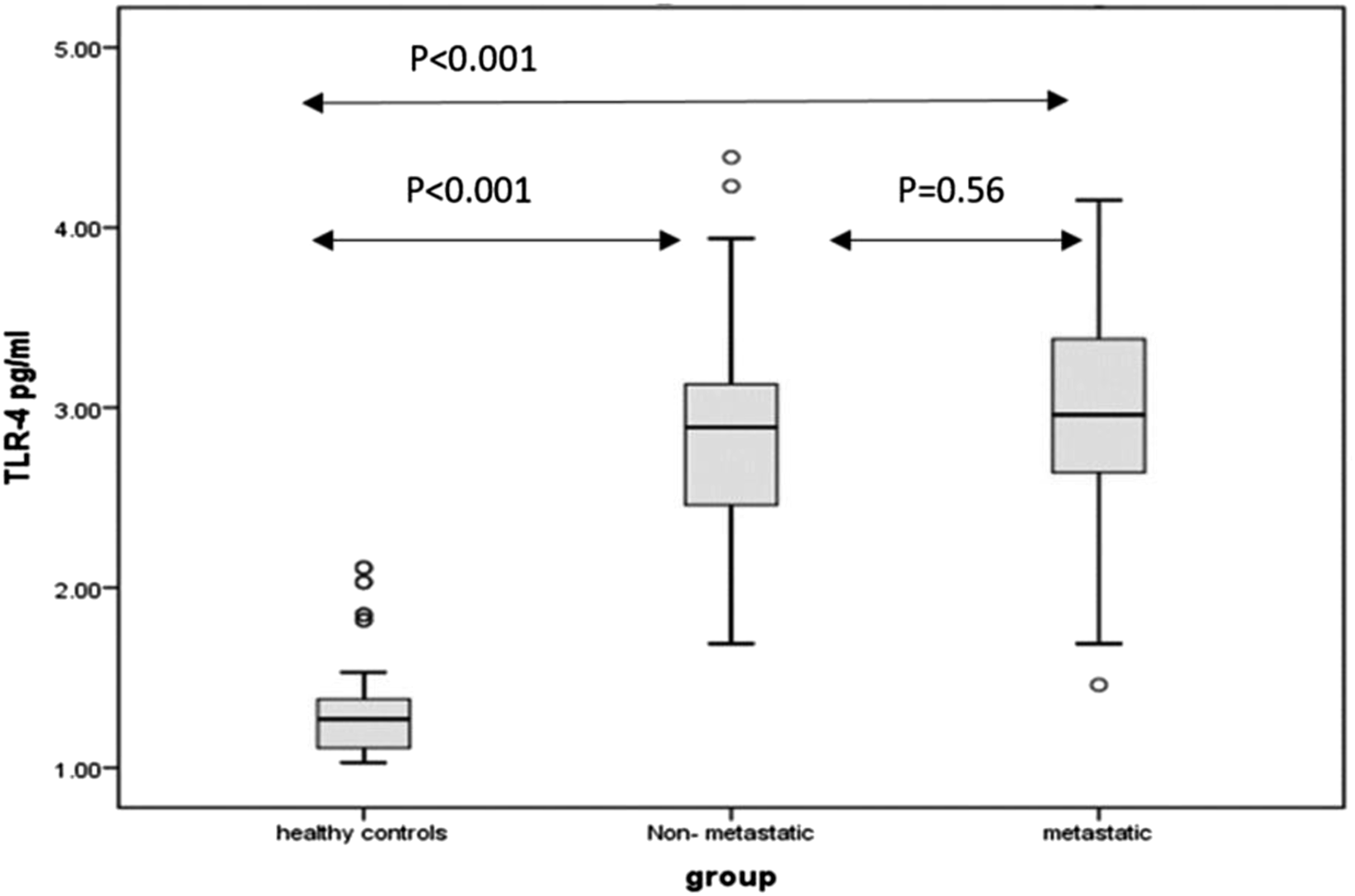
Box and whisker plots representing serum levels of sTLR4 in the non-metastatic, metastatic, and healthy control groups. The horizontal line in the boxes denotes the median value (50th percentile), while the box contains the 25th to 75th percentiles of dataset. The black whiskers mark the 5th and 95th percentiles, and values beyond these upper and lower bounds are considered outliers, marked with black dots.
A highly significant increase was observed in the serum level sTLR9 in non-metastatic (423.65 ± 114.55) and metastatic groups (493.62 ± 153.07) compared to healthy controls women (310.20 ± 52.57). Furthermore, serum sTLR9 was significantly increased among metastatic patients compared to non-metastatic group (P = .02) (Table 3, Figure 2). Box and whisker plots representing serum levels of sTLR9 in the non-metastatic, metastatic, and healthy control groups. The horizontal line in the boxes denotes the median value (50th percentile), while the box contains the 25th to 75th percentiles of dataset. The black whiskers mark the 5th and 95th percentiles, and values beyond these upper and lower bounds are considered outliers, marked with black dots.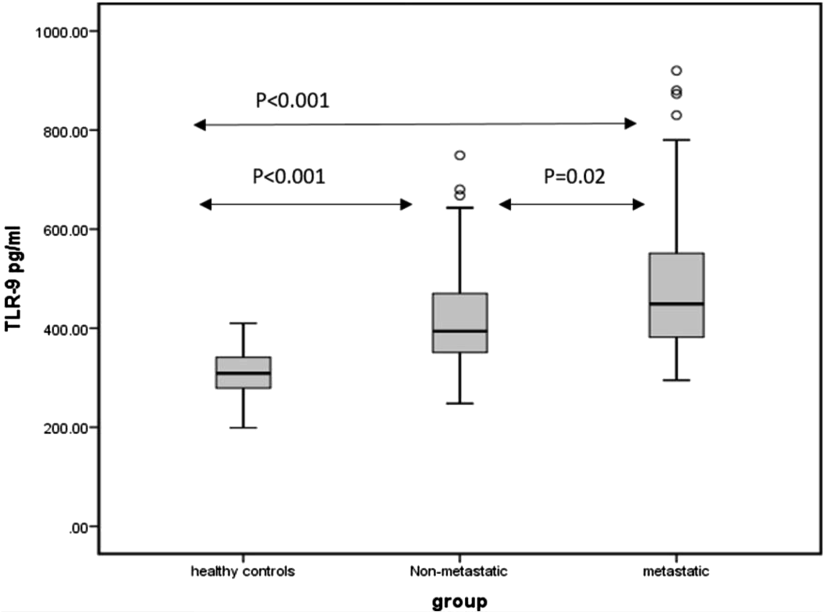
Serum levels of sTLR9 and sTLR4 were still significantly associated with presence of breast cancer after adjusting of: age, co-morbidities, and menopausal status in a multiple logistic regression model (P = <.001).
Diagnostic Significance of sTLR4 and sTLR9 in Breast Cancer Patients
Receiver Operating Characteristic Curve of Serum Toll-Like Receptors 4 and 9 Between Studied Groups.


Receiver operating characteristic curves for prediction capacity of sTLR4 and sTLR9. (A) Non-metastatic vs healthy controls. (B) Metastatic vs healthy controls. (C) Non-metastatic vs Metastatic. sTLR, soluble toll-like receptor; AUC, area under curve.
Comparable results were noticed in ROC curve analysis of serum sTLR9 values of non-metastatic breast cancer and normal healthy females. The area under curve for serum sTLR9 to predict non metastatic breast cancer was determined at 335 ng/mL (AUC: .829 [95% CI, .757-.902], P = <.001) (Table 4). This cut-off value had a sensitivity of 80.0% and a specificity of 70.0% (Figure 3A). Moreover, the AUC for serum sTLR9 to predict metastatic breast cancer reached (0. 918 [95% CI, .871–.964], P = <.001) with 87% sensitivity and 81% specificity at cut-off point = 355 ng/mL (Figure 3B).
Both serum sTLR4 and sTLR9 levels showed a poor predicting value in differentiation between metastatic and non-metastatic breast cancer patients. AUC= (0. 579 [95% CI, .477–.681], 0. 633 [95% CI, .534–.731], respectively) (Figure 3C).
Correlation Between Serum sTLR4 and sTLR9 Expression and Clincopathological Characteristics of Patients With Breast Cancer
Association of Serum Toll-Like Receptor 4 and Basic Characteristics of the Studied Groups.

Discussion
TLRs may play dual roles in human cancers. 9 TLRs play a critical role in tumor cell proliferation, resistance to apoptosis, cell invasion, and metastasis by activating the production of interleukins, tumor necrosis factor-alpha (TNF-α), nuclear factor-kappa Beta (NF-κβ), and metalloproteases and integrins.30,31 TLRs are highly expressed in breast cancer cells, and their activation can induce aggressive tumor behavior, cell proliferation, cell invasion, cell migration, and metastasis.29,32 Soluble TLRs (sTLRs) are considered to be negative regulators of TLR signaling. 33
The current study aims to evaluate the serum levels of sTLR4 and sTLR9 as a potential diagnostic biomarker in metastatic and non-metastatic breast cancer patients and to assess these serum levels as endogenous negative regulators of TLR4 and TLR9 signaling in patients with breast cancer. Moreover, it also aims to investigate their association with different clinicopathological parameters of these patients.
The current study revealed an increase in the serum levels of sTLR4 in non-metastatic (2.96 ± 1.04) and metastatic (3.20 ± 1.21) breast cancer groups in comparison to their levels among the healthy control group (1.32 ± .27). Our results are consistent with El-Kharashy et al., 29 who demonstrated a significant increase in serum sTLR4 in patients with both non-metastatic (1945.2 ± 1709.53 pg/mL) and metastatic breast cancer (7800.1 ± 13 041.28 pg/mL), compared with the control group (1106.8 ± 108.32 pg/mL; P = .0001).
Our results show that serum sTLR4 expression was significantly higher among breast cancer patients with regional lymph nodes involvement compared to patients without lymph nodes involvement (3.15 ± 1.22 vs 2.74 ± .49, P = .04). The same results was found by Zandi et al. 34 who stated that TLR4 expression was up-regulated both at mRNA and protein levels and significantly correlated with the high incidence of lymph node metastasis. Also, Yang et al. 21 found significantly higher levels of TLR4 in malignant breast cancer especially in patients with positive LN involvement.
A dual role of TLR4 was observed in the malignant cell environment; TLR4 has a role in inducing immune response to eradicate malignant cells via the recognition of their DAMPs, 35 while the overexpression of TLR4 associating with pathways such as TGF-β signaling and TP53 was demonstrated to be important in malignant cells invasiveness and metastasis.9,36
For instance, overexpression of TLR4 was demonstrated to have a positive correlation with breast cancer metastasis and was associated with large tumor size, distant metastasis, and recurrence upon investigating 74 patients with breast cancer tumors. 5 Furthermore, TLR4 is responsible for the pro-inflammatory microenvironment of tumors by inducing the production of pro-inflammatory cytokines from breast cancer cells. 37 Moreover, TLR4 was also demonstrated to elevate metastasis of breast cancer through Akt/GSK3β/β-catenin-dependent pathway. 38
The soluble form of TLR4 has been shown to exert inhibitory activity on TLR signaling. One possible mechanism is forming a complex of sTLR4 and MD2 that may block the interaction between membrane-bound TLR4 and its ligands. 39
The current study revealed an increase in the serum levels of sTLR9 in non-metastatic (423.65 ± 114.55) and metastatic (493.62 ± 153.07) breast cancer groups in comparison to their levels among the healthy control group (310.20 ± 52.57). Furthermore, the results showed a statistical significance in sTLR9 within the non-metastatic and metastatic breast cancer groups (P = .02).
Our findings are in harmony with González-Reyes et al. 5 who showed an increase of TLR3, TLR4, and TLR9 expression with breast cancer cells. Also, it is concurrent with Sandholm et al. 27 who demonstrated that female breast cancer patients have higher circulating levels of TLR9 compared to healthy controls. Berger et al. 40 reported that TLR9 expression promoted cell migration, cell invasion and aggressive tumor behavior in breast cancer cell line. These are in agreement with the current study that demonstrated an increase in sTLR9 level in the metastatic group vs non-metastatic group. TLR9 protein expression has been detected both in the epithelial breast cancer cells and the fibroblast-like cells associated with breast tumors. 26
The prognostic significance of TLR9 in cancers showed a bimodal pattern. TLR9 overexpression was associated with poor survival in glioma, prostate cancer, and esophageal adenocarcinoma while, in TNBC or renal cell carcinoma, low TLR9 expression upon diagnosis predicts poor prognosis.28,41-45 Also, it was demonstrated that TLR9 expression is associated with poor differentiation in breast and ovarian cancer specimens and its overexpression and stimulation with hypomethylated DNA increase the migratory capacity of cancer cell lines. 40
Regarding the association of TLR4 and TLR9 with clinicopathological characteristics, there was no association between the two TLRs in both non-metastatic and metastatic groups and clinicopathological parameters except that patients with metastatic axillary lymph nodes express higher levels of TLR4 in their serum (P < .001). This significant association is homogenous with Yang et al. 46 who showed that overexpression of TLR4 in human breast cancer tissue was correlated with lymph node metastasis.
Also, Leppänen et al. 47 showed homogenous results with our current study as there was no association between TLR2, TLR4, and TLR9 expression and tumor stage, tumor size, or tumor necrosis in pancreatic cancer. Contradictory to our results, El-Kharashy et al. 29 observed a significant positive correlation between sTLR4 and PR expression in breast cancer patients this difference could be explained by fact that 96.5% of our patients were positive for PR expression. Further, our study is contradictory with Qiu et al. 48 who concluded that lymph node metastasis in breast cancer patients was associated with a significant high TLR9 expression (P < .001). Besides, TLR9 expression was significantly higher in patients with large tumor size (P = .040) and advanced pathological stage (P = .006).
In the present study, the results of the ROC curve for sTLR4 and sTLR9 revealed a poor predicting value between patients with non-metastatic and metastatic breast cancer which is in agreement with El-Kharashy et al. 29 who showed that ROC curve analysis for sTLR4 was similar between both metastatic and non-metastatic breast cancer patients.
Higher levels of sTLR4 and sTLR9 may act as endogenous negative regulators that could be of prognostic and therapeutic value, counteracting tumor immune evasion mediated by tumor cell TLRs signaling resulting in the production of the pro-inflammatory cytokines. These factors result in tumor cell resistance to natural killer cell attack and evasion from immune surveillance. 30 These observations are in agreement with those of Huang et al. 49 who found that TLR4 expression may contribute to tumor cell immune evasion, since blocking the TLR4 pathway using small inhibitory RNA or TLR4 inhibitory peptides delays tumor growth and prolongs the survival of tumor-bearing mice.
Limitation
The study limitation was that the levels of the studied biomarkers were only detected by ELISA technique due to limited budget.
Conclusion
This study results show elevated levels of TLR4 and TLR9 in both non-metastatic and metastatic breast cancer indicating that these receptors may be critical to the development of breast cancer. Higher levels of sTLR4 and 9 have prognostic significance and suggest that these markers may represent new therapeutic targets in breast cancer. The promising results that were shown warrant using more advanced techniques to validate sTLR4 and sTLR9 as breast cancer diagnostic and prognostic biomarkers. Further studies on the TLRs expression in tumor context may help to better understand the process that links inflammation and cancer, as well as to assess the biological and clinical importance as a promising target for personalized immunotherapy in patients with breast cancer.
Footnotes
Acknowledgments
The authors would like to express their thanks and appreciation to Baheya Research Center administrative coordinator, Ms. Doaa Elsayed Mostafa Abo-Kresha for her effort to provide us with all needed data and make the path clear to implement this project.
Authors’ Contributions
All authors equally contributed in conducting this study and the manuscript was written and reviewed by Inas Moaz, Fayrouz A. Fouad, Hossam Elmasry, Heba El Batal, Merhan Fouad, and Mahmoud M. Kamel. All authors read and approved the final manuscript.
Declaration of Conflicting Interests
The author(s) declared no potential conflicts of interest with respect to the research, authorship, and/or publication of this article.
Funding
The author(s) received no financial support for the research, authorship, and/or publication of this article.
Ethical Statement
Consent to Participate
All participants provided written informed consent prior to study commencement.