
Abstract
A 16-mo-old, pregnant, Nigerian Dwarf doe was presented to the veterinary hospital after being struck by a farm truck. A piece of tissue was found on the ground next to the goat after impact. The doe was painful on abdominal and perineal palpation, but abdominal radiographs and ultrasound did not reveal fractures or soft tissue herniations. The accompanying piece of tissue was a blind-ended sac covered with fecal material, most suggestive of the cecal apex. The goat declined to lateral recumbency, and due to the poor prognosis, the owners elected euthanasia. Postmortem examination identified free feces throughout the abdominal cavity, a complete, circumferential laceration through the mid-body of the cecum with an absent apex, and a full-thickness rectal tear at the anal orifice. Histologic examination of the accompanying piece of tissue was confirmatory of the cecal apex, and the anorectal tear had acute hemorrhage most consistent with trauma. Our report highlights the unique case presentation of traumatic cecal transection with transanal evisceration through an anorectal tear, a combination of injuries not previously reported in people or animals, to our knowledge. Pregnancy is a predisposing factor to rectal injuries in humans and veterinary species.
A 19-kg, 16-mo-old, pregnant, Nigerian Dwarf doe was presented to the Texas A&M Veterinary Medical Teaching Hospital (VMTH; College Station, TX, USA) a few hours after being struck by a farm truck fully loaded with sand. Immediately post-impact, the goat was laterally recumbent, and a piece of tissue was found on the ground nearby. The doe had been healthy prior to the trauma. While initially able to stand at the referring veterinarian’s clinic, the patient’s condition rapidly declined to full lateral recumbency with obtundation on presentation to the VMTH. Bloody feces coated the perineum, and, due to pain upon abdominal and perineal palpation, a rectal examination was not performed. No tears in the anal orifice or prolapsed fetal membranes were visible on external examination. Abdominal radiographs and focused assessment with sonography in trauma ultrasound did not identify any fractures or body wall herniations, respectively. A cesarean section was not attempted, and the doe was euthanized due to poor prognosis and financial constraints.
The entire animal and the tissue found on the ground were submitted for autopsy. No perineal or anal perforations were noted externally. A large amount of hemorrhage was within the deep gluteal and epaxial muscles overlying the left hip luxation of the left sacroiliac joint. Free feces were visible throughout the abdominal cavity and were concentrated around a complete, circumferential laceration through the mid-body of the cecum; the apex was absent, with mild hemorrhage and edema of the remaining cecal margins (Fig. 1A). The body of the cecum was within the caudodorsal abdomen, near the pelvic canal entry. A moderate amount of clotted blood tracked between the descending colon and the uterus into the pelvic canal. A 3-cm-long, full-thickness tear (Fig. 1B) was in the ventral aspect of the rectum at the anal orifice, creating an ~2-cm perforation of the proximal margin of the anus. The uterus was in the right caudoventral abdominal quadrant and contained a single male fetus of ~3-mo gestation.
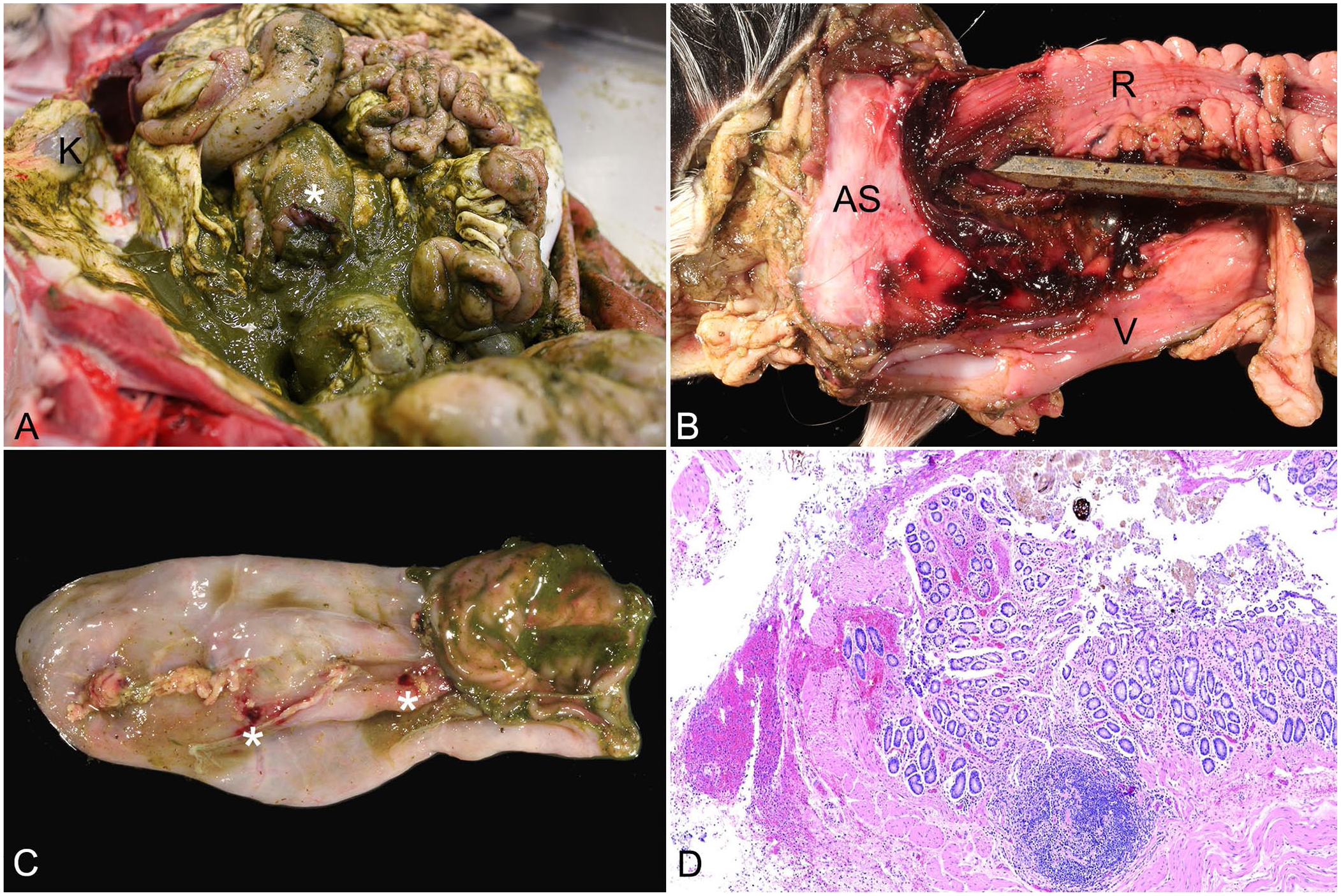
Gross and histologic images of traumatic cecal transection and anorectal rupture in a goat.
The separately submitted tissue (Fig. 1C) consisted of a 5 × 3 × 3-cm blind-ended, thin-walled pouch lined by fecal-covered epithelium that was consistent with the cecal apex. The cecal branches of the cecal vein were severed close to the serosa with minimal hemorrhage. No foreign bodies or anatomic abnormalities were noted in the digestive tract.
Major organs were sampled, fixed in 10% neutral-buffered formalin, and select tissues sectioned and embedded in paraffin for routine slide preparation with H&E staining. The cecal apex was histologically unremarkable, with minimal hemorrhage at the margins. The transected cecal body had evidence of regional peritonitis with acute hemorrhage, necrosis, and neutrophils containing mixed, intracytoplasmic, bacterial populations. The margins of the anorectal perforation had acute transmural hemorrhage and minimal necrosis near lymphoid nodules (Fig. 1D), but without inflammation or other structural abnormalities. No other significant histologic findings were discovered.
This unique case of a cecal transection, an anorectal tear, and transanal evisceration of the transected cecal segment is centered around a traumatic injury. We retrieved no cases of cecal transection with transanal evisceration in ruminants or humans in a search of Google Scholar, PubMed, CABI Direct, Web of Science, and Scopus, using search terms “transanal evisceration” or “traumatic evisceration,” “rectal tear” or “anorectal tear,” “intestinal transection” or “traumatic intestinal rupture” or “intestinal amputation”, and “goat” or “ruminant” as of 2024 Dec 10, suggesting that this condition has not been reported in ruminants or humans.
Intestinal transection, anorectal tears, and transanal evisceration of intact bowel segments have been documented and described in animals and humans independently, with various causes and predisposing factors. Transanal bowel evisceration is a rare complication of abdominal trauma and rectal prolapse in people.6,11 Reasons for traumatic evisceration in people can include blunt forces, impalement, and suction injuries. 11 The small intestine is most frequently involved. Chronic constipation, conditions resulting in increased abdominal pressure, and anatomic variations of the pelvic floor can all cause rectal prolapse. 5 Pregnancy in women is a predisposing factor for rectal prolapse, with several case reports of prolapse occurring in late gestation.7,14,15 However, no cases have been documented of rectal prolapse secondary to pregnancy that progressed to transanal bowel evisceration. In select cases of transanal evisceration, strangulation and rupture of the eviscerated bowel can occur, but complete transection and propulsion of bowel segments outside the body has not been reported in people. 6
Intestinal transection is very rare in people. Abdominal trauma can result in transection of the human appendix and subsequent appendicitis, with bicycle handlebar injuries being a leading cause in children.8,12 Evisceration of the appendix is a rare complication of abdominal surgical drain placement, without reports of spontaneous or iatrogenic transanal appendiceal evisceration in the literature. 10 Anorectal tears occur rarely, but more frequently than intestinal transection or transanal bowel evisceration, with common causes including parturition, penetrating trauma, or iatrogenic trauma.9,13 Causes of rectal tearing in ruminants include lacerations due to fetal malposition during parturition, mating mishaps, congenital malformations, and iatrogenic trauma. Chronic rectal prolapse can predispose to eventual rupture of the rectum or tearing of the anus in both humans and animals.3,6 Anal rupture with small bowel evisceration was reported secondary to rectal prolapse and dystocia in a young doe, but intestinal rupture or involvement of the cecum was not noted. 3 Intestinal evisceration is more commonly observed with umbilical, scrotal, or inguinal herniation in livestock species. Prolapse of the uterus through a vaginal tear has also been reported as a consequence of dystocia or blunt force trauma during pregnancy in goats.2,4
Unique features of our report include spontaneous, traumatic rectal rupture without preceding prolapse, and complete transection with evisceration of the cecal apex. Pregnancy appears to have predisposed the doe to this cataclysmic injury, as pregnancy predisposes people and animals to rectal injuries. We theorize that air trapped in the cecal apex during the moment of vehicular impact and the anatomical placement of the cecum in the pelvic canal due to the gravid uterus may have provided the necessary force to completely sever and propel the cecal apex out of the anorectal rupture. Impact and displacement of the left sacroiliac joint may have also created sudden, guillotine-like pressure on the cecal apex contributing to cecal evisceration, similar to cases of appendix transection.1,8 Although the anorectal tear was distal to the left sacroiliac joint, the regional pressure from joint displacement may have contributed to the acute tear. There was no histologic evidence of prior rectal disease or injury that could predispose to wall compromise and rupture; thus, the tear must have occurred due to the unique combination of acute injuries. Evisceration of a transected bowel segment through an anorectal tear can be considered a severe sequela of trauma in pregnant animals.
Footnotes
Acknowledgements
We thank Dr. Jeanine Messak for her clinical contributions to this case.
Declaration of conflicting interests
The authors declared no potential conflicts of interest with respect to the research, authorship, and/or publication of this article.
Funding
The authors received no financial support for the research, authorship, and/or publication of this article.