
Abstract
The Vetscan Imagyst system (Zoetis) is a novel, artificial intelligence–driven detection tool that can assist veterinarians in the identification of enteric parasites in dogs and cats. This system consists of a sample preparation device, an automated digital microscope scanner, and a deep-learning algorithm. The EasyScan One scanner (Motic) has had good diagnostic performance compared with manual examinations by experts; however, there are drawbacks when used in veterinary practices in which space for equipment is often limited. To improve the usability of this system, we evaluated an additional scanner, the Ocus 40 (Grundium). Our objectives were to 1) qualitatively evaluate the performance of the Vetscan Imagyst system with the Ocus 40 scanner for identifying Ancylostoma, Toxocara, and Trichuris eggs, Cystoisospora oocysts, and Giardia cysts in canine and feline fecal samples, and 2) expand the assessment of the performance of the Vetscan Imagyst system paired with either the Ocus 40 or EasyScan One scanner to include a larger dataset of 2,191 fecal samples obtained from 4 geographic regions of the United States. When tested with 852 canine and feline fecal samples collected from different geographic regions, the performance of the Vetscan Imagyst system combined with the Ocus 40 scanner was correlated closely with manual evaluations by experts. Sensitivities were 80.0‒97.0% and specificities were 93.7‒100.0% across the targeted parasites. When tested with 1,339 fecal samples, the Vetscan Imagyst system paired with the EasyScan One scanner successfully identified the targeted parasite stages; sensitivities were 73.6‒96.4% and specificities were 79.7‒100.0%.
Fecal examination for the detection of gastrointestinal parasitism in dogs and cats is an important component of routine veterinary care. 7 Although various techniques can be used to examine fecal samples for evidence of parasitic infection, centrifugal or passive fecal flotation followed by microscopic examination for parasite stages, such as eggs, oocysts, and cysts, is a standard practice in most veterinary clinics. However, the precision of these traditional fecal examinations can vary widely and be affected by procedural differences and experience levels of the personnel conducting the tests, possibly leading to poor recovery and errors of identification.1,7,12,22 To improve the accuracy, reliability, and consistency of fecal examinations in veterinary clinical settings, innovative computer technology–driven algorithms have been developed.8,17,26,27,33,34 Among these developments, the Vetscan Imagyst (Zoetis) is an artificial intelligence (AI)-based system that can be utilized in veterinary clinics to detect eggs, oocysts, and cysts of common parasites in canine and feline fecal samples. The Vetscan Imagyst enables veterinary clinics to provide a consistent fecal examination method that is not affected by an examiner’s level of experience.26,27,41
The Vetscan Imagyst system has a consistent ability to detect and correctly identify the eggs of Ancylostoma, Toxocara, Trichuris, and taeniid tapeworms, as well as Cystoisospora oocysts and Giardia cysts in fecal samples from dogs and cats.26,27 The system consists of 3 key components: a sample preparation device, an automated digital microscope scanner, and an AI-based data analytical algorithm using a deep-learning convolutional neural network (CNN). Studies with the Vetscan Imagyst system have shown high sensitivity (75.8–100.0%) and specificity (93.1–100.0%) across 6 targeted parasites compared to conventional centrifugal fecal flotation examinations performed by parasitologists.26,27 The capabilities of the Vetscan Imagyst system can be improved and expanded for additional parasites with supplemental training, as mentioned in previous studies.26,27
The EasyScan One scanner (Motic) has exhibited high image quality and fast scanning time, resulting in the successful identification of various forms of targeted parasites.26,27 However, the relatively large physical dimensions of the EasyScan One scanner, together with the requirement for a physical connection to a computer, may be problematic at many veterinary clinics in which physical space for laboratory equipment is at a premium. Additionally, the requirement for a slide tray for loading the prepared fecal slide into the scanner was considered cumbersome by some users. To address these concerns, we sought an alternative scanner based on smaller physical dimensions, high image quality, optics, and practicality and usability in the veterinary clinical setting. The Ocus 40 scanner (Grundium) met these specifications and was thus selected for further evaluation with the Vetscan Imagyst system.
In studies of Vetscan Imagyst performance, 188 canine and 112 feline fecal samples had been examined.26,27 Given that the collection of these samples was limited to central Oklahoma, the potential impact of variations in fecal components in different geographic regions was not addressed, even though certain gastrointestinal parasites, as well as microscopic plant pollens and seeds that may resemble parasite eggs, oocysts, and cysts, can vary across geographic regions.23,35 Thus, one of our aims was to overcome this limitation by extending the fecal sampling to other geographic regions. We tested the Vetscan Imagyst system using a large number of canine and feline fecal samples collected from 4 different geographic regions in the United States.
Our study objectives were 1) to qualitatively evaluate the performance of the Vetscan Imagyst system with 2 commercial scanners—Ocus 40 and EasyScan One—for correctly identifying Ancylostoma, Toxocara, and Trichuris eggs, Cystoisospora oocysts, and Giardia cysts in feces of dogs and cats, and 2) to assess the performance of the Vetscan Imagyst system among multiple users, using a large number of fecal samples collected from 4 different geographic regions.
Materials and methods
Technical specifications of Ocus 40 and EasyScan One scanners
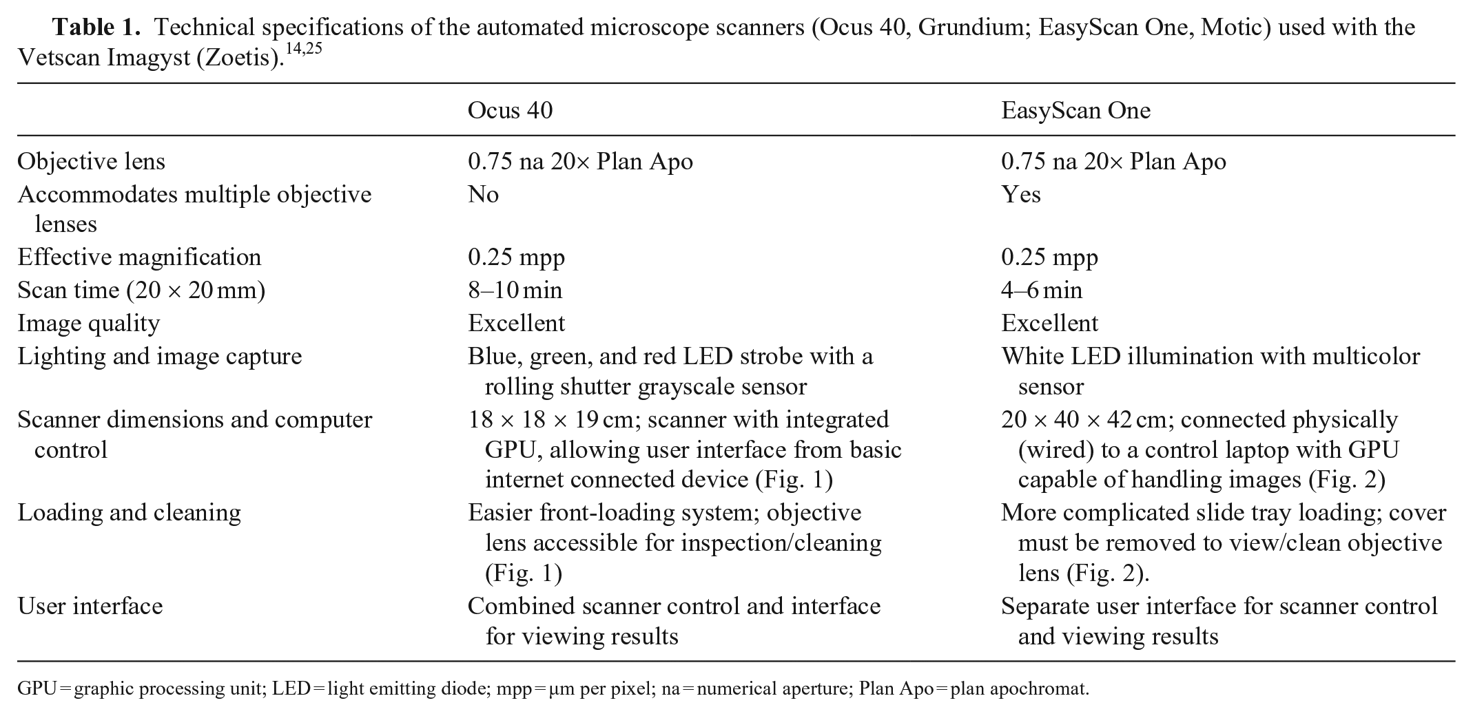
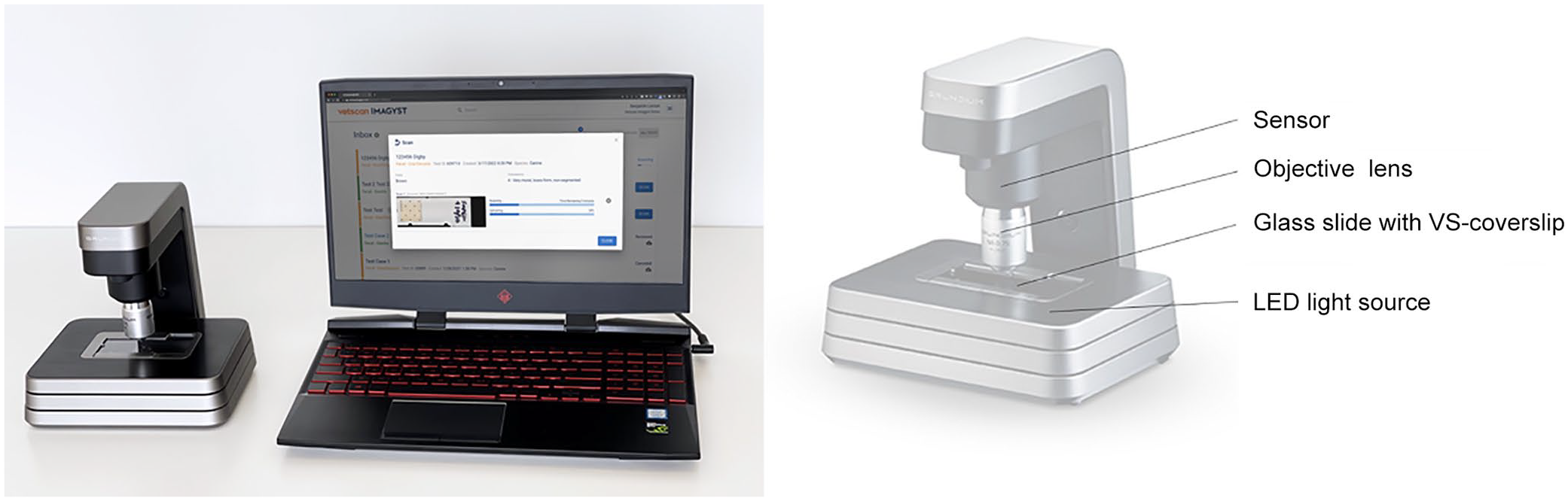
The Ocus 40 and EasyScan One scanners capture images with an effective magnification of 0.25 µm per pixel (mpp), have similar objective lenses (0.75 numerical aperture, 20× plan apochromat) and comparable scanning times for a 20 × 20 mm area, and provide high-quality images (Table 1). The Ocus 40 scanner uses a multi-color strobe of light-emitting diodes (LEDs), wherein several images are captured with blue, green, and red LED flashes; these images are assembled into a single image for analysis. 31 In contrast, the EasyScan One scanner uses a single 10-watt white LED light and captures a single multicolor image. 25 The Ocus 40 scanner is more compact (18 × 18 × 19 cm; Fig. 1) 14 than the EasyScan One scanner (20 × 40 × 42 cm; Fig. 2). 25 Additionally, the Ocus 40 scanner has a simpler front-loading system, allowing users to place a slide directly on the stage and inspect and clean the objective lens quickly and easily (Fig. 1). In contrast, the EasyScan One scanner requires a slide tray to load a sample slide, and the cover that encloses the objective lens must be removed to allow inspection and cleaning (Fig. 2).
GPU = graphic processing unit; LED = light emitting diode; mpp = µm per pixel; na = numerical aperture; Plan Apo = plan apochromat.
The Ocus 40 scanner (Grundium) has dimensions of 18 × 18 × 19 cm, and contains an integrated graphic processing unit, allowing a user interface with a basic internet-connected device (laptop shown for scale).
The EasyScan One scanner (Motic) has dimensions of 20 × 40 × 42 cm, and must be connected physically (wired) to a control laptop with a graphic processing unit capable of handling images.
To qualify for integration with the Vetscan Imagyst system, a scanner must be compatible, allowing the Vetscan Imagyst system to configure settings and control the scanner through an application programming interface (API). Assessment with the Vetscan Imagyst system has shown the EasyScan One scanner to be moderately configurable, allowing partial control with the API. In contrast, the Ocus 40 scanner was highly configurable, allowing a higher level of control with the API. The Ocus 40 scanner also includes an integrated graphic processing unit (GPU) and a network interface, allowing it to be used as a standalone device that users can interface with and control from any computer with an internet connection. The EasyScan One scanner, on the other hand, must be physically connected to a computer containing a GPU that is capable of rapid image capture.
Subjectively, the quality of images produced by both scanners is excellent: clear, sharp, well-focused, high resolution, low noise, and limited distortion. However, there are noticeable color distinctions in images generated by the Ocus 40 and EasyScan One scanners. Factors that can influence image colors include saturation, intensity, and contrast; different post-processing corrections can influence these color variations.4,10,11 The Ocus 40 scanner employs a multi-color flash of LEDs in which the individual flashes are captured using a rolling shutter grayscale sensor. 36 To get an accurate color presentation of the combined grayscale images that represent captured color channels, the Ocus 40 scanner utilizes a patented color correction method, 30 which results in more image information per pixel with greater depth and detail for the same resolution sensor. Additionally, the Ocus 40 scanner uses a unique image-stitching technique; at least 3 source images are composited to make an image while proposing a candidate transformation for each image pair and solving edge weights with an optimization algorithm to determine plausibility of the transformations. Generally, this procedure is thought to provide better quality images compared to techniques that have only one image to leverage for matching features and edges to stitch individual adjacent fields of view. 29
Fecal sample collections, pre-screening, and labeling
Fecal sample collection and the Vetscan Imagyst evaluation were conducted from August 2019 to September 2021 at 3 centers: Oklahoma Animal Disease Diagnostic Laboratory (OADDL; Oklahoma State University, Stillwater, OK, USA); Auburn University–College of Veterinary Medicine (AU-CVM; Auburn, AL, USA); and Cornell University–College of Veterinary Medicine (CU-CVM; Ithaca, NY, USA). Additional fecal sample collection was coordinated at the Zoetis Reference Laboratory (Mukilteo, WA, USA), and fecal samples collected at this location were mailed to and assessed at the 3 centers. Most fecal samples originated locally from OK, AL, NY, and WA. Our study did not require regulatory review or approval by Institutional Animal Care and Use Committees because animals were not handled either directly or indirectly.
Fecal samples submitted from dogs and cats undergoing routine fecal examinations were processed using the modified Wisconsin fecal examination technique with sugar solution (specific gravity: 1.25‒1.27) or zinc sulfate solution (specific gravity: 1.18), 39 without prior randomization. Fecal samples (≥ 1 g) containing any targeted parasites (Ancylostoma, Cystoisospora, Giardia, Toxocara, and/or Trichuris) and fecal samples (≥ 5 g) that were not observed to contain any parasites were included in the study. All fecal samples were stored at 4°C and examined within 14 d of the submittal date.
Assessment of Vetscan Imagyst system performance
The EasyScan One scanner was utilized in previous studies and from August 2019 to June 2020 in our study; we evaluated the Ocus 40 scanner from July 2020 to September 2021. A deep-learning CNN was applied to the Vetscan Imagyst system as described previously.26,27 The EasyScan One scanner was combined with the algorithm v.3033, which we also utilized in our previous study and had been largely developed and trained using images generated by the EasyScan One scanner. 27 For the Ocus 40 scanner, we used a new version of the algorithm, v.8293, which was specifically designed and adapted to the characteristics of images produced by the Ocus 40 scanner.
Fecal examination slides were prepared using the Vetscan Imagyst sample preparation device according to the instructions described previously26,27 or in the Vetscan Imagyst Quick Start Guide. 42 Samples evaluated for Giardia were prepared in zinc sulfate solution (specific gravity: 1.18–1.20), and the rest of the samples were prepared in sugar solution (specific gravity: 1.28–1.30; Zoetis data on file). Slides were then analyzed by the Vetscan Imagyst system and manually by experienced laboratory personnel. Fecal slide examination results were compared, and the sensitivity and specificity of the Vetscan Imagyst algorithm paired with the Ocus 40 and EasyScan One scanners were calculated.
Statistical analysis
A fecal sample was scored as “positive” if one or more of the targeted parasites were detected by an expert veterinary parasitologist (OADDL: R. Scimeca, S.E. Little, Y. Nagamori; AU-CVM: B. Blagburn, L.A. Starkey; CU-CVM: D.D. Bowman, A. Lucio-Forster). When multiple targeted parasites were present in a sample, samples were counted as positive for more than one analysis; some samples were counted as negative for more than one analysis. Using positive and negative determinations from both the expert and Vetscan Imagyst (negative corresponding to a count of 0, positive corresponding to counts ≥ 1), 2 × 2 contingency tables were constructed for each targeted parasite by scanner and sample preparation types. Using expert determination as the gold standard, sensitivity, specificity, and 95% Clopper–Pearson exact CIs were estimated from the proportions of samples, determined to be true-positive (TP) and true-negative (TN) among the positive (TP + false-negative [FN]) and negative (TN + false-positive [FP]) samples, respectively. In addition to Clopper–Pearson exact CIs, Jeffrey 95% CIs were constructed. R v.4.2.1 (https://www.r-project.org/) was used for the statistical analyses.
Results
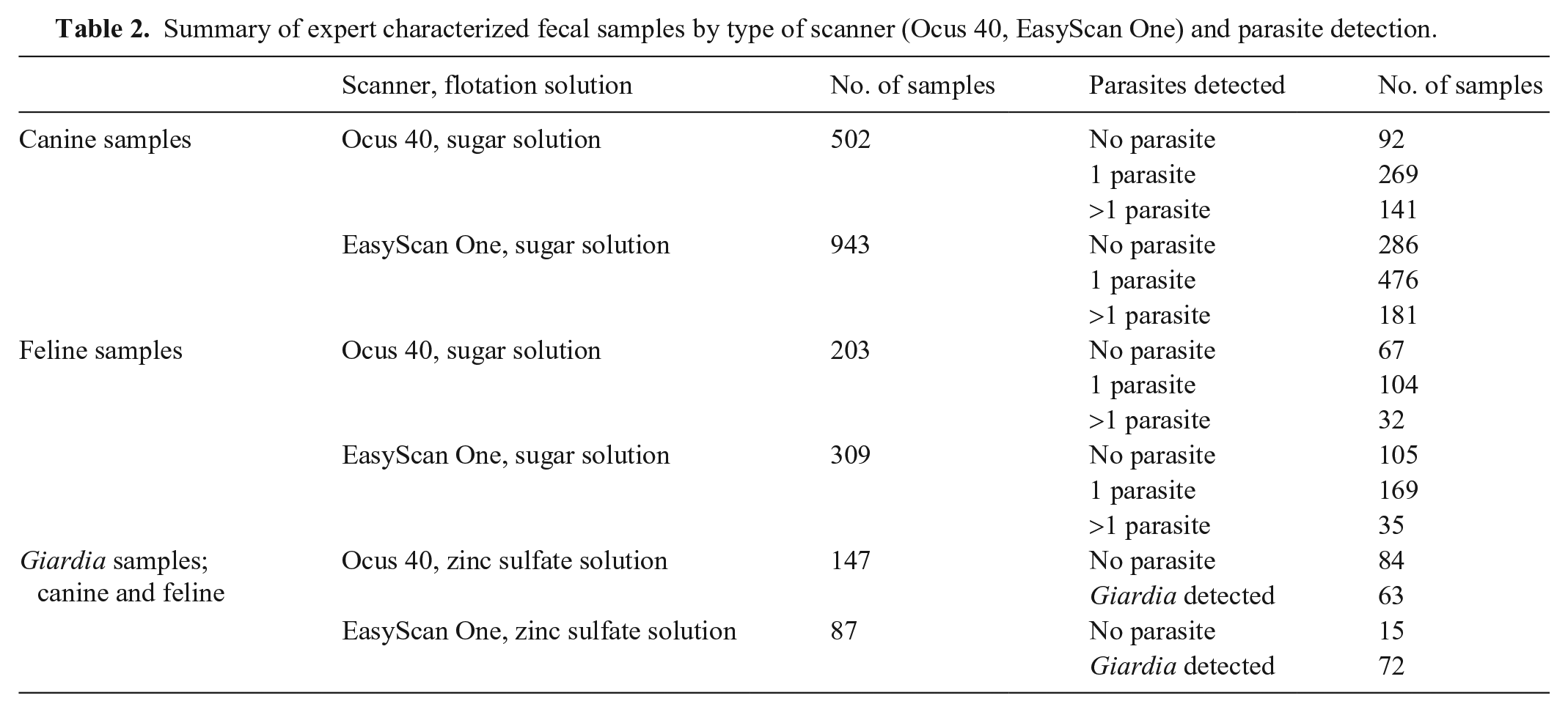
We examined 2,191 fecal samples for the detection of canine and feline Ancylostoma, Toxocara, Trichuris eggs, Cystoisospora oocysts, and Giardia cysts. Of the 2,191 fecal samples, 852 were evaluated by the Ocus 40 scanner paired with the v.8293 algorithm, and 1,339 were evaluated by the EasyScan One scanner paired with the v.3033 algorithm (Table 2).
Summary of expert characterized fecal samples by type of scanner (Ocus 40, EasyScan One) and parasite detection.
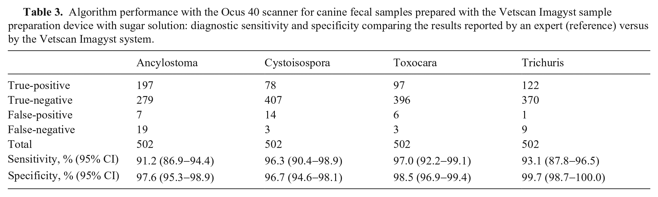
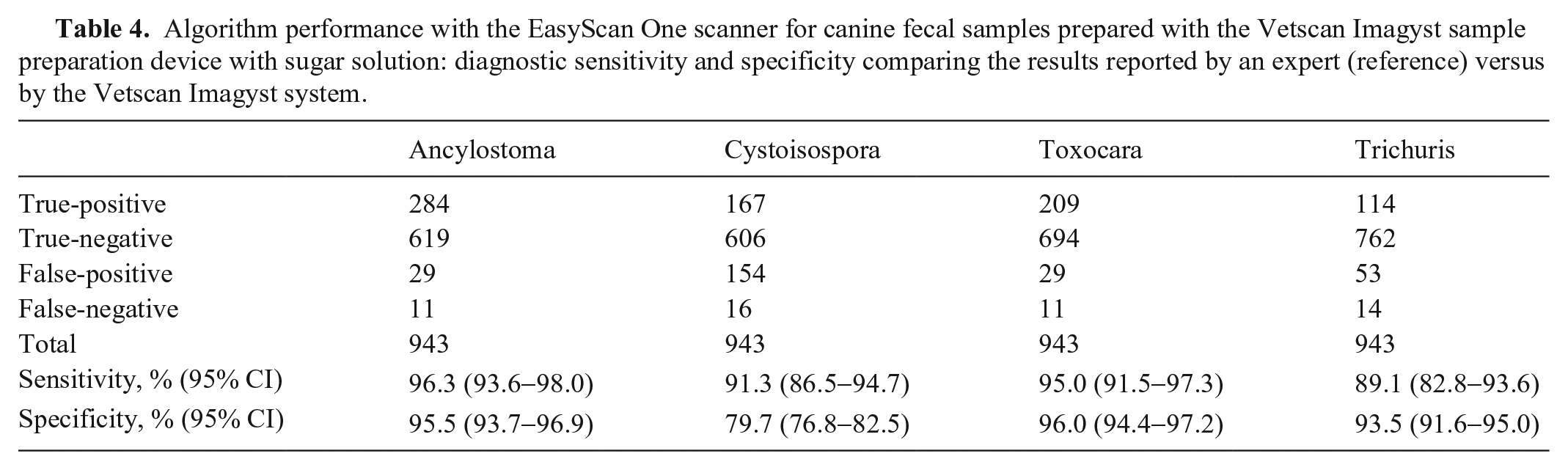
For Ancylostoma, Toxocara, and Trichuris eggs, and Cystoisospora oocysts in canine fecal samples, the diagnostic sensitivity and specificity (DSe and DSp, respectively) of the Vetscan Imagyst system with the Ocus 40 scanner compared with the experts’ assessments were 91.2‒97.0% and 96.7‒99.7%, respectively (Table 3). The DSe and DSp of the Vetscan Imagyst system with the EasyScan One scanner compared with the experts’ assessments were 89.1‒96.3% and 79.7‒96.0%, respectively (Table 4).
Algorithm performance with the Ocus 40 scanner for canine fecal samples prepared with the Vetscan Imagyst sample preparation device with sugar solution: diagnostic sensitivity and specificity comparing the results reported by an expert (reference) versus by the Vetscan Imagyst system.
Algorithm performance with the EasyScan One scanner for canine fecal samples prepared with the Vetscan Imagyst sample preparation device with sugar solution: diagnostic sensitivity and specificity comparing the results reported by an expert (reference) versus by the Vetscan Imagyst system.
For the same targeted parasites in feline fecal samples, the DSe and DSp of the Vetscan Imagyst system with the Ocus 40 scanner compared with the experts’ assessments were 80.0‒93.5% and 93.7‒100.0%, respectively (Table 5). With the EasyScan One scanner, the DSe and DSp of the Vetscan Imagyst system compared with the experts’ assessments were 91.4‒96.4% and 92.7‒98.2%, respectively (Table 6).
Algorithm performance with the Ocus 40 scanner for feline fecal samples prepared with the Vetscan Imagyst sample preparation device with sugar solution: diagnostic sensitivity and specificity comparing the results reported by an expert (reference) versus by the Vetscan Imagyst system.
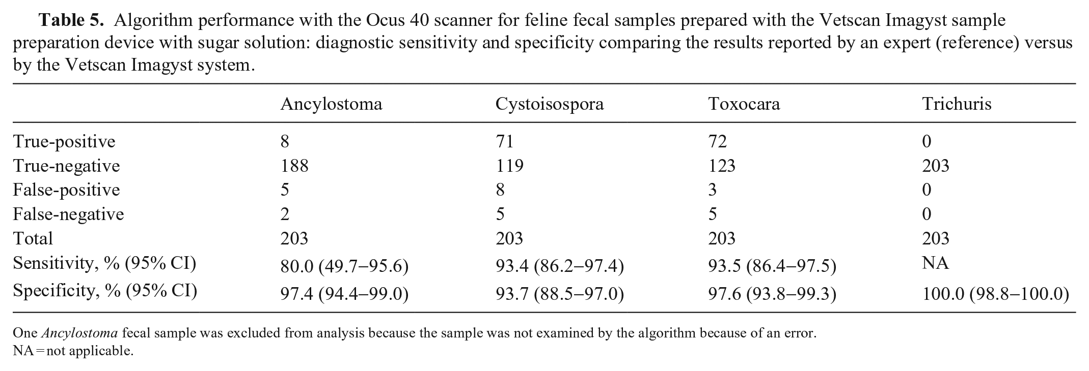
One Ancylostoma fecal sample was excluded from analysis because the sample was not examined by the algorithm because of an error.
NA = not applicable.
Algorithm performance with the EasyScan One scanner for feline fecal samples prepared with the Vetscan Imagyst sample preparation device with sugar solution: diagnostic sensitivity and specificity comparing the results reported by an expert (reference) versus by the Vetscan Imagyst system.
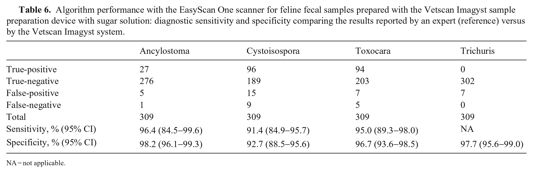
NA = not applicable.
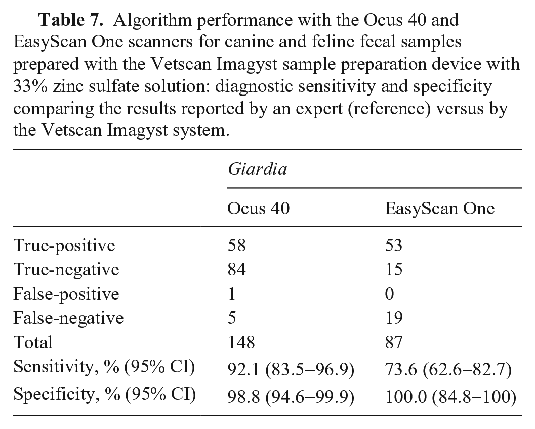
For the Giardia analysis, results of canine and feline fecal assessments were combined because of a recording error. The DSe and DSp of the Vetscan Imagyst system with the Ocus 40 scanner for Giardia detection were 92.1% and 98.8%, respectively. With the EasyScan One scanner, the DSe and DSp of the Vetscan Imagyst for Giardia cysts were estimated as 73.6% and 100.0%, respectively (Table 7).
Algorithm performance with the Ocus 40 and EasyScan One scanners for canine and feline fecal samples prepared with the Vetscan Imagyst sample preparation device with 33% zinc sulfate solution: diagnostic sensitivity and specificity comparing the results reported by an expert (reference) versus by the Vetscan Imagyst system.
Although the number of FP results decreased with the Ocus 40 scanner combined with the v.8293 algorithm, the most frequently observed FP result throughout the study was Eimeria oocysts misclassified as Cystoisospora oocysts. Additionally, during the evaluation of the EasyScan One scanner combined with the v.3033 algorithm, some other FP results were noticed: yellow debris confused with Trichuris eggs, end-on or tilted Ancylostoma eggs misclassified as Cystoisospora oocysts, and various plant materials misidentified as eggs of Ancylostoma, Toxocara, and Trichuris, as well as Cystoisospora oocysts.
Occasionally the Vetscan Imagyst system detected and classified targeted parasites successfully even when the human eye missed them at the initial manual reading. Specifically, the Vetscan Imagyst system correctly reported 2 cases of Ancylostoma eggs, 11 cases of Cystoisospora oocysts, 3 cases of Giardia cysts, 3 cases of Toxocara eggs, and 1 case of Trichuris eggs that had evaded human detection during their examinations. For most of these cases, there were very few parasite forms, generally only 1 or 2 eggs, oocysts, or cysts, observed on the entire slide. In these cases, a boarded diagnostic parasitologist (Y. Nagamori) re-reviewed the whole slide images to determine if these cases were TPs or FPs.
Discussion
The pairing of the Ocus 40 microscope scanner with the Vetscan Imagyst system resulted in high sensitivities and specificities for Ancylostoma, Toxocara, and Trichuris eggs, Cystoisospora oocysts, and Giardia cysts in feces of dogs and cats. DSe and DSp observed for targeted parasites in canine fecal samples were comparable between the Ocus 40 and EasyScan One scanners, with mostly overlapping 95% CIs. Specificities for Cystoisospora oocysts (96.7%; 95% CI: 94.6–98.1%) and Trichuris eggs (99.7%; 95% CI: 98.7–100.0%) recorded with the Ocus 40 scanner were higher than specificities for Cystoisospora oocysts (79.7%; 95% CI: 76.8–82.5%) and Trichuris eggs (93.5%; 95% CI: 91.6–95.0%) recorded with the EasyScan One scanner, with no overlap of 95% CIs. These increases in specificity may be explained by differences in the development and presentation of colors in images and the process of generating images by the Ocus 40 scanner given that the color variations can be pivotal for the deep-learning object detection algorithm because its performance is affected by colors and other details, including texture, edges, shape, and size of the objects.4,10,11 Additionally, the new algorithm, v.8293, reconciled well with the Ocus 40 scanner, and the good compatibility of the algorithm and scanner may have enabled the Vetscan Imagyst system to identify Cystoisospora oocysts and Trichuris eggs more accurately.
In feline fecal samples, diagnostic sensitivities and specificities to detect Ancylostoma, Toxocara, and Trichuris eggs, and Cystoisospora oocysts demonstrated by the Ocus 40 and EasyScan One scanners were comparable with overlapping 95% CIs (Tables 5, 6). The sensitivity for Ancylostoma eggs (80.0%; 95% CI: 49.7–95.6%) recorded with the Ocus 40 scanner was slightly lower than that of other targeted parasites. This result was very likely because only 10 positive fecal samples were available. In addition, the 2 FN samples contained only 2–3 visibly deteriorated Ancylostoma eggs on the entire fecal slide. Specificity for Trichuris eggs was 100.0% (95% CI: 98.8–100.0%) with the Ocus 40 scanner and 97.7% (95% CI: 95.6–99.0%) with the EasyScan One scanner. We did not assess sensitivities for Trichuris eggs in feline fecal samples given the lack of TP samples, which reflects the low prevalence of this infection in cats in most of the United States.2,6,13,39 However, Trichuris infection in cats is frequently reported in St. Kitts, the West Indies, 21 and several different species of Trichuris have been described from the Caribbean and Central and South America, including T. felis, T. campanula, and T. serrata.3,32 Additionally, Trichuris has been found in Florida, USA. 13 Further study is required to confirm the capability of detecting and identifying feline Trichuris eggs using the Vetscan Imagyst system.
Higher sensitivity for Giardia cysts was observed with the Ocus 40 scanner (92.1%; 95% CI: 83.5–96.9%) than with the EasyScan One scanner (73.6%; 95% CI: 62.6–82.7%). This increase in sensitivity might be the result of the difference in color presentation and production process of images between the 2 scanners and the different compatibility of the algorithm versions applied to the 2 scanners. Giardia is a commonly detected gastrointestinal parasite in dogs and cats12,15,16,18,19,24,28,37,38; however, Giardia is considered one of the more challenging gastrointestinal parasites to detect by fecal examination given its small size, transparency, and intermittent shedding of the cysts and trophozoites.20,39,40 Additionally, application of an optimal fecal examination method is essential to detect Giardia cysts; a centrifugal fecal flotation using 33% zinc sulfate solution is a recommended technique, and recovery of this parasite from fecal samples can be difficult with suboptimal methods, such as a passive flotation technique with sodium nitrate solution.2,39 Given that Giardia infection can be subclinical for many patients, it is important to conduct a fecal examination when new animals are introduced to homes, whether or not animals have clinical signs.5,9,39 As a supplemental detection test in conjunction with conventional fecal examinations, Giardia-specific coproantigen detection assays are commercially available to facilitate the detection of Giardia infection, primarily for clinical patients.5,9,39 A centrifugal fecal flotation test with 33% zinc sulfate solution is a recommended follow-up test after the completion of treatment because Giardia antigens may continue to be excreted in feces after parasite elimination.5,9,39 The increased sensitivity for detection of Giardia cysts by the Vetscan Imagyst system with the Ocus 40 scanner combined with 33% zinc sulfate solution can provide accurate, consistent detection of Giardia infection in dogs and cats.
A limitation of the Giardia analysis in our study was a relatively low number of TN samples (n = 15) for the Easy Scan One scanner assessment. This limitation could have influenced the DSp for Giardia (100.0%; 95% CI: 84.8–100.0%) that was demonstrated by the EasyScan One scanner, although the specificity of Giardia observed in the previous study by the same scanner was similarly high (97.0%; 95% CI: 90.8–99.4%). 27
Using the EasyScan One scanner with the Vetscan Imagyst system, the diagnostic sensitivities for identifying Ancylostoma, Toxocara, and Trichuris eggs, Cystoisospora oocysts, and Giardia cysts in canine and feline fecal samples were 73.6‒96.4% in our present study and 75.8‒100.0% in our 2 previous studies.26,27 Similarly, the diagnostic specificities for Ancylostoma, Toxocara, and Trichuris eggs, and Giardia cysts in canine and feline samples aligned among the 3 studies: 79.7‒100.0% in our present study and 93.1‒100.0% in the previous 2 studies.26,27 The specificity associated with detection of Cystoisospora oocysts in canine samples, however, was lower in our present study (79.7%; 95% CI: 76.8–82.5%) compared to the specificity of 93.1% (95% CI: 88.6–96.2%) in our previous study. 27 This notable difference could have been the result of the different sample size; we tested 943 canine fecal samples in our present study, but only 200 fecal samples (104 canine and 96 feline fecal samples) in our previous study. 27
Our statistical evaluations were limited by previously defined ground truth values. Given that the manual examination conducted by laboratory personnel was considered ground truth, even when the Vetscan Imagyst system correctly identified targeted parasites, some results were categorized as FPs because human examiners missed them at the initial manual reading. If these parasites had been detected by the examiners during the initial reading, the sensitivity and specificity values of the Vetscan Imagyst system could have been better than the calculated values in our study. With continuous guided algorithm training, learning, and development, the diagnostic performance of the Vetscan Imagyst system to detect gastrointestinal parasites in feces is expected to improve even further over time.
Footnotes
Acknowledgements
We thank Emily Looper, Tracey Land, Jamie Butler, and Joy Bowles for assistance in preparation of the fecal samples and examination of the flotation slides. We also thank all of the personnel who were involved in collecting the fecal samples from dogs and cats for our study. Writing and editorial assistance were provided by Litto Communications.
Declaration of conflicting interests
Travis Cree, Michael Loenser, Cory Penn, Austin Rhodes, and Richard Goldstein are employees of Zoetis. Benjamin Larson is an employee of Techcyte. Yoko Nagamori was an employee of Oklahoma State University during our study and is now an employee of Zoetis. Ruth Hall-Sedlak was an employee of Zoetis when our study was conducted and is now an employee of Amgen.
Funding
Our study was funded by Zoetis.