
Abstract
Glutamate dehydrogenase (GLDH), a key enzyme in amino acid oxidation and urea production, is mainly derived from the liver and its activity may increase with hepatocellular necrosis. Feline hyperthyroidism is associated with elevated serum activities of various enzymes, but the pattern of serum GLDH activity has not been reported, to our knowledge. Feline clinical biochemistry results from 2 commercial diagnostic laboratories were reviewed retrospectively to assess changes in serum GLDH activity in cats with significantly elevated serum total T4 concentrations, which is highly suggestive of hyperthyroidism. A total of 2,773 records were analyzed, of which 2,370 (85%) had normal total T4 (≤50 nmol/L) and 403 (15%) had increased total T4 (≥60 nmol/L) concentrations. Among cats with an increased total T4 concentration, 26.5% had increased serum GLDH activity. All cats with increased GLDH activity also had increased serum ALT activity. In 42.9% of cats, ALT activity was increased, but GLDH activity was normal. In 30.5% of cats, both serum GLDH and ALT activities were within RIs. The fold-increase of GLDH activity was almost half of the ALT fold-increase. Although serum GLDH activity increased in some cats with hyperthyroidism, serum ALT activity increased more frequently and to a greater extent.
Glutamate dehydrogenase (GLDH; EC 1.4.1.3) is a mitochondrial enzyme that catalyzes the oxidative deamination of L-glutamate to α-ketoglutarate using NAD(P)+ as a coenzyme. 11 GLDH, a key enzyme in amino acid oxidation and urea production, 7 is a relatively liver-specific enzyme located in the mitochondrial matrix of cells. Given its remote intracellular concentration and large size, release of GLDH as a result of cellular damage is delayed, making it a specific marker of cell necrosis. 7 Although GLDH is present in other tissues, such as kidneys and nervous tissue, the liver is the main source of blood GLDH activity. 7
Hyperthyroidism is the most common endocrine disorder in cats, 4 and measurement of the blood total T4 concentration has become the screening test of choice for the diagnosis of feline hyperthyroidism, given that >90% of hyperthyroid cats will have a total T4 concentration that is clearly high. 8 Elevations in the serum activities of alanine aminotransferase (ALT), aspartate aminotransferase (AST), alkaline phosphatase (ALP), and lactate dehydrogenase (LDH) are among the biochemical alterations observed most commonly in cases of feline hyperthyroidism 9 ; up to 90% of hyperthyroid cats are reported to have increased liver enzyme activities, 4 and >50% have increased serum ALT activity. 2 To our knowledge, the changes of serum GLDH activity with feline hyperthyroidism have not been described. Therefore, we assessed the patterns of serum GLDH activity in cats with normal and elevated serum total T4 concentrations.
Elevations of serum liver enzyme activities are well documented in cats with hyperthyroidism. Ultrasonographic abnormalities of liver parenchyma are not typically detected, liver dysfunction is not apparent, and liver enzyme activities usually return to normal after control of hyperthyroidism. 1 Interestingly, despite several studies focusing on clinicopathologic changes accompanying feline hyperthyroidism, changes in serum GLDH activity have not been reported in cats with hyperthyroidism. This may be because ALT has traditionally been recognized as the main indicator of hepatocellular injury in cats, 12 and because of the relatively limited use of GLDH in small animal practice.
A 2-y retrospective search was performed using the Laboratory Information Management Systems at Finn Pathologists (Harleston, Norfolk, UK) and Axiom Veterinary Laboratories (2020.01.01–2021.12.31; Harleston, Norfolk, UK); 2,949 cats with available results for serum total T4 concentration, as well as serum GLDH and ALT activities, were included. Feline RIs used were 7.5–55.0 nmol/L for serum total T4 concentration, 18.0–77.0 IU/L for serum ALT activity, and 0.0–10.0 IU/L for GLDH activity (at 37°C) at both sites.
Identical methodologies were used at both laboratories. The serum total T4 concentration was measured with a human thyroxine enzyme immunoassay (DRI thyroxine assay; Thermo Fisher) that has been validated internally for use in cats. Serum ALT activity was measured with a kinetic ultraviolet assay approved by the International Federation for Clinical Chemistry (Alanine aminotransferase; Beckman Coulter). Serum GLDH activity was measured with an optimized standard method according to the recommendations of the Deutsche Gesellschaft für Klinische Chemie (Glutamate dehydrogenase; Randox). All analyses were performed on automated chemistry analyzers (AU680; Beckman Coulter). Daily maintenance was performed as per manufacturer’s recommendations, and within-run quality control is performed at both laboratories. The analytical performance of these analyte tests is also assessed by participation in an external analytical quality assurance scheme (RIQAS; Randox).
Cats with severe elevations of serum enzyme activities (i.e., >20× the upper limit of the RI) were excluded from our study, given that it was considered likely that they had significant comorbidities that could bias the liver enzyme activity results. It has been suggested that cats with hyperthyroidism that have marked elevations of serum liver enzyme activities are likely to have concurrent hepatobiliary disease. 1 Given the 5% analytical imprecision for the measurement of blood total T4 concentration at 50 nmol/L (internal data) and the possibility of high-normal total T4 concentrations in cats with mild hyperthyroidism, 4 cats were considered more likely to be normothyroid when the total T4 concentration was ≤50 nmol/L and more likely to be hyperthyroid when the blood total T4 concentration was ≥60 nmol/L. Therefore, records in the intermediate range (50 < T4 < 60 nmol/L) were excluded, as well as cats with partial results.
Statistical analyses were performed using Prism v.9.2.0 (GraphPad). The nonparametric Mann–Whitney and Kruskal–Wallis tests were used to assess differences between 2 groups or multiple groups, respectively. Correlation was assessed using the nonparametric Spearman coefficient.
A total of 2,949 records contained results for total T4, ALT, and GLDH at both laboratories; 2,773 records remained after applying the exclusion criteria. Of these, 2,370 (85%) had normal serum T4 concentrations (≤50 nmol/L), and 403 (15%) had increased serum T4 concentrations (≥60 nmol/L). The age (x̄ ± SEM) of cats with increased total T4 concentrations (15 ± 0.1 y) was significantly higher (p < 0.0001) than the age of cats with normal serum T4 (13 ± 0.1 y).
Serum ALT activity was significantly higher (p < 0.0001) for cats with increased serum T4 concentrations (157 ± 8 IU/L) compared to those with normal T4 concentrations (84 ± 3 IU/L; Fig. 1A). Similarly, GLDH activity was significantly higher (p < 0.0001) in cats with increased T4 concentrations (11 ± 0.8 IU/L) than those with normal T4 concentrations (8 ± 0.4 IU/L; Fig. 1B).
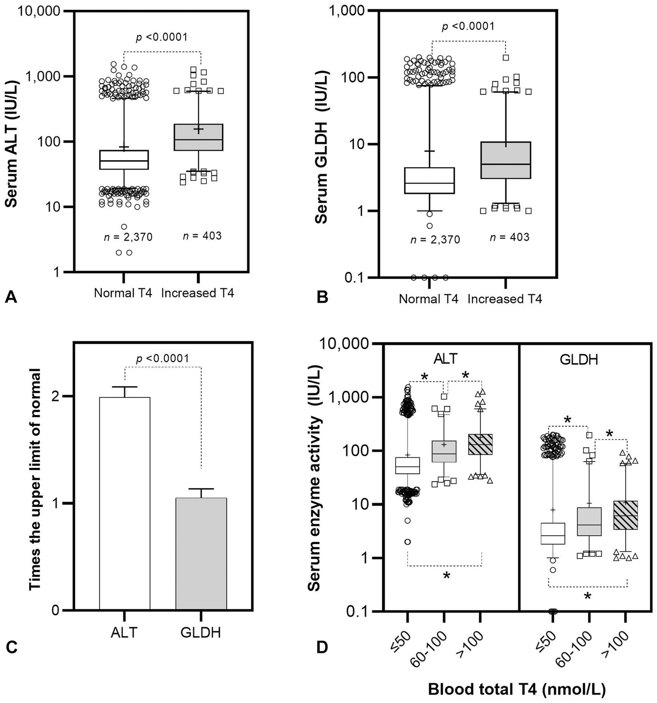
Serum activities of glutamate dehydrogenase (GLDH) and alanine aminotransferase (ALT) in cats with normal and increased serum T4 concentrations. Box and whisker plots (A, B, D) show 2.5th–97.5th percentiles and x- as “+”. The bar graph C shows the x- and SEM.
The extent of fold-increase (times the upper limit of normal) for ALT (2 ± 0.1) activity was higher (p < 0.0001) than that of GLDH (1 ± 0.08) activity in cats with increased serum total T4 concentrations (Fig. 1C). There was significant correlation (p < 0.0001) between serum ALT and GLDH activities (Spearman r = 0.78).
Among cats with increased total T4 concentrations, 107 (26.5%) cats had serum GLDH activity above the RI. All cats with increased GLDH activity also had increased serum ALT activity. In 173 (42.9%) cats, ALT activity was increased, but GLDH activity remained within the RI. In 123 (30.5%) cats, serum GLDH and ALT activities were within their RIs.
In cats with increased serum total T4 concentrations, the serum activities of ALT and GLDH correlated poorly with serum total T4 concentration (Spearman r = 0.27 and 0.22, respectively). However, increased serum ALT activity was strongly correlated with increased serum GLDH activity in cats with increased serum T4 concentrations (Spearman r = 0.78).
When total T4 concentrations were divided in 3 groups labeled as normal (T4 ≤ 50 nmol/L; n = 2,370), moderately increased (60 ≤ T4 ≤ 100 nmol/L; n = 191), and markedly increased (T4 > 100 nmol/L; n = 212), the serum activities of ALT and GLDH in each group were significantly different (p < 0.05; Fig. 1D). However, although serum GLDH activity in cats with markedly elevated blood T4 concentrations was significantly higher (p = 0.0395) than GLDH in cats with moderately elevated blood T4 concentrations, the means for both groups were the same (11 IU/L).
Hyperthyroidism is a disease of middle-aged and older cats, with mean age at the time of diagnosis of 12–13 y. 5 In our study, the mean age of cats with increased blood T4 was 15 y (±0.08). Although cats with increased blood total T4 concentrations were significantly (p < 0.0001) older than cats with normal T4 concentrations (13 ± 0.1 y), we cannot draw meaningful conclusions about the age profiles of these cats. Cats were grouped based on their blood total T4 concentrations irrespective of the time of initial diagnosis and other factors (e.g., treatment status, treatment compliance, comorbidities); age profile results are likely skewed.
Despite the good correlation between serum ALT and GLDH activities in cats with increased blood total T4 concentrations, only 26.5% had both ALT and GLDH activities above normal limits; 42.9% of cats had increased ALT activity and normal GLDH activity. Additionally, there were no cats with increased GLDH activity without a concurrent increase in ALT activity, and the extent of fold-increase was significantly higher for ALT activity than for GLDH activity. This preferential and more appreciable rise in the serum activity of ALT in cats with elevated blood T4 concentrations may reflect its subcellular localization. ALT is found mainly in the cytoplasm of hepatocytes, whereas GLDH is more protected in the mitochondria and is released predominantly with more severe, irreversible cell injury. 6
The 69% of cats with increased blood T4 concentrations that had elevated serum ALT activity in our study appears lower than what is typically reported for hyperthyroid cats, 10 although a similar percentage has been reported in one study examining a small group of hyperthyroid cats. 1 Our study shows an association between the level of increase in blood T4 concentration and serum ALT and GLDH activities. Cats with markedly increased blood T4 had significantly higher serum enzyme activities than cats with moderate elevations in blood T4 concentrations. This finding suggests that there is some association between the extent of blood T4 concentration increase and the rise in serum ALT and GLDH activities.
In the absence of further investigations, the exact etiology of liver enzyme changes in feline hyperthyroidism remains unknown, although various mechanisms (e.g., malnutrition, direct toxic effects of thyroid hormones, congestive heart failure) have been suggested. 1 In rats, severe hyperthyroidism has been shown to induce mitochondria-mediated apoptosis in hepatocytes. 13 In humans, the patterns of liver histopathologic changes associated with hyperthyroidism have been investigated, 3 but similar studies are lacking in cats. Histologic examination of liver was not performed in our study, and therefore it is not possible to associate the observed enzyme activity changes in cats with increased blood T4 with potential morphologic changes of the liver parenchyma. However, liver biopsy is an invasive process that is not performed routinely in cats with suspected hyperthyroidism.
A major limitation of our study is that clinical information was not recorded for subject cats. It was not known if cats with increased blood T4 concentrations had signs compatible with hyperthyroidism, if they were truly hyperthyroid, and if they were being treated. Methimazole, for example, which is commonly used to treat feline hyperthyroidism, is known to cause liver toxicity and increased serum activities of liver enzymes. 10 It can be safely assumed, however, that most cats in the increased blood total T4 concentration group were hyperthyroid at the time of sampling, given that elevated blood total T4 concentration indicates hyperthyroidism with very high specificity. 10
Additionally, it is likely that during the 2-y period studied, some cats were sampled on more than 1 occasion. It is even possible that a patient who may have responded temporarily to therapy and relapsed subsequently could appear in both the normal T4 and elevated T4 concentration groups. Intra-individual replicate samples should not affect the results of our study, given that, irrespective of the clinical context, increased blood total T4 concentrations are typically indicative of hyperthyroidism, which can have an effect on serum liver enzyme activity.
Footnotes
Acknowledgements
I thank Lewis Gallagher from IT support at Axiom Veterinary Laboratories for his help with obtaining the required data for this study.
Declaration of conflicting interests
The author declared no potential conflicts of interest with respect to the research, authorship, and/or publication of this article.
Funding
The author received no financial support for the research, authorship, and/or publication of this article.