
Abstract
Coccidioidomycosis is a fungal disease caused by Coccidioides immitis or Coccidioides posadasii. We searched the records of the California Animal Health and Food Safety Laboratory from 1990 through 2020 for cases of coccidioidomycosis in horses. The selection criteria for these cases were: 1) live-born horses submitted for autopsy, and 2) a diagnosis of coccidioidomycosis was established, regardless of cause of death. During that time, 19,054 horses were received, and 26 cases (0.14%) of coccidioidomycosis were diagnosed in horses, of which 19 (73%) cases had pneumonia and/or pleuritis with or without lesions in other organs, and 7 (27%) cases had lesions only in organs other than the lungs (nasal mucosa, spleen, thoracic lymph nodes, heart, pericardial sac, liver, kidney, mediastinum, and/or mesentery). Pneumonia was diagnosed as the cause of death in 1,838 (9.64%) of the horses received; Coccidioides spp. was the cause of pneumonia in 19 (1.0%) of these animals. Horses have been reported to have low susceptibility to coccidioidomycosis, and the severity and chronicity of the disease can be variable. Lesions in our cases consisted of multifocal-to-coalescing pyogranulomas with intralesional fungal spherules. Coccidioidomycosis must be considered a differential diagnosis in cases of persistent cough, chronic weight loss, fever, and cases with a travel history to, or living in, a region considered endemic for coccidioidomycosis. Coccidioides spp. infection should also be considered when pyogranulomatous inflammation is found within lung, spleen, nasal mucosa, and lymph nodes of horses.
Coccidioidomycosis, commonly referred to as San Joaquin Valley fever or simply valley fever, is a fungal disease caused by Coccidioides immitis or Coccidioides posadasii. 18 Both species cause disease that is indistinguishable clinically and pathologically, and are endemic in the sandy and silty areas of the west and southwest United States, northern Mexico, parts of Central and South America, and Australia.2,4,20 Coccidioides spp. are dimorphic fungi in the Ascomycota phylum and are related to Histoplasma capsulatum and Blastomyces dermatitidis. 16 Coccidioidomycosis is the second-most common fungal disease in humans, and it also occurs frequently in dogs, llamas, and alpacas. 8 Other species, including, but not limited to, non-human primates, cows, cats, and horses may also be infected by Coccidioides spp.5,7,9,13
Coccidioidomycosis is contracted by inhalation of C. immitis or C. posadasii arthroconidia. Once in the warm environment of the lungs, the arthroconidia undergo thermal transition to the infectious spherule form. 11 This transition was demonstrated when a temperature of 37°C initiated the thermal transition in vitro. 6 Additionally, leukocyte contact contributes to spherule formation. 3 As the spherules mature, they grow to >200 µm, undergo endosporulation, and later rupture spreading endospores in the lung. The endospores eventually develop into other endospore-filled spherules completing the life cycle and, frequently, disseminating to other organs.8,11 Direct inoculation of arthroconidia through the skin can also occur, but is uncommon,1,10,15 and this inoculation typically does not result in systemic disease.
In most mammals, lesions of coccidioidomycosis may be limited to the lungs, in which they vary from focal to multifocal, and typically result in a granulomatous-to-pyogranulomatous inflammatory response. However, systemic lesions that result from hematogenous spread of the endospores from the lung to other organs may also occur. Rare cases have also been described in which systemic disease occurs with no pulmonary lesions, and it has been postulated that these cases probably reflect resolution of the initial lung lesion rather than an extrapulmonary route of infection. 8
The scientific literature on coccidioidomycosis in horses is limited, and to our knowledge, the pathology of the diseases in this species has not been described thoroughly. We present here a retrospective study of 26 cases of coccidioidomycosis in horses, including the organ distribution and description of the lesions, and we review the literature on coccidioidomycosis in horses.
We searched the records of the California Animal Health and Food Safety (CAHFS) laboratory system for all equine autopsies performed between January 1, 1990 and January 31, 2020. We reviewed all of the equine autopsies and those with a diagnosis of pneumonia and/or coccidioidomycosis that were submitted during that period. The selection criteria for coccidioidomycosis cases were: 1) live-born horses submitted for autopsy for which 2) a diagnosis of coccidioidomycosis was established, regardless of cause of death. In all cases, routine autopsies had been performed, and samples had been collected from liver, lung, kidney, spleen, skeletal muscle, skin, adrenal gland, small intestine, colon, and/or brain, fixed in 10% neutral-buffered formalin for of 24 h to 1 wk, and processed routinely for the production of 4.0-µm thick H&E sections. Selected sections from most cases were also stained with Gomori methenamine silver, periodic acid–Schiff, and/or Giemsa stains. All of the sections from coccidioidomycosis cases were reviewed by one of the authors (M. Macías-Rioseco). The diagnosis of coccidioidomycosis was based on the presence of pyogranulomatous inflammation with intralesional 20–200-µm spherules with a double refractile, hyaline wall, which occasionally contained 2–5-µm endospores.
Coccidioidomycosis cases were classified according to the number of organs affected and the distribution of lesions. Cases in which more than one organ was affected were classified as disseminated coccidioidomycosis. Cases with only one organ affected were classified as localized coccidioidomycosis. Coccidioidomycosis was considered the cause of death when the coccidioidomycotic lesions were the only, or the main, lesions found on postmortem examination, and the lesions were severe enough to impair the functionality of the organ(s) affected.
Several other laboratory tests had been performed in most animals, including aerobic and anaerobic bacterial culture, fecal flotation, and heavy metal screen. These tests were performed following the standard operating procedures of the CAHFS laboratory system.
During the study period, 19,054 live-born horses were received for autopsy in 3 of the CAHFS laboratories (Tulare, Davis, San Bernardino). All of the cases originated from within California. Coccidioidomycosis was diagnosed in 26 (0.14%) of the live-born horses received during this period. The age of the affected horses was 1-mo to 20-y-old. Fourteen of the horses were female and 12 were male. Affected breeds included 14 Quarter Horses, 8 Thoroughbreds, 1 Warmblood, 1 Paint, 1 American Miniature, and 1 mixed-breed.
Coccidioidomycosis was the cause of death in 11 of 26 (42%) horses; the coccidioidomycotic lesions were considered an incidental finding in the remaining 15 horses. Of the 26 live-born horses with coccidioidomycosis, 19 (73%) had pneumonia and/or pleuritis. Of these, 12 (63%) had only pneumonia and/or pleuritis, and 7 (37%) had pneumonia as well as infection in at least one other organ. The remaining 7 horses (27%) had Coccidioides spp.–associated lesions identified only in an extrapulmonary site, including nasal mucosa, spleen, thoracic lymph nodes, heart, pericardial sac, liver, kidney, mediastinum, and/or mesentery. No gross lesions were reported in the bones of any animal, although no information about examination of bones was available for most cases (Table 1). No information about treatment was available in the clinical history of any of the affected horses.
Distribution of lesions in 26 horses with coccidioidomycosis received at the California Animal Health and Food Safety laboratory system, 1990–2020.
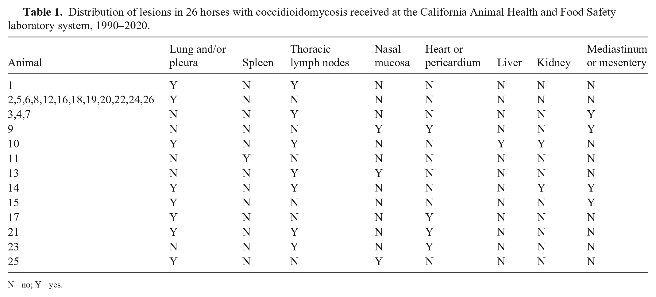
N = no; Y = yes.
Of the 19,054 live-born horses received at CAHFS during the study period, 1,838 (9.3%) had pneumonia as the cause of death. The most prevalent etiologic class for pneumonia was bacterial (n = 815; 44.3%), followed by aspiration and immune-mediated (n = 219; 11.9%), fungal (n = 69; 3.8%), viral (n = 54; 2.9%), parasitic (n = 43; 2.3%), and many not determined or unknown causes (n = 638; 34.7%), Of the 1,838 pneumonia cases, 19 (1%) were diagnosed as coccidioidomycosis. Other fungal causes of pneumonia included Aspergillus fumigatus and zygomycetes. All gross lesions of coccidioidomycosis in the affected organs of live-born horses were similar and consisted of multifocal-to-coalescing, nodular, firm, pale-tan to pale-yellow, pyogranulomas; the severity of lesions varied between cases (Figs. 1, 2). Many larger nodules also contained central friable regions of necrosis.
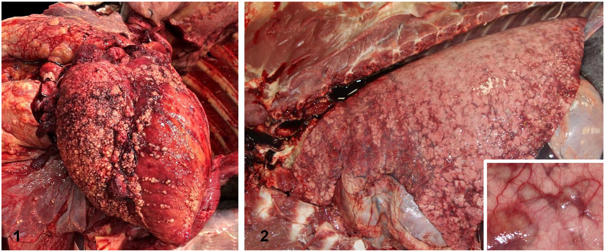
Coccidioidomycotic lesions in horses.
Microscopically, lesions of coccidioidomycosis were similar regardless of the organ affected and consisted of pyogranulomatous inflammation primarily (Fig. 3), characterized by numerous epithelioid macrophages and fewer foreign-body and Langhans giant cells surrounding aggregates of neutrophils (Fig. 4). Often, central areas of necrosis were present with or without mineralization. Smaller numbers of lymphocytes, plasma cells, and areas of fibroplasia surrounded the pyogranulomas. Throughout the pyogranulomatous inflammation, and often in regions of central necrosis, were variable numbers of round, 20–200-µm, fungal spherules (Fig. 5). The spherules had a 2–4-µm thick, refractile, hyalinized, double-contour wall, and central loose fibrillar basophilic material, as well as 5–7-µm rounded endospores. The spherules were observed frequently in the cytoplasm of giant cells (Fig. 5). Most lesions in the affected tissues had surrounding regions of edema, congestion, and hemorrhage.
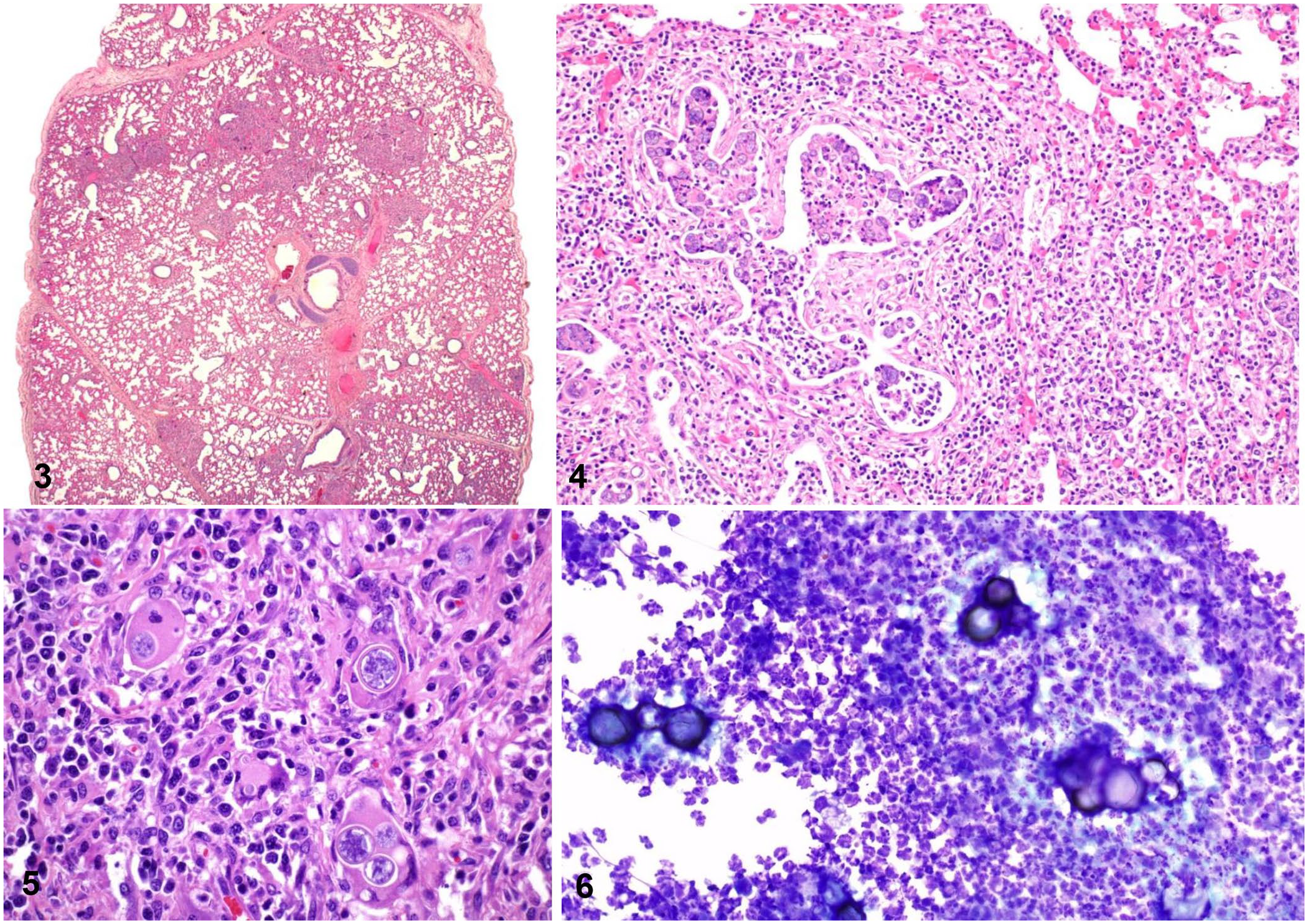
Microscopic pulmonary lesions from horses with coccidioidomycosis.
Based on our findings, coccidioidomycosis is rare in horses (0.14% of total live-born equine cases received at CAHFS over 30 y). Given that incidental lesions were common in our series, the prevalence of these lesions in the overall equine population of California is likely higher, because many horses with subclinical lesions would not be examined postmortem. This supposition is supported by a study that evaluated the seroprevalence of antibodies against C. immitis in healthy Arizona horses and found a positive titer in ~4% of horses. 13 Breed predilection, specifically Arabians, and sex predilection for mares have been suggested.12,23 However, we identified no sex or age predilection in our case series. Quarter Horses were overrepresented in the coccidioidomycosis group in our study (14 cases; 54%), but they were also an overrepresented group in the overall study population. Arabian horses were a fairly small proportion of the total horses received for autopsy in our study population, which may indicate why there were none in our infected group.
The respiratory system (lung and/or pleura) of horses was affected most commonly, encompassing 73% of coccidioidomycosis cases (19 of 26 of the total cases, 10 of which were the cause of death and 9 of which were incidental). Disseminated disease, which also included a pulmonary component, was identified in 7 horses (27% of cases) as the cause of death. Our results are consistent with findings in other species, 8 indicating that the respiratory system is the main route of entry for Coccidioides spp. in horses.
Bacterial infection was the most common cause of pneumonia (44.3%) in the population of horses received during the study period. Fungi were an uncommon etiology (3.8% of pneumonias), but of all the fungal pneumonias, the proportion caused by Coccidioides spp. (27.5%) was higher than that caused by other fungi. This higher proportion of Coccidioides spp. pneumonias is suspected to be a result of the more arid environment in which our equine population lived, which supported Coccidioides spp. over other species of fungi that may prefer more humid environments.
Horses have low susceptibility to coccidioidomycosis. 24 However, some breeds, specifically Przewalski and Arabian horses, as well as immunocompromised horses of any breed, are reported to be more susceptible.22,24 Also, some pregnant mares, likely as a result of their immunocompromised state, develop coccidioidomycosis leading to abortion, and may develop mastitis.21,23 When horses develop clinical disease, severity and course can be quite variable; therefore, treatment of coccidioidomycosis varies from supportive to more intensive combined medical and surgical interventions depending on the severity and the course of the disease. Medical treatments for chronic disease can last months, be costly, and when horses develop severe clinical signs, they commonly die or are euthanized. 19
A report from California and Arizona indicated that the main clinical complaint in horses with coccidioidomycosis was chronic weight loss and persistent cough 24 ; ~60% of the abnormalities registered on clinical examination were related to the respiratory tract, and all studied cases had serology positive for the pathogen. In our series of cases, the clinical history was variable. The literature available on coccidioidomycosis in horses indicates that rhinitis, dermatitis, and osteomyelitis occur at a higher prevalence than found in our study.12,24 This discrepancy is likely explained by the fact that most cases of equine coccidioidomycosis reported in the literature are based on clinical and not on postmortem studies, as in our study. The reported prevalence of coccidioidomycosis in humans and mice depends on other risk factors including genetic susceptibility, level of exposure, other diseases, immunosuppression, and environmental conditions.18,20 Most cases of rhinitis, dermatitis, and osteomyelitis are treatable, whereas pulmonary coccidioidomycosis carries a poor prognosis, although there are rare reports of successful treatment. 14 Severe pneumonia and disseminated coccidioidomycosis typically progress rapidly and are more likely to cause the demise of the animals, increasing the chances that postmortem examinations are performed. The gross and microscopic findings of multifocal pyogranulomatous disease with variable amounts of necrosis are similar to the disease processes identified in various other species infected with Coccidioides spp. 8
Except in rare instances, human and animal infections are acquired from inhalation of the arthroconidia, the highly infectious environmental form of Coccidioides spp. 15 A few human cases have been reported after exposure to live or dead animals.10,17 In these cases, the spherule and endospore form has been implicated in human disease after exposure to infected tissues or fluids.
Postmortem diagnosis of coccidioidomycosis is usually based on gross and microscopic examination, particularly the observation of the typical spherules in affected tissues. Diff-Quick (MilliporeSigma) staining of tissue smears allows for a quick diagnosis if spherules are observed (Fig. 6). However, confirmation of the species of Coccidioides spp. should be based on molecular methods and/or fungal culture. In our series of cases, the species was not determined, and fresh tissues were not available for culture or molecular tests. We cannot confirm, therefore, if C. immitis or C. posadasii, or both, were present in the tissues of our horses.
Although coccidioidomycosis is rare in horses and appears to be an incidental finding in many cases, pulmonary lesions were the most common findings in our study population, indicating the importance of including coccidioidomycosis as a differential diagnosis when persistent cough, chronic weight loss, and fever are observed in horses. Our results are consistent with the finding in other species that the respiratory system is the main portal of entry for Coccidioides spp. in horses.
Footnotes
Acknowledgements
We thank Mrs. Lucy Gomes for her valuable help in the data compilation.
Declaration of conflicting interests
The authors declared no potential conflicts of interest with respect to the research, authorship, and/or publication of this article.
Funding
The authors received no financial support for the research, authorship, and/or publication of this article.