
Abstract
An 8-y-old jenny was presented because of anorexia and mild depression. The jenny had weaned her colt 10 d before the admission. Upon arrival at the University of Illinois Veterinary Teaching Hospital, the heart rate was elevated, and the right udder was painful and swollen on palpation. Milk stripping of the affected side revealed purulent content; the contralateral udder had normal-appearing milk. Cytology of mammary gland secretions from the affected side revealed a large number of hypersegmented reactive neutrophils with phagocytized bacteria. Complete blood count, serum chemistry, and fibrinogen were within normal limits. A diagnosis of clinical mastitis was made, and the jenny was started on a 5-d course of broad-spectrum antibiotics, a non-steroidal anti-inflammatory, hydrotherapy, and milk stripping. Clinical signs reduced over time, and the cure was attained by 96 h post-admission. Aerobic culture and subsequent MALDI-TOF MS analysis identified a bacterium of the Streptococcus genus but not the species. Whole-genome analysis was performed, and 16S rDNA sequencing and analysis determined that our isolate 20-37394 clustered with 2 other Streptococcus strains (27284-01 and 28462). Single-nucleotide variations and phylogenetic tree analysis revealed that Streptococcus 20-37394 had 96.8% and 94.9% identities to Streptococcus strains 27284-01 and 28462, respectively; therefore, the bacteria isolated in our case was deemed as a new Streptococcus species.
Mastitis is a significant disease affecting domesticated mammals.11,14 Dairy cows and goats are particularly prone to develop mastitis given the high storage capacity of the udder, the proximity of the teats to the ground, the frequent manipulations of the udder during milking, and lactostasis in dry-off.16,20 Conversely, equids are thought to be less susceptible to develop mastitis given the smaller storage volume of the udder and the anatomically secluded location of the gland. 11 In recent years, donkey milk has become a popular product for infants allergic to cow’s milk 8 ; to meet this demand, donkey dairy farms have become more common in Europe. Despite numerous lactating dairy donkeys in Europe, clinical mastitis has not been described in this species, to our knowledge. The low incidence of mastitis in donkeys could also be attributed to the extremely high concentrations of lysozyme (3,750 mg/mL) in milk compared to cows (0–0.9 mg/mL).19,22 This enzyme is a potent natural antimicrobial playing a paramount role in the innate immune response of the udder.17,18
In mares, bacteria cultured in association with clinical mastitis include Escherichia coli, Pseudomonas spp., Staphylococcus spp., and Streptococcus spp., with the latter predominant.4,14 Staphylococcus spp. were reported to be the cause of subclinical mastitis in 3 dairy jennies in Southern Italy 7 ; however, the exact role of S. aureus in subclinical mastitis in donkeys is unclear. Additionally, there are no other reports of subclinical or clinical mastitis in donkeys.
In horses, clinical mastitis can occur during lactation, dry-off, or peripartum, and can affect barren mares and pre-pubertal fillies. 4 A perceived high incidence of equine mastitis has been reported between May and September in the northern hemisphere, 14 a time window that coincides with a high population of insects in the United States. 14 In addition, post-weaning lactostasis predisposes mares to ascending infections because the papillary muscle is stretched under the pressure of the accumulated milk, and flies are attracted to the dripping milk. 11 We describe here a case of clinical mastitis in a donkey and the isolation of a novel Streptococcus sp. associated with mastitis.
An 8-y-old Mammoth jenny (365 kg body weight) was presented to the University of Illinois Veterinary Teaching Hospital (Urbana, IL, USA) with anorexia and apathy. The owner reported that the jenny had weaned a colt 10 d before arrival. The jenny had been bred 8 mo before presentation. At admission, a general physical examination was unremarkable (temperature 37.5°C; reference interval [RI]: 36.5–37.7°C; respiratory rate 20 breaths/min, RI: 13–31 breaths/min) except for an increased heart rate (68 beats/min, RI: 31–53 beats/min), 2 and an abnormal right-side udder (Fig. 1). Upon close examination, the right-side udder was warm and swollen; the left half was normal (Figs. 1, 2). The secretions of the right udder had a purulent appearance; the left udder secretion had a normal milk appearance (Fig. 3). Complete blood cell count, serum chemistry, and fibrinogen were unremarkable (Suppl. Table 1).2,3 A milk sample was harvested aseptically and cultured aerobically and anaerobically. Transabdominal ultrasonography revealed a singleton fetus with a normal heartbeat of 80 beats/min and no signs of placentitis or impending abortion. Collectively, the clinical findings were consistent with clinical mastitis. While waiting for culture and susceptibility test results, the donkey was started on a 5-d course of standard mastitis treatment for horses, 4 namely a broad-spectrum antimicrobial (sulfamethoxazole–trimethoprim, 30 mg/kg, PO, q24h), non-steroidal anti-inflammatory drug (flunixin meglumine, 1.1 mg/kg, IV, q24h), hydrotherapy (15 min, q12h), and milk stripping (1–3 times/d).
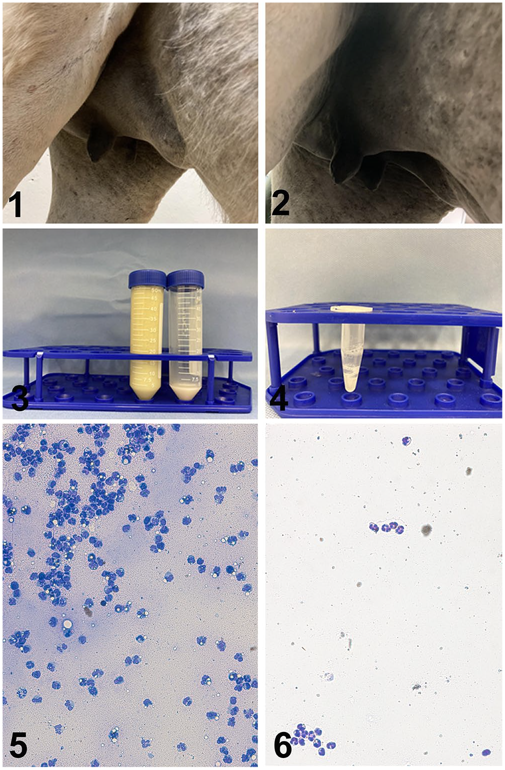
Mammary gland, mammary gland secretion, and cytologic preparation from a jenny with clinical mastitis.
Milk stripping was performed 3 times/d in the first 3 d, then once a day for 2 d, and milking was discontinued at 5 d post-admission. During each sampling, milk features such as volume, color, and density were assessed, and smears were prepared for cytologic evaluation. Slides were stained with Romanowski stain and evaluated under a microscope (BX51; Olympus). Neutrophils were counted in 5 random fields at 40× magnification, and the average number of neutrophils was calculated. In addition, quantitative assessment of the neutrophils in the mammary gland secretions was carried out after isolation by Percoll gradient centrifugation. 15 Briefly, the lipidic portion of the milk was discarded after centrifugation (600 × g, 15 min, 4°C), and the pellet was subsequently washed twice (300 × g, 10 min, 4°C; 200 × g, 15 min, 4°C) and then resuspended in 3 mL of PBS at 4°C. Samples were mixed with 1.5 mL of gelatin (0.5 mg/mL Dulbecco PBS) and loaded above 3 mL of Percoll solution with 1.092 g/L and at 1.071 g/L density in a 15-mL conical tube. Centrifugation was carried out at 400 × g for 25 min at 4°C, and the middle phase containing neutrophils was harvested and pooled in a clean tube. After adding 1 mL of PBS at 4°C, the sample was centrifuged at 200 × g for 10 min at 4°C. The pellet was resuspended in 2 mL of autologous plasma. An aliquot of the purified solution with neutrophils was mixed 1:1 with trypan blue and loaded into a disposable hematocytometer. A single operator manually counted the neutrophils and determined the concentration based on the dilution factor. 15
For the microbiologic analyses, the milk sample was seeded onto Columbia blood agar (Remel), Columbia nalidixic acid agar (Remel), MacConkey agar (Remel), Brucella agar pre-reduced (Remel), reduced Brucella laked blood agar with kanamycin and vancomycin (Remel), and pre-reduced agar with phenyl ethyl alcohol (Remel). For the aerobic culture, media were incubated up to 48 h at 37°C in a standard air incubator with 5% CO2; for the anaerobic culture, media were incubated at 37°C for 5 d in an anaerobic chamber (Bactron). The isolate was further characterized by Gram staining, the catalase test, the ability to grow in a 40% bile-enriched medium (Remel) after aerobic incubation at 37°C for 48 h, the ability to hydrolyze esculin (Remel), and the ability to grow in a brain-heart infusion broth with 6.5% NaCl (Thermo Fisher) after aerobic incubation at 37°C for 72 h. The susceptibility test was performed using a direct colony apposition method for the determination of the minimum inhibitory concentration (MIC) following standard methodology. 5 The antimicrobial susceptibilities reported have been included in categories based upon the most recent Clinical Laboratory Standards Institute (CLSI) Guidelines for Veterinary Antimicrobial Susceptibility Testing. 6 The isolate was assessed with a matrix-assisted laser desorption/ionization time-of-flight mass spectrometer (MALDI-TOF MS; Bruker).
Whole-genome sequencing (WGS) of the bacterial isolate was performed (Nextera XT library preparation kit, MiSeq reagent micro kit v2 [300 cycles], MiSeq; Illumina). Raw FastQ files were assembled using SPAdes assembler. Single-nucleotide variation (SNV) and phylogenetic tree analysis of the whole genome were performed using kSNP3.0, and the 16S phylogenetic tree analysis was conducted on MEGA 7.026 (https://www.megasoftware.net/).1,9,12
The day after admission and throughout the hospitalization period, vital parameters remained within normal limits. The donkey started eating normally immediately after initial treatments. The right-side udder decreased in size, temperature, and pain by 72 h post-admission. By 96 h post-admission, both halves of the udder were symmetrical (Figs. 1, 2). The volume of the milk secretion drastically decreased from 55 mL at admission to 1.5 mL at 96 h post-admission (Figs. 3, 4); the milk had been grossly abnormal at admission, with abundant purulent flakes (Fig. 3). By 96 h post-admission, the secretion of the affected side had no gross abnormalities compared to the unaffected side (Fig. 4). A smear of mammary gland secretions from the affected side showed abundant debris, neutrophils, and macrophages, with phagocytized bacteria and rare epithelial cells (Fig. 5). The number of neutrophils decreased over time (Figs. 5, 6; Suppl. Fig. 1). By 96 h post-admission, the neutrophil counts were identical between the 2 sides. The isolation of neutrophils from the mammary gland secretions revealed a pattern similar to the cytology, and neutrophils decreased by 96 h to the same numbers of the healthy side (Suppl. Fig. 1). The healthy side had a moderate elevation of number of neutrophils that decreased within 48 h.
The aerobic culture of the secretion from the affected side revealed the growth of a single type of bacteria with numerous colonies of a Streptococcus-like organism. Gram-staining revealed the presence of gram-positive cocci. The isolate was catalase negative, could hydrolyze esculin, but could not grow in the presence of 40% bile or 6.5% NaCl. Based on these results, the isolate was identified as a Streptococcus spp. The organism was not identified to species level by MALDI-TOF MS. The MALDI-TOF MS analysis was performed twice and yielded scores of 1.56 and 1.49, which suggest unreliable identification. 13 No anaerobic bacteria were isolated.
Susceptibility testing performed with the most common antimicrobials used in horses revealed that the isolate was susceptible to 9 antimicrobials (ampicillin, azithromycin, ceftiofur, chloramphenicol, erythromycin, imipenem, penicillin, tetracycline, sulfamethoxazole–trimethoprim); sulfamethoxazole–trimethoprim was used for treatment (Suppl. Table 2). No interpretation was possible regarding the susceptibility results for 10 of 20 antimicrobials tested (cefazolin, ceftazidime, clarithromycin, doxycycline, gentamicin, oxacillin, rifampin, ticarcillin, ticarcillin–clavulanate). The isolate was deemed resistant to amikacin and enrofloxacin.
WGS was applied to the donkey milk Streptococcus isolate 20-37394, and the raw sequences and 16S were deposited in GenBank (SRR13755881 and MW662649, respectively). Phylogenetic analysis of 16S rDNA sequences of various Streptococcus species showed that our Streptococcus 20-37394 isolate clustered with 2 Streptococcus strains: strain 27284-01 isolated from an equine fetus in the United States, and strain 28,462 isolated from an equine tracheal sample in the United Kingdom (Fig. 7). The species was not determined of either of these equine Streptococcus isolates (27284-01 and 28462). SNV and phylogenetic tree analysis also revealed that 20-37394 closely correlated with the isolate 28,462 but was distantly related to other Streptococcus species, including S. dysgalactiae, S. equi subsp. zooepidemicus, S. equi subsp. equi, S. iniae, and S. pyogenes (Suppl. Fig. 2). Further 16S rDNA sequence identity comparison showed that isolate 20-37394 had 96.8% and 94.9% identity to 28,462 and 27284-01, respectively (Fig. 7). These data strongly suggest that our donkey milk isolate 20-37394 is a new Streptococcus species.
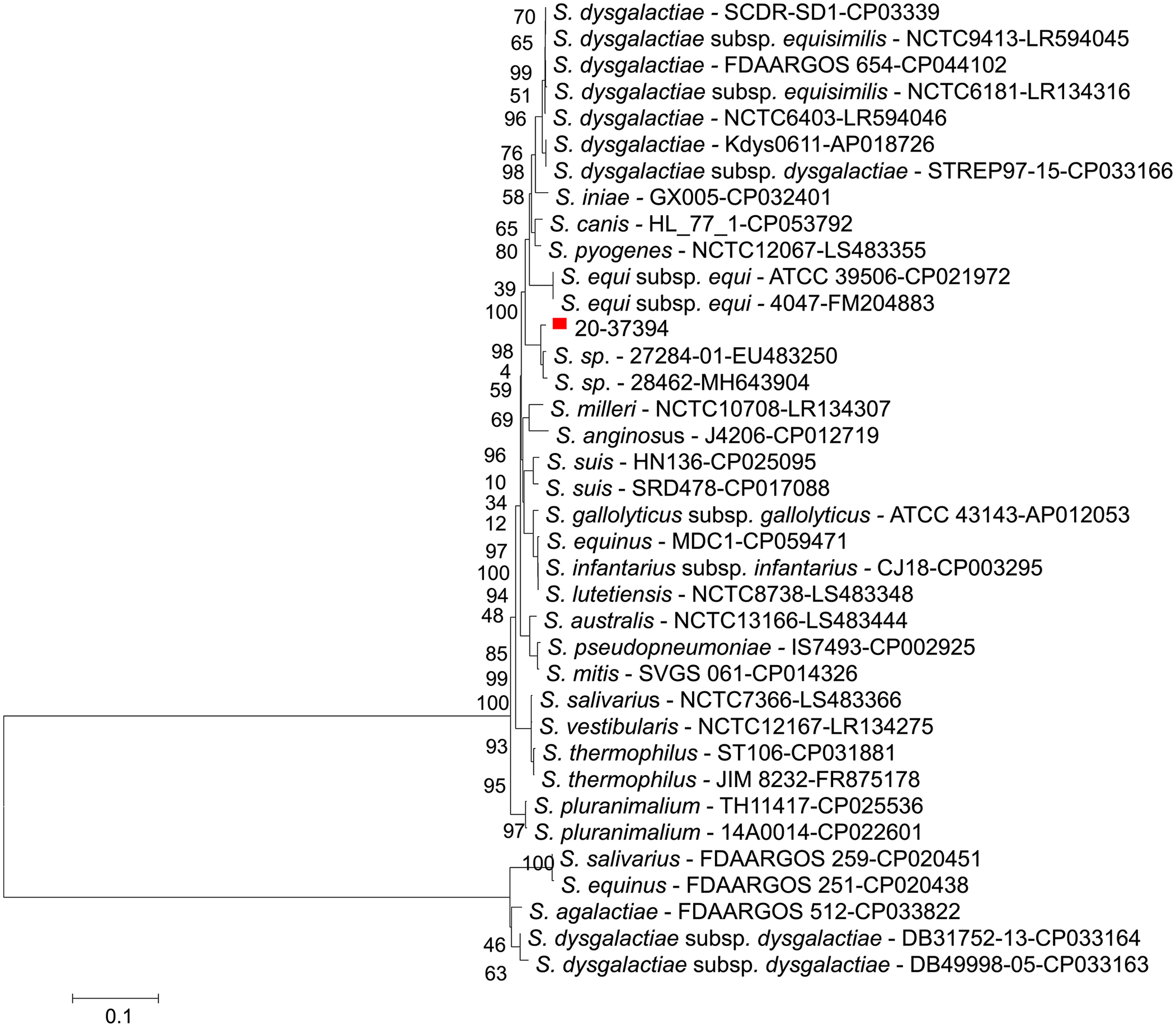
Phylogenetic analysis of 16S rDNA sequence of different Streptococcus species including the donkey milk Streptococcus isolate 20-37394 from our study marked with a solid red square.
A recent history of weaning has been identified as a risk factor for mastitis in horses, 14 and this is consistent with the history of the present donkey that had weaned her colt 10 d before hospital admission. In addition, the jenny was presented in July, which is the time of year coinciding with peak insect populations in Illinois, and was the time reported most commonly for the occurrence of equine mastitis in the United States. 14
It is interesting that the clinical signs of mastitis in our case resembled equine mastitis. 4 Anorexia and depression are clinical signs commonly observed in mares with mastitis, 3 and those were the reasons that the owner sought veterinary attention for his donkey. Moreover, the jenny responded quickly to the standard treatment for equine mastitis 4 ; the clinical signs subsided completely by 96 h post-admission. It is possible that this rapid response to treatment was the result of the jenny receiving veterinary care early in the disease process, given that the owner brought her in for evaluation as soon as the clinical signs were noted. Mastitis can occur unilaterally or bilaterally, and sometimes only one lobule is affected. 14 In our case, only one side was affected, but both lobules were affected. Although mastitis can be associated with leukocytosis and hyperfibrinogenemia in mares,4,14 not all mares have those findings, which is consistent with our case.
Our case also suggested the potential usefulness of cytologic evaluation of neutrophils in donkey mastitis; cytologic smears in conjunction with milk appearance were used to monitor the effect of treatment. In a previous report, 33% of the mares had bacteria present in their cytologic evaluations of mammary gland secretions. 14 Similarly, the jenny had bacteria identified on smears of mammary gland secretions from the date of admission to 72 h.
Genomic analyses revealed a unique etiology: an novel Streptococcus species. In comparison, standard methods for bacteriologic identification could only suggest that the isolate was an α-hemolytic Streptococcus spp., but not an Enterococcus spp. Analyses of the core genome SNVs and 16S rDNA gene both indicated that the organism was a previously unidentified Streptococcus species. WGS has been used frequently to characterize bacterial isolates given a higher level of discriminatory power than traditional typing methods, such as multi-locus sequence typing, pulsed-field gel electrophoresis, and 16S rDNA sequencing. 10 Both SNV and phylogenetic analysis of WGS data revealed that our isolate 20-37394 is closely related to the equine Streptococcus strain 28,462. However, given that the 16S rDNA sequence similarity between strain 28,462 and our isolate 20-37394 was <97%, this suggests that the 2 isolates are different Streptococcus species and reveals that 20-37394 could be a new Streptococcus species. 21
Streptococcus spp. have been identified as the main bacteria associated with clinical mastitis in mares.4,12 In cows, Streptococcus dysgalactiae and Streptococcus uberis are the most common environmental streptococci associated with mastitis. 20 Staphylococcus aureus, Streptococcus equisimilis, and Streptococcus equi subspecies equi have been grown in cultures of milk from clinically healthy donkeys.18,19 The Streptococcus isolate found in our study may have been an opportunistic environmental infection associated with lactostasis post-weaning, or an infection that resulted from an overgrowth of the mammary gland microbiome. However, the origin of the infection was not determined.
Supplemental Material
sj-pdf-1-vdi-10.1177_10406387211027306 – Supplemental material for A novel Streptococcus species causing clinical mastitis in a pregnant donkey
Supplemental material, sj-pdf-1-vdi-10.1177_10406387211027306 for A novel Streptococcus species causing clinical mastitis in a pregnant donkey by Giorgia Podico, Sarah M. Gray, Leyi Wang and Igor F. Canisso in Journal of Veterinary Diagnostic Investigation
Footnotes
Acknowledgements
We thank the owner of the donkey for trusting us with the care of his animal and for consenting to the publication of this manuscript.
Declaration of conflicting interests
The authors declared no potential conflicts of interest with respect to the research, authorship, and/or publication of this article.
Funding
Sequencing was funded in part by the Food and Drug Administration Veterinary Laboratory Investigation and Response Network (FOA PAR-17-141) under grants 1U18FD006673-01.
Supplemental material
Supplemental material for this article is available online.
References
Supplementary Material
Please find the following supplemental material available below.
For Open Access articles published under a Creative Commons License, all supplemental material carries the same license as the article it is associated with.
For non-Open Access articles published, all supplemental material carries a non-exclusive license, and permission requests for re-use of supplemental material or any part of supplemental material shall be sent directly to the copyright owner as specified in the copyright notice associated with the article.