
Abstract
Foot-and-mouth disease (FMD) is a highly contagious aphthoviral infection of cloven-hoofed animals, inducing vesiculopustular stomatitis, pododermatitis, and thelitis. Vesicular fluid represents a major pathway of virus excretion, but bovine milk is another important source of virus shedding. We describe here the time course of FMD virus (FMDV) excretion in the milk and characterize associated lesions in the mammary gland. Three dairy cows were infected by nasopharyngeal instillation of FMDV and monitored over 12 d. Autopsy was performed at the end of the study, and specimens were collected for histopathology, IHC, and RT-qPCR. All 3 cows developed fever, drooling, vesiculopustular stomatitis, interdigital dermatitis, and thelitis. FMDV RNA was detectable in whole milk until the end of the trial, but only transiently in saliva, nasal secretions, and blood serum. Although histology confirmed vesiculopustular lesions in the oral and epidermal specimens, the mammary glands did not have unequivocal evidence of FMDV-induced inflammation. FMDV antigen was detectable in skin and oral mucosa, but not in the mammary gland, and FMDV RNA was detectable in 9 of 29 samples of squamous epithelia but only in 1 of 12 samples of mammary gland.
Foot-and-mouth disease (FMD) is of global veterinary importance given its high contagiousness and the large variety of possible hosts among cloven-hoofed domestic and wild animals. The etiologic agent is FMD virus (FMDV; Picornaviridae, Aphthovirus), of which there are 7 known serotypes: O, A, C, Asia 1, SAT1, SAT2, and SAT3, with numerous subtypes.10,26 Although outbreaks of FMD usually cause relatively low mortality, morbidity, in contrast, is quite high because of the ease of direct or indirect transmission.2,17,18 Large amounts of virus are produced in the typical vesicular lesions of non-haired skin (e.g., interdigital clefts, coronary bands) and mucous membranes of the oral cavity. 26 The interaction of FMDV capsid proteins with at least 4 epithelial integrins (αvβ1, αvβ3, αvβ6, αvβ8) plays an important role in cellular invasion of the virus, explaining its epithelial tropism. 29 In addition to the fluid released from bursting vesicles, the virus is shed to the environment via all secretions and excretions of an infected animal. 10 Initial infection and primary replication of FMDV in the bovine host occurs in the nasopharynx, as was shown as early as 1981, 12 and confirmed by more recent studies.5,25
The mammary gland, or, to be exact, its secretory epithelium, has been considered an important site of secondary virus replication since the 1970s. Virus was detected in milk by isolation in cell culture, 11 as well as in secretory epithelium via immunofluorescence and electron microscopy. 6 Detection of FMDV or FMDV RNA in milk and the udder can precede the appearance of clinical signs such as oral aphthae or pedal vesicles.3,22,26 The relatively high and long excretion of FMDV RNA in milk qualifies pooled milk samples as an ideal matrix for epidemiologic surveillance.4,9,22 We are aware of only 2 consecutive experimental studies considering FMDV serotype O–induced mastitis using histopathology and scanning electron microscopy.7,8 More recent studies focus mainly on bulk milk samples for surveillance purposes, or on possible FMDV contamination of food (i.e., dairy products or meat).4,15,19,23,27 Therefore, with a pilot study, we wanted to characterize the excretion of FMDV in milk and describe the lesions of FMD with special regard to teats and mammary gland parenchyma as potential sources of viral excretion and shedding.
Our experiment was performed under veterinary BSL4 conditions at the Friedrich-Loeffler-Institut (Greifswald-Insel Riems, Germany), in compliance with national animal welfare regulations. The animal experimental protocol was approved by the State Office for Agriculture, Food and Fisheries of Mecklenburg-Vorpommern (7221.3-1-026/17). We infected 3 Holstein dairy cows, aged 2.4, 2.3, and 8.4 y, by nasopharyngeal instillation of 2 mL of homogenized vesicular epithelium collected from cattle infected with FMDV serotype A/IRN/22/2015. 21 The clarified homogenate had been diluted to a titer of 5.7 log10 TCID50/mL with sterile cell culture medium. For the inoculation, the animals were deeply sedated with xylazine (0.2 mg/kg body mass, IM). After the virus had been deposited in the nasopharynx of the recumbent animals through the nares and nasal cavities using a 3-mL syringe and a 30-cm length of 2-mm diameter flexible plastic catheter, the sedation was reversed with atipamezole (0.05 mg/kg, IM). The animals were monitored daily, and samples of blood, saliva, milk, as well as nose swabs and sloughed vesicular epithelium were collected over a period of 12 d. The animals were euthanized at 12 d post-infection (dpi), and autopsy was performed, including standardized sampling of muzzle, oral mucosa, tongue, interdigital skin, teats, mammary gland, heart, lung, spleen, liver, kidney, rumen, and jejunum for histopathology, immunohistochemistry (IHC), and further molecular examinations (Suppl. Table 1).
FMDV IHC was performed using the avidin-biotin-peroxidase complex method (Vectastain Elite ABC kit; Vector) with a hematoxylin counterstain. After antigen retrieval with citrate buffer (pH 6, microwave 700 W, 20 min), tissue sections were incubated with an in-house polyclonal rabbit serum against FMDV strain A Iran 97 for 16 h overnight at room temperature, followed by incubation with a biotinylated goat anti-rabbit IgG (H+L) secondary antibody (BA-1000, Vector; 1:200) for 30 min at room temperature and development with 3-amino-9-ethylcarbazole (AEC; Dako AEC substrate chromogen ready-to-use; Agilent) for 10 min. Known positive samples of a bovine tongue and interdigital skin with vesiculopustular FMD lesions were used as a positive control; negative control sections were incubated with normal rabbit serum (1:2,000) instead of the primary antibody.
RNA was isolated from saliva, nasal secretions, and blood serum (NucleoMag VET kit, Macherey-Nagel; KingFisher magnetic particle processor, Thermo Fisher). Formalin-fixed (FF) specimens of oral, dermal, and mammary gland tissue were homogenized in TRIzol reagent, and RNA was extracted (RNeasy kit; Qiagen) from the aqueous phase obtained after the addition of chloroform. Whole milk was mixed with TRIzol LS and the aqueous phase was used for RNA extraction (NucleoMag VET kit) as described previously. 1 FMDV genome in the samples was quantified by reverse-transcription quantitative PCR (RT-qPCR; AgPath-ID one-step RT-PCR kit; Applied Biosystems) with a previously published primer pair and probe targeting the 3D coding region, 13 and a β-actin internal control. 28
Although all cows exhibited characteristic lesions, there was individual variability in the lesion profile and viral topography (Suppl. Fig. 1). Although the clinical evaluation was limited to the externally visible lesions, gross and histologic examination revealed consistent lesions on the teats and to a variable extent on the muzzle and mouth as well as the interdigital skin. Even though antigen could only be detected immunohistochemically in 4 lesions, RT-qPCR confirmed the presence of FMDV in 3 of 4 lesions, and additionally in 3 more locations.
All animals developed clinical signs typical for FMD. By 3 dpi, all cows had epithelial lesions in the mouth, and cows 2 and 3 had additional lesions on the feet. Lesions on all 4 feet of the animals as well as lesions on the teats of cows 2 and 3 were evident by 4 dpi. Animal 1 developed lesions on the teats by 6 dpi.
At autopsy, lesions were not evident in the abdominal organs. Pustules, vesicles, erosions, and ulcers were present in the oral mucosa, mainly on the tongue of cows 1 and 2 and the hard palate of cow 2, as well as on the muzzle of all 3 cows and the interdigital spaces of cows 1 and 3. In cow 3, a lesion was also present on 1 coronary band. The most evident lesions were seen at the apices of the teats in all 3 cows. There were sharply demarcated, gray-beige to tan, 2–6 mm diameter, crusty lesions around the opening of the streak canal, interpreted as epidermal erosions (Suppl. Fig. 2).
The histologic findings consisted of acantholytic epithelial cells with formation of intraepithelial vesicles or erosions, especially on the tongue. Because of secondary mechanical alteration and bacterial infection, intralesional bacteria were present in some areas. The main findings included dermatitis with erosions and ulcers (on the muzzle of all 3 cows; in the interdigital spaces of cows 1 and 3), glossitis (cow 1), and thelitis (all 3 cows; Fig. 1A). The mammary gland parenchyma did not show consistent signs of degeneration, necrosis, or inflammation (Fig. 1B), with the exception of a sample from cow 2 exhibiting minimal, subacute, oligofocal, suppurative mastitis. Although FMDV was detectable immunohistochemically in the FMD lesions of the teats (cows 1, 3; Fig. 1C), tongue (cow 1), and interdigital skin (cows 1, 3), there were no detectable lesions or antigen present in mammary gland parenchyma (Fig. 1D).
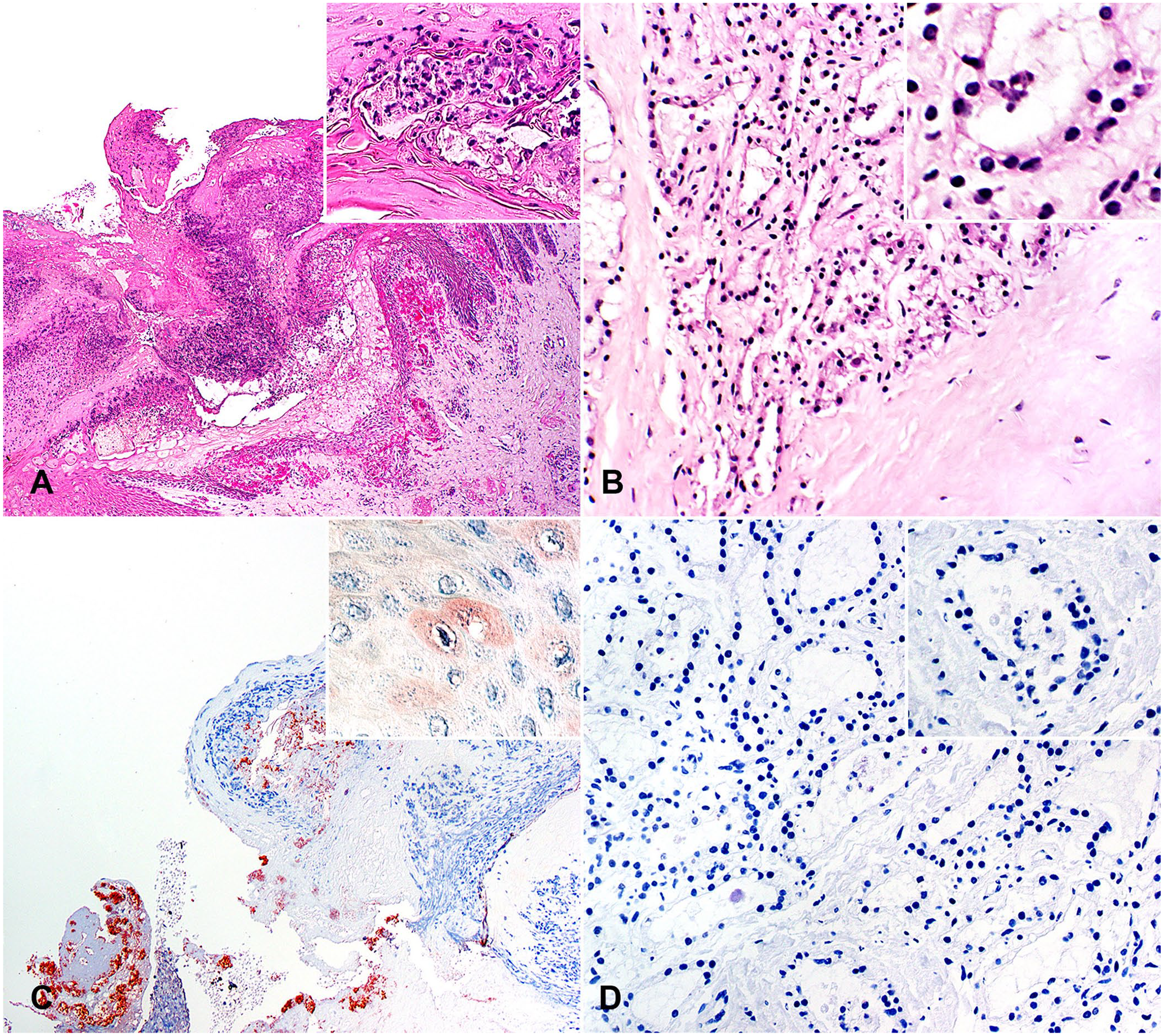
Histologic and IHC findings in teat skin and mammary gland parenchyma in lactating cows 12 d post-infection (dpi) with foot-and-mouth disease virus (FMDV).
FMDV genome was present in samples of whole milk from 3 dpi until the end of the trial in moderate copy numbers in all 3 cows (Suppl. Fig. 3). In contrast, FMDV genome was detectable at 1–3 dpi until 10–11 dpi in saliva, at 1–3 dpi until 7–10 dpi in nasal secretions, and at 2–3 dpi until 5–7 dpi in serum, and was no longer detectable in any of these matrices by 12 dpi (Suppl. Fig. 4).
In the FF tissues, FMDV RNA was detected in 9 of 29 samples from lesions on the teats, in the interdigital skin, and in the oral mucosa, but in only 1 sample of the mammary gland of 1 cow of the 12 samples of mammary gland (4 per cow) examined in total. The β-actin internal control was negative in 13 of the 41 samples examined, but 3 of those 13 were positive for FMDV RNA.
Although there was ongoing detection of viral RNA in milk and residual lesions in the skin, including on the teats and oral mucosa, no specific FMD lesions were present in the mammary gland parenchyma, and FMDV RNA was only detected in 1 of 12 specimens. Thus, we did not see in our study the viral replication described previously in the bovine mammary gland. 6 One reason for this difference could be the route of infection, which was via intramammary instillation in the earlier studies but via nasopharyngeal instillation in our experiment,6,11 which is considered a more natural way of infection.5,20 Transient increase in the cellular content of the milk as well as typical clinical findings suggestive of acute, catarrhal mastitis were reported in the inoculated quarter after experimental intramammary FMDV infection.10,11 Mastitis in association with natural FMDV infection is considered to be part of the generalized disease and is often the result of secondary bacterial infection further to lowered defense mechanisms (e.g., related to FMD lesions of the teats).10,16,30 Another group of researchers was able to detect necrotizing mastitis and FMDV antigen via immunofluorescence, mainly in sloughed mammary gland epithelium, in cows experimentally infected by aerosol as well as by mammary inoculation, or a combination of both.6,7 Those findings were corroborated via scanning electron microscopy in a later similarly designed study by the same group. 8 There also seems to be a difference in the amount of virus in the milk, dependent on virulence, infection dose, or virus strain, as was shown in other studies,3,11,22 which used FMDV O1 BFS 1860 (lineage O/EURO-SA/O1), O/UKG/34/2001 (lineage O/ME-SA/PanAsia), and A22 Iraq 24/64 (lineage A/ASIA/A22). It is possible that the virus isolate in our experiment (A/IRN/22/2015, lineage A/ASIA/G-VII) has reduced tropism for or restricted replication in mammary gland tissue. It is unknown whether the virus was of the same lineage in a retrospective study of an FMDV serotype A outbreak in a dairy farm in Iran during 2014, 2 in which there was an unusually low impact on milk production.
We used RT-qPCR to test secretions and excretions, but RT-qPCR has limitations when testing FF samples. Viral detection within FF samples is generally considered to be less sensitive compared to native samples because of degradation of RNA by formalin, 24 which therefore carries a high risk of false-negative results. Even slight contamination of the specimen with formalin severely inhibits RNA extraction. 14 Although the bovine udders in our study were examined closely at autopsy, the screened tissues only represent a tiny fraction of the mammary gland, which could also be a reason for our inability to detect FMDV in the mammary tissue.
It remains unclear why viral RNA was still present in the milk when blood, saliva, and nasal secretions no longer contained any detectable FMDV RNA, and why we did not find any FMD lesions or FMDV antigen in the mammary tissue. Apart from the possibility of having tissue type–dependent, restricted FMDV antigen and RNA expression per cell in mammary gland compared to squamous epithelium, alternative sources of FMDV contaminating the milk, such as the consistently observed dermal teat lesions, should be considered. In any case, we conclude that generalized FMD and virolactia do not necessarily lead to fulminant viral replication and mastitis in the bovine mammary gland.
Supplemental Material
sj-pdf-1-jvd-10.1177_10406387211022467 – Supplemental material for After nasopharyngeal infection, foot-and-mouth disease virus serotype A RNA is shed in bovine milk without associated mastitis
Supplemental material, sj-pdf-1-jvd-10.1177_10406387211022467 for After nasopharyngeal infection, foot-and-mouth disease virus serotype A RNA is shed in bovine milk without associated mastitis by Marcel Suchowski, Michael Eschbaumer, Jens P. Teifke and Reiner Ulrich in Journal of Veterinary Diagnostic Investigation
Footnotes
Acknowledgements
We thank Holger Freese, Christian Loth, Silvia Schuparis, and Anja Schulz for technical support.
Declaration of conflicting interests
The authors declared no potential conflicts of interest with respect to the research, authorship, and/or publication of this article.
Funding
The authors received no financial support for the research, authorship, and/or publication of this article.
Supplemental material
Supplemental material for this article is available online.
References
Supplementary Material
Please find the following supplemental material available below.
For Open Access articles published under a Creative Commons License, all supplemental material carries the same license as the article it is associated with.
For non-Open Access articles published, all supplemental material carries a non-exclusive license, and permission requests for re-use of supplemental material or any part of supplemental material shall be sent directly to the copyright owner as specified in the copyright notice associated with the article.