
Abstract
We assessed the causes of polyserositis in pigs, categorized by causative agents and ages of animals affected. In a 3-y study, 246 pigs from 80 different farms with recurrent problems of polyserositis, in a high-density breeding area, were submitted for autopsy; 154 pigs with typical fibrinous serosal lesions were sampled for further bacterial and viral investigation. The most common gross lesions were pleuritis and pericarditis (141 of 154; 92%). The animals most affected were weaned pigs (139 of 154; 90%). Haemophilus parasuis and Mycoplasma hyorhinis were the most common bacteria detected and were present at the same rate (85 of 154; 55%). Other bacteria isolated were Streptococcus sp. (44 of 154; 29%), Pasteurella multocida (21 of 154; 14%), Escherichia coli (19 of 154; 12%), Actinobacillus pleuropneumoniae (7 of 154; 5%), and Trueperella pyogenes (4 of 154; 3%). Porcine reproductive and respiratory syndrome virus (PRRSV; 119 of 154; 77%) predominated among the viruses detected, followed, with lesser prevalence, by porcine circovirus 2 (40 of 154; 26%) and swine influenza A virus (19 of 154; 12%). Bacterial coinfection and coinfection of bacteria and viruses were common (128 of 154; 83%). A strong positive correlation was found between coinfection by H. parasuis and M. hyorhinis and also by H. parasuis with PRRSV.
Polyserositis is a widespread and important disease in Italian pig farms. The associated economic loss is significant and is mainly the result of culling. 4 Polyserositis is typically characterized by fibrinous or fibrinopurulent pleuritis, pericarditis, or peritonitis, frequently associated with arthritis and meningitis. Four- to 8-wk-old pigs are typically the most affected animals and, to a lesser extent, 3- to 4-mo-old pigs. 2 Piglets are at risk, especially when reared in a contaminated environment. Polyserositis has typically been the result of infection with Haemophilus parasuis (Glässer disease), with impact on pig production varying based on different serovars, and on environmental and immunologic factors. 1 However, studies have highlighted the importance of other microorganisms such as Mycoplasma hyorhinis, Streptococcus suis, Escherichia coli, and porcine reproductive and respiratory syndrome virus (PRRSV; Betaarterivirus suid).8,11 Viruses that can cause viremia, immunosuppression, such as porcine circovirus 2 (PCV-2), or respiratory disease, such as swine influenza A virus (IAV), could be involved and should, therefore, be investigated further.
We examined 246 dead pigs from 80 different farms from the Po Valley region, in northern Italy, from various production systems (including farrow-to-finish, farrow-to-wean, and multiple-site herds) and with a history of recurrent problems of polyserositis. We only included pigs that were not vaccinated or had no history of treatment for H. parasuis. Viral status of the farms was unknown.
Animals were submitted to the Diagnostic Laboratory of Animal Diseases of the Istituto Zooprofilattico Sperimentale della Lombardia ed Emilia Romagna (IZSLER; Brescia, Italy) for autopsy. Only those animals with fibrinous or fibrinopurulent pleuritis, pericarditis, or peritonitis were enrolled in the study. Swabs from the pleura, pericardium, or peritoneum, and tissue from the lung, spleen, kidney, and brain, were collected from each pig. Organs were pooled, homogenized (10% w/v in modified Eagle medium), and then analyzed for pathogens.
We performed direct culture for non-target agents, such as common bacteria, rather than the more sensitive PCR methods, which can detect nucleic acid from nonviable organisms from past or chronic infections not related to polyserositis. For fastidious microorganisms, such as H. parasuis and M. hyorhinis, in view of the low sensitivity of culture, we used PCR. Bacteria were isolated from tissues and swabs in pure cultures on solid agar plates (Gassner agar and 5% blood agar, and with Staphylococcus aureus as feeder organisms in a microaerophilic condition) at 37°C for 24–48 h. Identification took place by standard phenotypic characterization including the API system. 3 For H. parasuis, we used an end-point PCR based on amplification of a species-specific 821-bp sequence of 16S ribosomal RNA. A real-time PCR (rtPCR) assay for the p37 protein was used for the detection of M. hyorhinis.5,10 IAV was detected by using a reverse-transcription rtPCR assay. 9 PRRSV was detected with the AgPath-ID NA and EU PRRSV multiplex reagent kit (Applied Biosystems), as specified by the manufacturer. PCV-2 was detected by rtPCR. 6
The prevalence of each infectious agent was calculated, and the binomial exact method was used to compute 95% CIs. The occurrence of coinfection, defined as the presence of at least 2 pathogens in the same animal, was also calculated. The association of pathogens, animal category, and coinfection were evaluated by a chi-square test (χ 2 ) or a Fisher exact test, as appropriate. Correlations between pathogens were calculated by the Phi index. A logistic regression model was performed to calculate the association between polyserositis and pathogen occurrence. For each test, p ≤ 0.05 was considered as statistically significant. All analyses were performed using R software (v3.3.1; https://www.r-project.org/).
Typical fibrinous lesions involving at least 2 serosae were found in 154 of 246 animals (63%). Pleuritis was present in 153 of 154 animals (99%), pericarditis in 141 of 154 animals (92%), and peritonitis in 108 of 154 animals (70%). The main association was between pleuritis and pericarditis (92%); lesions in all 3 serosae were present in 96 of 154 animals (62%).
Regarding the age groups of animals, 90% of pigs with lesions were weaned pigs (139 of 154), 9% were finishers (14 of 154), and 0.6% were piglets (1 of 154). The distribution of lesions in different serosae was not associated with pig age categories (data not shown).
The most common pathogens detected were PRRSV (77%), M. hyorhinis (55%), and H. parasuis (55%; Table 1). Coinfection was detected in 128 of 154 animals (83%). The most frequent coinfection was by PRRSV and bacteria, particularly H. parasuis and/or M. hyorhinis (Table 2).
Prevalence of infectious agents detected among 154 pigs with polyserositis.
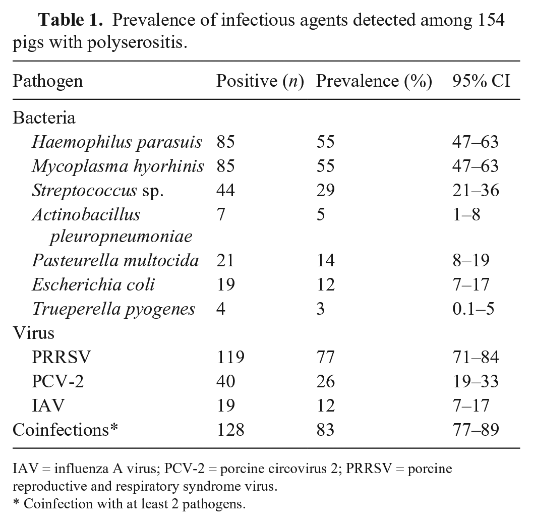
IAV = influenza A virus; PCV-2 = porcine circovirus 2; PRRSV = porcine reproductive and respiratory syndrome virus.
Coinfection with at least 2 pathogens.
Prevalence of infection by multiple pathogens in 154 pigs with polyserositis.
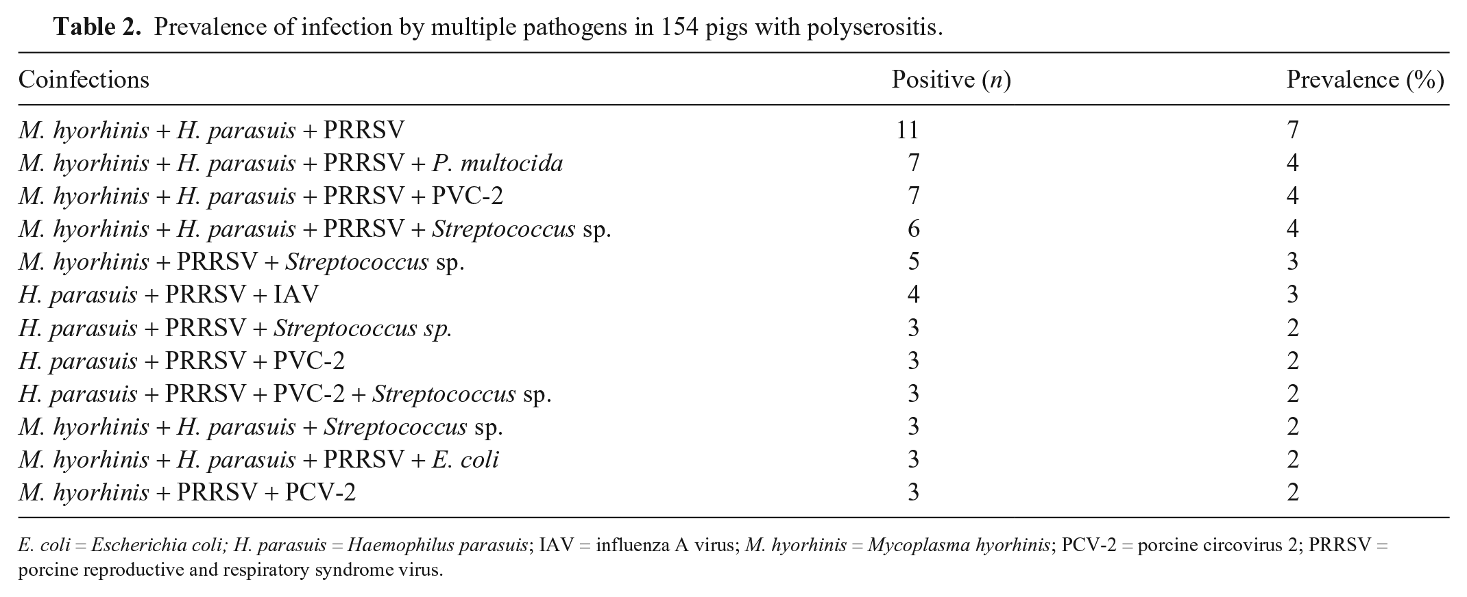
E. coli = Escherichia coli; H. parasuis = Haemophilus parasuis; IAV = influenza A virus; M. hyorhinis = Mycoplasma hyorhinis; PCV-2 = porcine circovirus 2; PRRSV = porcine reproductive and respiratory syndrome virus.
Pathogens were equally distributed among the pig age categories (data not shown) with the exception of A. pleuropneumoniae (APP; p = 0.02) which was more commonly detected in finishers than in weaned pigs. The probability of detection of APP was 9 times more likely in finishers than in weaners (odds ratio [OR]: 9.2; 95% CI: 1.6–47).
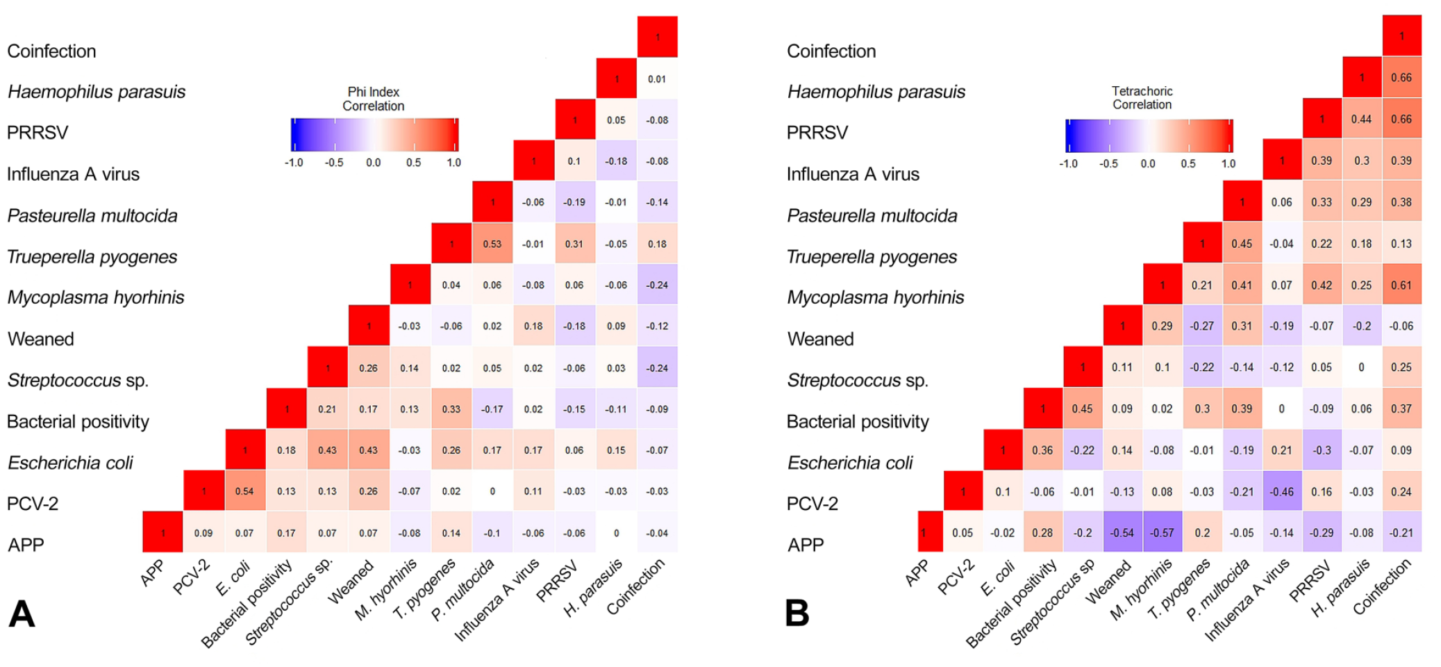
Phi and tetrachoric correlation matrixes were calculated for 153 pigs (the piglet was excluded) among 13 factors (M. hyorhinis, H. parasuis, PRRSV, IAV, PCV-2, Trueperella pyogenes, APP, E. coli, Streptococcus sp., Pasteurella multocida, coinfection, weaned category, and bacterial positivity) (Fig. 1). Results were similar for both matrixes. Positive associations were statistically significant between H. parasuis, M. hyorhinis, and PRRSV, with a medium-strong value; associations were weaker for H. parasuis and P. multocida, and for H. parasuis and APP. Negative associations were evident for APP and M. hyorhinis, and for APP and weaning pigs.
Correlation matrix results were confirmed by means of a Fisher test; there was a statistically significant association of H. parasuis with each of M. hyorhinis, E. coli, P. multocida, PRRSV, and IAV (Table 3). In particular, the probability of infection with the above agents was 3-fold if H. parasuis was present. The only exception is E. coli for which there was a weaker probability. A significant association of H. parasuis infection was not observed with Streptococcus sp., APP, T. pyogenes, or PCV-2.
Association of H. parasuis with other bacterial and viral pathogens.
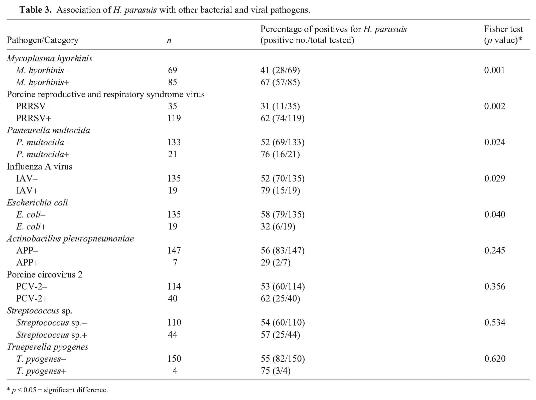
p ≤ 0.05 = significant difference.
For pathogens with a positive association with H. parasuis infection, the OR was estimated using logistic regression (Table 4). The probability of finding H. parasuis in coinfection with another pathogen was more than 20 times higher than finding H. parasuis alone. Conversely, the probability of observing E. coli infections decreased when H. parasuis was detected.
Odds ratio estimated for H. parasuis by considering the positive factors from the Fisher test (Table 3). Baseline is the presence of the considered factor.
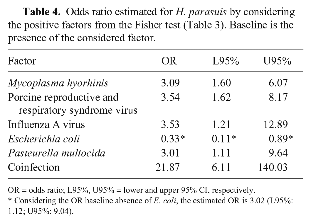
OR = odds ratio; L95%, U95% = lower and upper 95% CI, respectively.
Considering the OR baseline absence of E. coli, the estimated OR is 3.02 (L95%: 1.12; U95%: 9.04).
Our findings confirmed a high infection rate of H. parasuis, M. hyorhinis, and high prevalence of PRRSV in coinfections (Table 2) in examined pigs, as reported by others.7,8 The prevalence of PRRSV in pigs in our study area in 2002–2016 was 27% (95% CI: 26–28%; IZSLER, unpublished data), which is much lower than its prevalence in our investigated cases of polyserositis. Our results support the concept that PRRSV, via its immunosuppressive effect, predisposes to bacterial infection, especially by the main agents of polyserositis (H. parasuis and M. hyorhinis). 12
PCV-2 is also an important agent that can induce immunosuppression, but PCV-2 was not detected significantly in our investigated area because of the common practice of vaccinating piglets. PCV-2 prevalence in 2002–2016 was 29% (95% CI: 26–32%; IZSLER, unpublished data), which was not statistically different (binomial p = 0.26) from the rate of polyserositis that we detected (26%; 95% CI: 19–33%).
Footnotes
Declaration of conflicting interests
The authors declared no potential conflicts of interest with respect to the research, authorship, and/or publication of this article.
Funding
The authors received no financial support for the research, authorship, and/or publication of this article.