
Abstract
In February 2015, we conducted a field study of causes of mortality of northern elephant seal (Mirounga angustirostris) pups on San Miguel Island, California. Autopsies were performed on 18 freshly dead pups. Ages of pups ranged from stillborn to 6–8 wk. Gross and histologic lesions included trauma (9 of 18 pups), multifocal necrotizing myopathy (8 of 18), starvation with emaciation (7 of 18), congenital anomalies (3 of 18), bacterial infections (3 of 18), and perinatal mortality (stillbirths and neonates; 2 of 18). Trauma and emaciation or starvation were the most significant contributors to death. Bacterial infections included hemolytic Escherichia coli isolated from the lungs of 2 pups with pneumonia. Additionally, non-hemolytic Streptococcus sp. and hemolytic E. coli were isolated from the liver of an emaciated pup that had mild multifocal suppurative hepatitis. Other lesions, including a previously described necrotizing myopathy, congenital anomalies, and bacterial infections, were detected concurrently in cases with starvation and/or emaciation or trauma.
Elephant seals of the genus Mirounga (Gray, 1927) are the largest pinnipeds in the world. There are 2 species in this genus: northern elephant seals, M. angustirostris (Gill, 1866), and southern elephant seals, M. leonina (Linnaeus, 1758). 10 Northern elephant seals occupy rookeries along the western coast of mid-Baja California, Mexico, to central California and forage in the eastern North Pacific up to the Aleutian Islands of Alaska. 17
Northern elephant seals were presumed extinct by 1892 as a result of commercial harvesting that began in the mid-1800s when it became more difficult to find whales to obtain blubber for rendering into oil. A small, residual breeding colony survived on Isla de Guadalupe, Baja California, Mexico and grew rapidly through the early 1900s following legal protection from further hunting. 21 To date, the northern elephant seal population is estimated to be 210,000–239,000 animals; ~179,000 animals inhabit California rookeries. 21 At least 10 northern elephant seal rookeries are in California; the largest is on San Miguel Island, which is the most northwestern of the 8 Channel Islands of southern California within the Channel Islands National Park. 21
Northern elephant seals recolonized San Miguel Island in the 1950s. Their population on San Miguel Island is currently increasing and estimated to be ~62,000 animals, based on counts through 2010, and alone produces ~40% of all elephant seal pups born in California. 5 Despite extensive studies addressing ecology and dynamics of northern elephant seal populations, few papers addressing causes of mortality in pups on rookeries have been published.14,18,19 We investigated causes of mortality in northern elephant seal pups in rookeries on San Miguel Island, which may also provide insights into the significance of pup loss on recruitment and population status.
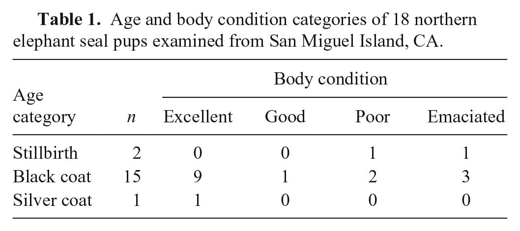
We conducted our study February 7–15, 2015, at 4 rookeries on San Miguel Island: West Cove, North West Cove, Adams Cove, and Judith Cove. Eighteen freshly dead pups (11 females and 7 males) were found within rookeries. Carcasses were transported ~30–50 m from the rookery edge for autopsy. Estimation of age of pups was based on previously described criteria. 19 Pups were categorized into 3 age groups: stillbirths, black-coated pups (1-d to 3-wk-old), and silver-coated pups (>3-wk-old). Body condition of pups was scored by mid-sternal blubber thickness and assigned to 1 of 4 categories: excellent (>15 mm of sternal blubber), good (15–10 mm), poor (<10–5 mm), and emaciated or starving (<5 mm) as described previously. 19
Samples from all major organs (brain, heart, lungs, liver, kidneys, tongue, stomach, small and large intestine, skeletal muscle, thymus, adrenal glands, thyroid glands, adipose tissue, skin, bone) were collected, placed in 4.5-L plastic bags, and taken to a small field station for further processing. Portions of these same tissues were preserved in 10% neutral-buffered formalin. Selected tissues (brain, liver, lung, kidney, heart, skeletal muscle, bile, adipose tissue) were frozen for ancillary laboratory studies. Formalin-fixed tissues were processed routinely, stained with hematoxylin and eosin, and reviewed by a board-certified pathologist (T.R. Spraker). Selected tissues were stained with special stains, including Gram (Brown–Hopps modification) and Gomori methenamine silver, for further study. 15 Frozen tissues (muscle, lung, liver, bile, kidney) were thawed, swabbed, and processed for routine microbiology at the Colorado State University Diagnostic Medicine Center (Colorado State University, Fort Collins, CO). Swabs were plated on trypticase soy agar with 5% sheep blood, MacConkey II, Columbia with 5% sheep blood for aerobic organisms, and CDC with 5% sheep blood (Center for Disease Control formulation). Brucella blood agar plates were used for anaerobes.
Body condition of pups varied from excellent to emaciated (Table 1). Body condition score did not appear to be correlated with the causes of death in individual pups. Based on autopsy findings, 6 general processes were evident: 1) blunt trauma, 2) emaciation, 3) myopathy, 4) inflammatory diseases, 5) perinatal mortality (stillbirths and neonates), and 6) congenital anomalies (Table 2; Supplementary Table 1).
Age and body condition categories of 18 northern elephant seal pups examined from San Miguel Island, CA.
Summary of gross lesions found in 18 northern elephant seal pups from San Miguel Island, CA, compared to body condition scores.
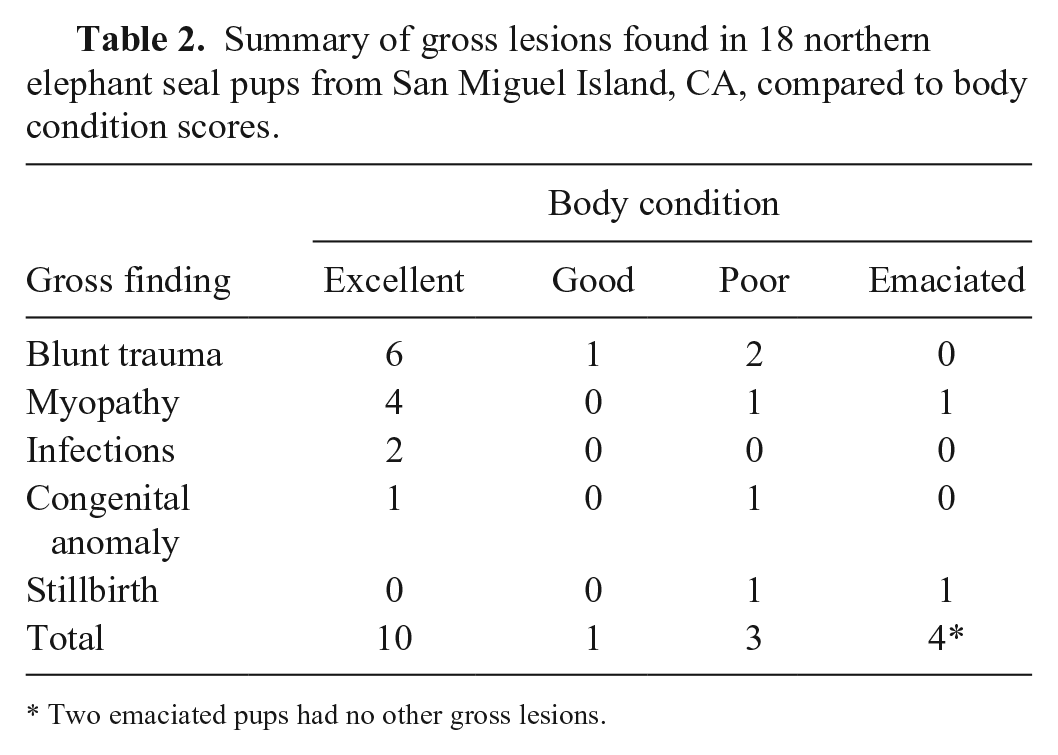
Two emaciated pups had no other gross lesions.
Lesions indicative of blunt force trauma were the most common gross findings, identified in 9 of 18 pups and within all body condition scores except for emaciated pups. The source of trauma was presumed to have occurred by conspecific adults. Sharp trauma or bite wounds were not found in any of the pups in our study. Abdominal trauma resulting in a fractured liver and hemoabdomen (6), trauma to the chest causing hemothorax (2), and head trauma with severe subdural hemorrhage (1) were documented (Supplementary Table 1). In 6 of 9 cases, other concurrent predisposing conditions (idiopathic myopathy [5] and omphalophlebitis with mild hydrocephalus [1]) were present. This myopathy was characterized grossly by pale streaking in musculature of the abdominal wall and, to a lesser extent, diaphragm and limbs, and was compatible with a previously described necrotizing myopathy in elephant seal pups. 20 Debilitation secondary to myopathy or umbilical infection and hydrocephalus could have impaired mobility and made affected animals more vulnerable to trauma. In the other 3 cases, traumatic injury was the sole finding.
Trauma has been commonly reported in most marine mammal populations and is a contributor to death or stranding in a large proportion of cases. 4 In a 2014 study addressing mortality in elephant seal pups on Año Nuevo State Reserve, California, trauma was reported in 16 of 21 of the pups examined; however, trauma was consider to be the primary cause of death in only 6 pups. 19 In a review of 1,277 northern elephant seal strandings (159 weaned black-coated pups, 901 silver-coated pups, 206 yearlings, 6 subadults, and 5 adults) on the central coast of California from 1992 to 2001, trauma was considered the primary cause of death in 43 seals (3%), and a secondary factor in 115 (9%). 2
Poor body condition and emaciation were found in 7 pups; 4 of these pups had sternal blubber thickness of <5 mm, and 3 pups had sternal blubber thickness of 5–10 mm. Additional indicators of emaciation noted histologically included parenchymal organ atrophy, absence of lipid in adipose tissues, and various degrees of lymphoid and thymic depletion. In a single case, emaciation was the only gross or histologic lesion of significance detected. Two cases of emaciation were stillbirths; one was small (girth:length = 55:96), and suppurative pneumonia was detected histologically in the second stillbirth. Additional lesions noted in 4 of the other emaciated pups included terminal bacteremia (1), the previous described myopathy 20 (2), omphalophlebitis (1), mild hydrocephalus (1), and mild subdural hemorrhage (1).
Emaciation or starvation is considered to be a common cause of death in pups on rookeries.8,9,11,14,18,19 In a study performed in 2012 on Año Nuevo, emaciation or starvation was reported in 29% of the pups examined. 19 In a similar study on San Benito Island, Mexico, wherein authors tagged and followed newborn pups, 8 of 12 marked pups found dead had been orphaned and starved. 18 In a report addressing mortality in stranded elephant seals, emaciation or starvation was considered to be the primary cause of the stranding in 724 seals (57%). 2
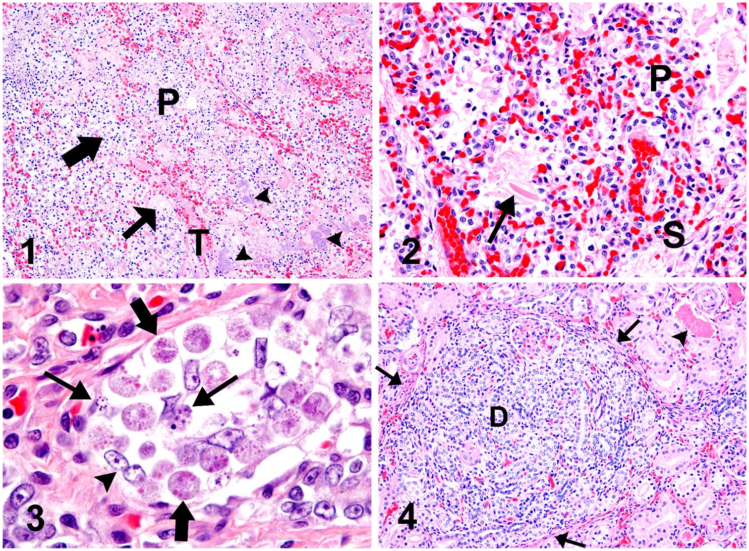
Gross evidence of infectious (presumed bacterial) diseases were found in 2 pups and included cases of suppurative bronchopneumonia and severe omphalophlebitis. Escherichia coli was isolated from the lung in the case of bronchopneumonia (Fig. 1). Culture confirmation was not done in the case of omphalophlebitis because of extensive environmental contamination. Histologic evidence of a bacterial infection was found in 2 other pups that had no gross lesions suggestive of an infectious condition. Pneumonia was detected in one stillborn pup, and hemolytic E. coli was isolated from the lung (Fig. 2). A severely emaciated pup had mild multifocal necrosuppurative hepatitis. Non-hemolytic Streptococcus sp. and hemolytic E. coli were isolated from the liver of this pup.
Lesions in northern elephant seal pups.
One pup in excellent body condition that presumably died as a result of severe blunt chest trauma had mild, multifocal gastritis histologically, with rare intraepithelial protozoa morphologically compatible with coccidia (Fig. 3). This was considered an incidental finding and did not contribute to the death of this pup. Similar organisms have been observed previously in the upper duodenum in 3 stranded elephant seal pups from the Marine Mammal Center, Sausalito, CA (K.M. Colegrove, pers. comm., 2018).
Infectious agents have been reported rarely in elephant seal pups living on rookeries. However, in one study on Año Nuevo, bacterial infections (infected bite wounds and pneumonia) were reported in 9 of 21 pups. 19 In a review of stranded elephant seals from the Maine Mammal Center, infectious agents including parasites and bacteria were considered to be the primary cause of stranding in 402 (31%) animals. 2
Three of the 18 pups in our study had congenital anomalies: hydrocephalus (2) and renal tubular dysplasia (1; Fig. 4) In the cases of hydrocephalus, concurrent lesions that were considered the immediate cause of death included head trauma and myopathy. 20 The renal cortical dysplasia was characterized by discrete foci in which tubules were closely spaced, tortuous, and irregular. Dysplastic foci were surrounded by a thin zone of fibroblasts and degenerate tubules filled with fibrin and cellular debris (Fig. 4); ~60% of the kidney cortex was affected. Other underlying or concurrent lesions were not detected in this case.
Numerous congenital anomalies have been reported in cetaceans and pinnipeds, but most have been reported in individual stranded animals and not from rookeries. 3 Congenital anomalies were reported in 7 of 21 elephant seal pups on the rookery at Año Nuevo. 19 In juvenile elephant seals stranded along the central California Coast, congenital anomalies have been documented in 5.2% of the animals autopsied.2,24
The congenital anomalies observed in our study (3 of 18 pups) were fewer compared to the study in pups from Año Nuevo in which 7 of 21 were reported; however, both of these studies report higher prevalence than the average rate of 1.0% in domestic animals 22 or wildlife. 25 One possible explanation for the apparently elevated prevalence of congenital anomalies could be the low genetic diversity documented in the elephant seal population, which may predispose these animals to congenital anomalies and infectious diseases.1,22
Perinatal mortality includes stillbirths (lungs not aerated; sinks in formalin) and neonatal pups (estimated to be <1-d-old). Two pups examined in our study had gross findings consistent with stillbirth. One case was emaciated and relatively small (girth:length = 55:96) and had no sternal blubber. Other gross or histologic lesions were absent. Based on size, a premature birth was considered and may have contributed to this stillbirth. The second stillbirth was considered to be a full-term pup. Histologically, this pup had pneumonia, and hemolytic E. coli was isolated from the lungs. Stillbirths on pinniped rookeries are common. In 2 previous studies addressing mortality in elephant seal pups, perinatal mortality was reported in 3 of 21 pups on Año Nuevo, 19 and in 1 of 21 pups examined on San Benito Island. 18 The perinatal mortality found in our study (11%; Supplementary Table 1) was considered to be similar to these previous studies.
There have been several long-term, population-scale, behavioral and ecological studies of elephant seals on Año Nuevo11,12,16 and San Miguel Island. 5 Multi-year population studies performed on Año Nuevo have shown that 31.5 ± 12.4% of the pups die each year before they depart to sea, 16 and the first-year mortality when these pups were at sea has been estimated to be 13–26%. 11 In these studies, causes of mortality were rarely addressed.
Authors reported on pup mortality on Año Nuevo and concluded that starvation and trauma often occurred together. 9 They described this set of conditions as “the trauma/starvation syndrome.” This syndrome begins with mother–pup separation, followed by abandonment and loss of protection by the female. This leads to pups incurring various degrees and types of trauma from subadult males and aggressive females; ultimately, the pup dies from trauma or starvation with trauma.8,9 Other researchers also confirmed that starvation and trauma were extremely common processes affecting pups on crowded elephant seal rookeries.14,18 However, these reports seemed to focus on gross evidence of trauma and starvation; infections, congenital anomalies, and other causes of pup mortality were not addressed. Histopathology or bacteriology were not reported in any of these studies. In a previous study on mortality of elephant seal pups on Año Nuevo 19 and in our study, autopsies were followed with histopathology and microbiology, which revealed additional lesions not evident on gross examination alone.
It is difficult to make reliable comparisons addressing causes of mortality in elephant seal pups in our study to previous investigations given the differences in methodology and extent and nature of testing done.14,18 However, combined data support several general conclusions: 1) emaciation or starvation is a commonly reported cause of death on rookeries and stranding of older pups; 2) trauma is a common finding on gross examination in starving and in clinically healthy pups, but there may be underlying conditions such as young age, infections, non-fatal bite wounds, or myopathy to name a few conditions that predispose pups to trauma; and 3) congenital anomalies (especially hydrocephalus) are more common in elephant seal pups than in domestic animals and other marine mammals, perhaps given the limited genetic diversity in elephant seals. 1
Population studies document that most elephant seal populations on the coast of California are steadily increasing. 13 However, several small elephant seal rookeries off the coast of Baja California, Mexico, located near fishing villages are reported to have been declining since the 1990s, possibly because of climate change. 6 The growing numbers of elephant seals inhabiting California rookeries benefit from legal protection from human harassment, and the seals still have a reliable, adequate, and nutritionally balanced food supply. Given that elephant seals forage on mesopelagic prey offshore in extremely deep waters (500–1,500 m) over a large region of the eastern North Pacific, their food base may not be as affected by commercial fishing and climate change as the food base for other marine mammals and birds that usually feed within the first 100–150 m of the water column, which is heavily fished.7,17,23
Supplemental Material
Supplemental_material – Supplemental material for Causes of mortality in northern elephant seal pups on San Miguel Island, California
Supplemental material, Supplemental_material for Causes of mortality in northern elephant seal pups on San Miguel Island, California by Terry R. Spraker, Tetiana A. Kuzmina and Robert L. DeLong in Journal of Veterinary Diagnostic Investigation
Footnotes
Acknowledgements
Our project was conducted under National Marine Mammal Laboratory, NOAA, Seattle, Washington, permit 16087-02. We thank Denise Bolte and Michael Russel for the microbial cultures; Todd Bass, Ana Mario Benavides, and Joe McDowell for the histologic sections and special staining of formalin-fixed tissues; Dr. Elena Salogni for reviewing this paper and giving us extremely useful suggested changes; and Iskra Majewski and Maddi Funk for photographic formatting and labeling. We thank Eugene T. Lyons for helping with the field work; Lyons died before this paper was written.
Declaration of conflicting interests
The authors declared no potential conflicts of interest with respect to the research, authorship, and/or publication of this article.
Funding
The authors received private financial support for the research. Publication of this article was supported by the Colorado State University Diagnostic Medical Center, College of Veterinary Medicine and Biomedical Sciences, Fort Collins, CO.
Supplementary material
Supplementary material for this article is available online.
References
Supplementary Material
Please find the following supplemental material available below.
For Open Access articles published under a Creative Commons License, all supplemental material carries the same license as the article it is associated with.
For non-Open Access articles published, all supplemental material carries a non-exclusive license, and permission requests for re-use of supplemental material or any part of supplemental material shall be sent directly to the copyright owner as specified in the copyright notice associated with the article.