
Abstract
The role of type A Clostridium perfringens in canine acute hemorrhagic diarrhea syndrome and foal necrotizing enteritis is poorly characterized. However, a highly significant association between the presence of novel toxigenic C. perfringens and these specific enteric diseases has been described. These novel toxigenic strains produce 3 novel putative toxins, which have been designated NetE, NetF, and NetG. Although not conclusively demonstrated, current evidence suggests that NetF is likely the major virulence factor in strains responsible for canine acute hemorrhagic diarrhea syndrome and foal necrotizing enteritis. NetF is a beta–pore-forming toxin that belongs to the same toxin superfamily as CPB and NetB toxins produced by C. perfringens. The netF gene is encoded on a conjugative plasmid that, in the case of netF, also carries another putative toxin gene, netE. In addition, these strains consistently also carry a cpe tcp-conjugative plasmid, and a proportion also carry a separate netG tcp-conjugative plasmid. The netF and netG genes form part of a locus with all the features of the pathogenicity loci of tcp-conjugative plasmids. The netF-positive isolates are clonal in origin and fall into 2 clades. Disease in dogs or foals can be associated with either clade. Thus, these are strains with unique virulence-associated characteristics associated with serious and sometimes fatal cases of important enteric diseases in 2 animal species.
Introduction
The genus Clostridium is part of the family Clostridiaceae that, to date, has 229 validly described species (https://www.dsmz.de/services/online-tools/prokaryotic-nomenclature-up-to-date). Approximately one-fifth are pathogenic to animals and humans, including Clostridium botulinum, Clostridium chauvoei, Clostridium novyi, Clostridium perfringens, Clostridium septicum, and Clostridium tetani. 57 The pathogenic clostridial species can be classified into 3 groups (enterotoxic, histotoxic, neurotoxic), based on their toxin activity and the target tissues. 60 Although the clostridial species are phylogenetically heterogeneous, the medically significant species are gram-positive, anaerobic, rod-shaped bacteria with the ability to form heat-resistant endospores.13,72
C. perfringens is the most frequently isolated clostridial species throughout the world. 34 This species is ubiquitous in the environment and in the intestine of mammals and birds. 34 In humans, C. perfringens can cause gas gangrene and gastrointestinal diseases. Although gas gangrene, also known as clostridial myonecrosis, has been recognized for over a century, the role of C. perfringens in gastrointestinal diseases (such as necrotic enteritis and food poisoning) took longer to recognize. C. perfringens was first identified as a cause of human food poisoning–associated enteritis in the 1940s. 35 Later, C. perfringens infection associated with enteritis necroticans (“Darmbrand”) was recognized after World War II in Germany. Various strains (toxinotypes) of C. perfringens cause significant diseases in domestic animals, particularly in food animals. These diseases include enteric syndromes such as avian necrotic enteritis, lamb dysentery, neonatal hemorrhagic or necrotizing enteritis, and ovine, caprine, and bovine enterotoxemia.
C. perfringens as a pathogen
C. perfringens is well known for its ability to express a wide variety of exotoxins or enzymes. The pathogenesis of C. perfringens diseases is directly associated with its prolific toxin-producing ability.54,58,61 The C. perfringens exotoxins are historically organized into 2 categories, major (mouse lethal) and minor (non-lethal) toxins. 6 The major toxins are termed alpha (CPA), beta (CPB), epsilon (ETX), and iota (ITX) toxins, which are encoded by cpa (also referred to as plc), cpb, etx, and iap/ibp, respectively. 53 Traditionally, the major toxins have provided the basis for classification of the individual strains into 5 toxinotypes (A–E). However, other toxins such as NetB, CPE, and NetF, have major roles in specific diseases. The toxinotyping system of C. perfringens has been modified, and 2 additional types, type F (CPE-producing strains; no production of CPB, ETX, and ITX) and type G (NetB-producing strains) were included. 62 The netF-positive strains were identified as a likely future candidate as an additional toxinotype, but it was judged that further experimental work was required to confirm such a designation. As new epidemiologic and experimental findings emerge, the current C. perfringens typing scheme will almost certainly be expanded to encompass the pathogenic diversity of this bacterium.
In addition to major toxins, C. perfringens produces numerous extracellular enzymes and minor toxins. Most, such as collagenase (κ-toxin), neuraminidase, caseinase (λ-toxin), deoxyribonuclease (η-toxin), hyaluronidase (μ-toxin), and urease, have individually only a minor role in the pathogenesis of C. perfringens diseases, but their cumulative effect is likely profound.58,59 The primary and cumulative role of these and other degradative enzymes is the generalized degradation of host tissues to provide the essential nutrients that the organism itself is incapable of producing. 59 However, further work is required to elucidate the contributions of these exoenzymes to C. perfringens virulence. For instance, it has been reported that C. perfringens exo-alpha-sialidase, NanI, is able to increase the binding capacity of some toxins, including CPB, CPE, and ETX, to the host cell membrane and therefore enhances their cytotoxicity. 73 In addition, it has been shown that CnaA (collagen-binding protein) plays an important role in colonization of netB-positive strains in chicken intestinal mucosa and that a cnaA mutant is unable to cause necrotic enteritis in an in vivo experimental infection. 78 Therefore, the focus in C. perfringens studies on its extracellular toxins and exoenzymes probably grossly underestimates the importance of adhesion to host structures, including intestinal mucins, in the pathogenesis of infection.
Newly described pore-forming toxins of C. perfringens
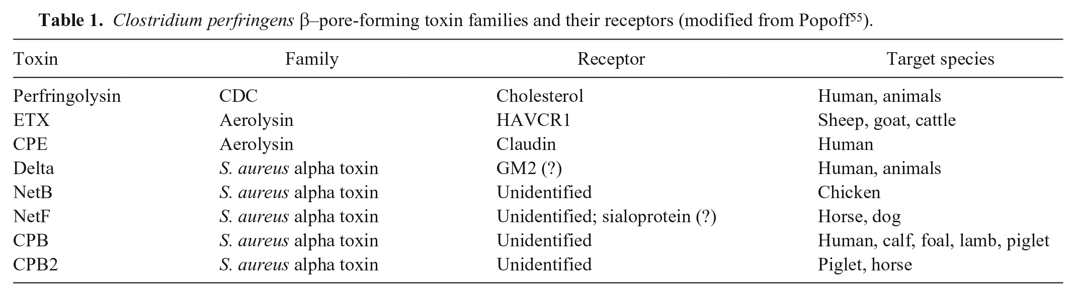
Pore-forming toxins (PFTs) are the largest family of bacterial cytotoxic proteins leading to the disruption of host plasma membrane integrity and eventually osmotic cell lysis. 19 Almost all C. perfringens PFTs belong to the β-PFT family. 55 All β-PFTs have a similar mechanism of action including secretion as soluble monomers that diffuse in the extracellular environment of bacterial cells followed by recognizing and binding to specific host cell surface receptors. Clustering of β-PFTs to the surface of target cells triggers a conformational change of a number of β-sheets of each monomer, creating a β-barrel structure that forms a pore in the lipid bilayer of the cell membrane. 18 Clostridial β-PFTs based on their structure can be organized into 2 main classes: a) thiol-activated cytolysins or cholesterol-dependent cytolysins (CDCs) and b) heptameric β-PFTs, which contains 2 subfamilies consisting of the aerolysin family and the Staphylococcus aureus alpha-toxin family. 7 Almost all CDCs bind to a single host cell surface receptor, cholesterol, whereas the heptameric β-PFTs recognize different types of receptors (Table 1). 55 Therefore, the heptameric β-PFTs are active on specific subsets of host cells.
Clostridium perfringens β–pore-forming toxin families and their receptors (modified from Popoff 55 ).
In 2015, a novel toxigenic type A C. perfringens was isolated from a series of fatal cases of 2 clinically important enteric diseases of animals, foal necrotizing enteritis and canine acute hemorrhagic diarrhea syndrome (AHDS). 37 These strains encode 3 new putative toxin genes. The newly described toxins were designated NetE (32.9 kDa), NetF (31.7 kDa), and NetG (31.7 kDa). Amino acid sequence analyses showed that these toxins belonged to the leukocidin/hemolysin superfamily and were members of the S. aureus alpha-toxin subfamily. 37 Multiple in vitro experiments were conducted to assess the role of these new toxins in producing cytotoxicity. A netF insertional inactivation mutant was shown to be no longer toxic for an equine ovarian (EO) cell line, whereas NetE and NetG mutants remained as toxic as the wild type. EO toxicity was restored by complementation in trans with the wild-type netF gene. Moreover, only antiserum against NetF toxin neutralized the cytotoxicity of wild-type NetF-producing supernatants at high titer. These data, together with conjugation and transformation experiments on these plasmids, clearly showed that NetF was responsible for cytotoxicity for EO cells. 37
NetF is a 305–amino acid protein, related to, but distinct from, other PFTs including C. perfringens NetB toxin (48% similarity), C. perfringens delta-toxin (39% similarity), CPB (34% similarity), and S. aureus alpha-toxin (30% similarity), respectively. 37 The cytotoxic activity of NetF was evaluated in vitro on different cell lines of various host origins. Only an EO cell line was notably susceptible to NetF, with toxicity for recombinant NetF of 5.0 ng/mL (half maximal effective concentration, EC50). Although the conclusion of these studies was that NetF is likely the major toxin in these strains, the unique high sensitivity of EO cells may be an artefact of this cell line, and the NetE and NetG toxins, which are expressed in vitro, may be equally or more important in vivo. However, without the high susceptibility of the EO cell line to NetF, these unique strains of C. perfringens might never have been discovered. The finding of the high susceptibility of EO cells might also suggest that a specific receptor on the host cell membrane is required for the cytopathic activity of NetF. The ability of NetF to form pores in biological membranes has been investigated, as well as the host cell surface receptor for NetF or some of its major characteristics; NetF is able to oligomerize and form pores with 6–8 subunits in biological cell membranes such as sheep erythrocytes and EO cells. 42 The defined size of the NetF pore was estimated to be ~4–6 nm. NetF pores are very similar in size to C. perfringens delta-toxin pores with a functional diameter of 5 nm, 33 but are larger than NetB pores (1.8 nm) and S. aureus alpha-toxin pores (2.8 nm).16,25 In addition, a cell surface sialoprotein(s) most likely plays a crucial role in binding or cytopathic activity of NetF. 42 However, further work is required to identify the host cell surface receptor(s) for NetF because identification could not only improve understanding of the mode of action and host-specificity of this toxin but it might also lead to the development of therapeutic or preventive approaches that target the activity of NetF.
General genome features of netF-positive strains
C. perfringens was the first member of the phylum Firmicutes that had its genome sequenced completely. 67 As of November 2019, 160 genome assembly and annotation reports were available publicly on GenBank for C. perfringens. The rapidly increasing availability of genome sequences of C. perfringens provides an essential resource for analysis and understanding of their strain-specific characteristics.
In 2016, 2 netF+ strains (JFP55 [an equine strain] and JFP838 [a canine strain]) were sequenced, and their genomes were completely closed and annotated. 38 The resulting genome assembly of JFP838 consists of 6 contigs including 1 chromosome (3,530,414 bp; 28.37% GC) and 5 plasmids: pJFP838A (404,512 bp), pJFP838B (66,958 bp), pJFP838C (72,750 bp), pJFP838D (48,597 bp), and pJFP838E (14,657 bp). The genome assembly of JFP55 yielded a complete chromosome (3,347,300 bp; 28.37% G+C) and 5 plasmids: pJFP55F (72,549 bp), pJFP55G (36,664 bp), pJFP55H (58,447 bp), pJFP55J (42,209 bp), and pJFP55K (14,060 bp). A total of 3,033 genes were predicted for JFP55, with 2,825 coding sequences, 94 transfer RNAs, and 30 ribosomal RNAs. For JFP838, 3,202 genes were identified, including 3,014 coding sequences, 92 transfer RNAs, and 30 ribosomal RNAs. These values do not include the contributions of the plasmids. 38
Comparative bioinformatic analysis of the chromosome of 2 netF-positive isolates with 3 reference strains including a soil isolate (strain 13), 67 a type A CPE-producing food poisoning isolate (NCTC 8797; SM101), 46 and a type A human gas gangrene isolate (ATCC 13124) 46 revealed a number of chromosomal regions unique to JFP55 and JFP838 but not found in the chromosome of reference strains. Some of these unique chromosomal regions were prophage-associated genes, but no known virulence factor was found. To date, there is no evidence that bacteriophage-derived integrases play any role in virulence of C. perfringens, unlike Escherichia coli. 41 A putative function could not be assigned for the majority of genes located on these genetic regions. Plasmid annotation indicated that both NetF-producing strains, JFP55 and JFP838, carry 3 plasmids in common, including a NetF/NetE toxins-encoding plasmid (pJFP55F, pJFP838C; ~73 kb), a CPE/CPB2 toxins-encoding plasmid (pJFP55G, pJFP838D; ~48 kb), and a putative bacteriocin-encoding plasmid (pJFP55K and pJFP838E; ~14 kb). Apart from the common plasmids, each strain carried 2 unique plasmids, one of which encoded the netG toxin gene. 38
Plasmids and virulence in C. perfringens
C. perfringens plasmids can be broadly grouped into 3 major classes based on their replication initiator: pCW3, pCP13, and pIP404.2,29,79 The pCW3-like plasmids are conjugative and have a common conjugation system, transfer of clostridial plasmids (tcp), and have been described in all toxinotypes of C. perfringens. These conjugative plasmids are relatively large and harbor at least one toxin or/and antibiotic-resistance gene. Comparative sequence analyses of pCW3-like plasmids demonstrated that all members of this family have a highly conserved backbone region (~35 kb) as well as a variable region. The core region contains genes mainly involved in replication, maintenance, and conjugation transfer (tcp locus); the variable region usually encodes genes that play a critical role in virulence of C. perfringens.2,51,81 It has been shown that the pCP13 plasmid is also conjugative, but that it has a novel conjugative system, pCP13 C. perfringens (pcp) transfer locus. 79 The pcp locus encodes a putative type 4 secretion system. The pCP13-like plasmids can harbor toxin genes such as cpb2 and bec (binary enterotoxin of C. perfringens).67,83 The third class, pIP404-like plasmids, contains relatively small plasmids that often carry the bacteriocin-encoding gene (bcn). 79
Recently, whole genome sequence analysis of 30 netF-positive strains demonstrated that these newly sequenced strains consistently carry the described common plasmid profile (pNetE/NetF, pCPE/CPB2, and pBCN), independently of source, geographic origin, and time. 40
The pNetG plasmid is found in about half of all netF-positive strains. Comparative analyses indicated that pNetE/NetF, pCPE/CPB2, and pNetG are tcp-conjugative plasmids (pCW3-like plasmid), and that the bacteriocin-encoding pBCN plasmid is a member of pIP404-like family. 17
The netF/netE and netG genes are parts of loci with all the features of pathogenicity loci of tcp-conjugative plasmids, which were designated as the NetF pathogenicity locus (35 kb) and the NetG pathogenicity locus (31 kb), respectively. 38 We define pathogenicity locus as a genetic region unique to a particular pathotype that contains one or more virulence genes and mobility-associated genes. 38 These defined pathogenicity loci are highly conserved in all NetF-producing strains.
The NetF pathogenicity locus encodes 32 genes in addition to netF. Demonstration of a genomic inversion in NetF locus of the JFP838 strain as well as the presence of multiple transposases at both termini of the locus suggests that this pathogenicity locus might have derived from a mobile element. It is likely that acquisition of the NetF locus on a conjugative plasmid was a key step in evolution of this specific pathotype of C. perfringens. It has been shown that horizontal gene transfer and mobile genetic elements play an important role in biology, evolution, and conversion of non-toxigenic to toxigenic strains of Clostridioides difficile as well as in C. perfringens.3,21,27,45
An interesting feature of the NetF pathogenicity locus is the number of genes predicted to be involved in virulence behavior. For instance, 2 putative cell surface genes and a sortase enzyme might contribute to adhesion. In addition, the gene encoding another putative PFT, NetE, was also found consistently in the NetF locus. NetE is expressed, but no evidence for its cytotoxic activity in in vitro conditions was found. 37 It may be that the putative toxin might be toxic to cells other than those of the cell lines tested, given that the originating “host” of these strains is still unclear. 14 It is also possible that NetE and NetF have synergistic action in promoting disease in vivo. Pro-inflammatory effects of NetE are yet another possibility, similar to that of the leukocidins of S. aureus. 1
Another intriguing feature of NetF-producing strains is that a cpe-bearing plasmid always coexists in these isolates. The advantage to these netF-positive strains of expressing 3 different β-PFTs as well as CPE is unclear, but these strains look highly armed; further work is required to clarify the significance of this configuration. There may be an interaction between these toxins to cause disease, as has been shown with CPB and CPE of type C human enteritis necroticans strains. 30
Clonality in netF-positive strains
A core genome multilocus sequence typing (cgMLST) scheme was established for C. perfringens, and clonal expansion of NetF-producing strains was explored using this approach. 40 The study showed that these isolates belong to 2 distinct clonal populations despite the fact that they come from different hosts and geographic sources. The similarity between dog- and foal-derived netF-positive isolates suggests that these animals may be infected by C. perfringens strains adapted to a common environmental or animal source rather than to the 2 individual host species in which these strains have been associated with unique diseases.
The genetic relatedness of 56 isolates of C. perfringens, including 32 netF-positive isolates, has been examined using a core-genome phylogenetic approach. 26 This work supports the result of the cgMLST study by also showing that netF+ isolates cluster into 2 clades. Apart from netF-producing strains, there is a clear general need to examine the genetic relatedness of a far larger collection of C. perfringens from different sources including diseased animals to understand the epidemiology and disease associations of the organism.
Diseases associated with NetF-producing strains
Foal necrotizing enterocolitis
Diarrhea is one of the most common infectious disease conditions requiring veterinary care of young foals. 82 Although most episodes of diarrhea are not life-threatening, a substantial subset requires intensive veterinary care at considerable cost to the owners. High mortality rates are seen in neonatal foals with necrotizing enterocolitis (NE) and, although the etiology of the NE is not determined in many cases, C. perfringens has been implicated as a primary pathogen.8,10,11,15,20,22–24,37,47,50,52,70,80
General clinical signs
NE associated with C. perfringens has been reported in foals of any age, yet most cases occur in foals < 10-d-old including in foals < 24-h-old.10,11,22,23,47,52,74,80 In early stages of the disease, foals may have anorexia, diarrhea, depression, and dehydration; however, many foals have colic that may or may not be associated with diarrhea. Diarrhea, when present, is often bloody. Hypomotility of the intestines or ileus may also be present. Signs of enterotoxemia, SIRS (systemic inflammatory response syndrome), and sepsis may accompany the clinical signs and are thought to be a major contributor to the mortality associated with NE.8,11 Rapid progression of the clinical disease or sudden death is common.
It is common for affected foals to have adequate transfer of passive immunity (IgG > 8 g/L [800mg/dL]). 11 Although this finding seems counterintuitive, foals with adequate passive transfer also acquire high levels of intestinal trypsin inhibitors from the mare’s colostrum.63,64 These inhibitors, along with low levels of endogenous trypsin production, increase the risk of disease caused by C. perfringens toxins in neonates because these toxins are otherwise normally susceptible to trypsin degradation in the gastrointestinal tract.8,31
Farms may have outbreak situations or solitary cases, and some farms may be affected in successive years.12,76 Transmission of C. perfringens is fecal–oral or from environmental contamination, including contamination of the mare’s udder. 74 Incubation of C. perfringens infections may be < 24 h, thus early neonates are at significant risk of disease. 66 Foals may continue to shed pathogenic bacteria in their feces after clinical resolution, although the exact length of time is unknown but is suspected to be 3–7 d. 66
Foals with NE associated with C. perfringens have consistent, although not pathognomonic, gross pathologic and histopathologic findings.10,20,22,23,52 Gross findings include segmental or diffuse hemorrhage and thickening of the intestinal mucosa (Fig. 1). 8 Mucosal lesions include ulcers and necrosis, which may lead to pseudomembrane formation. Luminal fluid is frequently hemorrhagic. Gross findings may be limited to a single segment of the small intestine (frequently the jejunum) or may concurrently affect the small intestine, cecum, and/or the large colon. Other organs may have lesions consistent with enterotoxemia or sepsis. The histopathologic features of disease include necrotizing or necrohemorrhagic enteritis, enterotyphlocolitis, or enterocolitis.8,11,20 Villi may be denuded, ulcerated, or necrotic, and there may be mucosal or submucosal thrombosis. Bacteria typical of C. perfringens morphology are noted on the intestinal surface, within the affected mucosal cells, and in large numbers adhered to the surface of ulcers.8,20 Mural edema and mild-to-moderate neutrophilic infiltrates are also noted.8,9
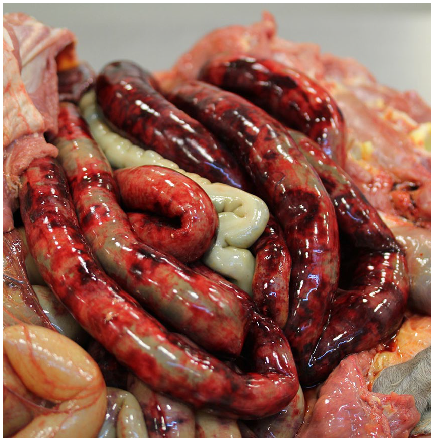
Severe necrohemorrhagic enteritis caused by a type A Clostridium perfringens netF strain in a 3-d-old Warmblood foal.
Role of NetF+ C. perfringens
The role of C. perfringens in foals with neonatal enterocolitis has been investigated worldwide.11,15,24,36,39,47–50,68,70 Although toxigenic type C and netF-positive type A C. perfringens strains may cause diarrhea of variable severity in young foals, C. perfringens type A has been identified commonly in the feces of non-diarrheic foals, especially in neonates.48,49,68,74 Shedding of type A C. perfringens in healthy foals is highest at 1–3 d, and ~64% of foals have detectable levels of shedding by 8–12 h of age. 74
Severe cases of C. perfringens–associated diarrhea in foals with hemorrhagic NE are most commonly associated with type C infections8,10,22,52; however, certain cases of C. perfringens type A–associated infections have considerable clinical and pathologic similarities, especially when CPE and CPB2 toxins are produced.20,75 It is important to note that occasionally CPE and CPB2 toxin–producing C. perfringens have been identified in healthy foals and are thus not always pathogenic.50,68,74
The potential importance of the novel PFT, NetF, has been identified with fatal NE of neonatal foals based on clinical and epidemiologic data. This novel toxin is related to the beta-toxin of type C strains, and affected foals have similar severe clinical signs and high mortality. In the first study investigating the new toxin, a significant association between detection of netF-positive type A C. perfringens in foals with fatal NE was identified when compared to foals with undifferentiated diarrheal illness, 37 whereas in adults with undifferentiated diarrheal diseases, the detection rate of netF in C. perfringens isolates was low. In another study, 26% of C. perfringens isolates recovered from 23 foals with enterocolitis in Kentucky were positive for netF, and the authors found evidence of previous exposure by broodmares to NetF as identified by high antibody titers. 39 These studies have identified the potential importance of NetF strains in severe or fatal enterocolitis in foals and thus advanced understanding of the role of at least a subset of type A strains in serious enteric disease in foals. NetF strains, similar to type C strains, do not appear to be adapted to the equine gastrointestinal tract given that no netF-positive strains of C. perfringens were identified in 137 fecal samples from 88 foals in Ontario, Canada. 14 Recently, one of the authors has identified cases of severe NE associated with C. perfringens possessing genes for netF in Alberta, Canada (Whitehead AE, unpublished observations).
A definitive clinical diagnosis of NE is difficult to attribute to C. perfringens given that it can be a normal gastrointestinal inhabitant. In cases in which toxigenic (netF-positive types A, C) C. perfringens is identified in sick foals, it remains unclear if this bacterium was the initial cause of diarrhea, or whether the C. perfringens has proliferated as a coinfection in response to intestinal damage caused by other agents, and, perhaps in the case of NetF strains, by inhibiting other bacteria through its bacteriocin. Regardless of the origin of the disease, a presumptive diagnosis of toxigenic C. perfringens NE is made based on signalment (usually neonatal or young foal), clinical signs, and PCR identification of toxin-producing genes (cpb or netF). Ideally, the detection of toxins by ELISA would also be performed, but the multiple clostridial toxin ELISAs are either not readily available or are unavailable. In fatal cases, postmortem examination can continue to support the presumptive diagnosis, especially if necrotizing enteritis is present in the small intestine and type C or NetF strains are identified directly or by PCR.
Prevention
Considerable work needs to be done to identify specific preventive strategies for C. perfringens–associated NE. Reducing the bacterial contamination of the environment may be important in reducing the occurrence of C. perfringens–associated NE; however, elimination of C. perfringens type A is not possible given its ubiquitous nature. 74 Foaling areas and stalls should be cleaned and disinfected using products labeled for Clostridium such as bleach or accelerated hydrogen peroxides. Routine disinfection of the stall doors (including latches), buckets, and cleaning equipment should also be undertaken. 32 If dogs are eventually shown to be an important reservoir of NetF C. perfringens, then keeping dogs (and their feces) away from foaling barns may turn out to be a simple and important preventive measure, given the rapid enteric colonization of foals by C. perfringens and their susceptibility to CPB and likely to NetF through the trypsin-inhibiting action of mare’s colostral milk.
Toxigenic strains of C. perfringens have been identified on mare’s teats and body. 74 Although there are no studies investigating preventive effects of decreasing bacterial load on the mare, many farms have instituted udder hygiene protocols before and after foaling. Personal hygiene of the animal handlers should include appropriate handwashing measures, use of disposable gloves, and appropriate personal protective equipment and biosecurity measures when working with foals with diarrhea. It is important to note that alcohol-based hand-cleaning products are ineffective for Clostridium spp.
An autogenous vaccine is available and is apparently effective for mare immunization in Kentucky for the prevention of type A C. perfringens enterocolitis in their foals. 75 This likely includes some NetF toxin because mares immunized with the bacterin toxoid have been shown to develop antibodies to NetF. 39 In addition, it has been shown that the C. perfringens strain (UK MF 05/00) used to make the autogenous bacterin toxoid vaccine encoded netF. 39 Hyperimmune plasma is available from horses immunized with types A, C, and D C. perfringens supernatants, as well as NetF, for the prevention or treatment of NE in foals, although there are no studies yet supporting its use.
Canine acute hemorrhagic diarrhea syndrome
In 1977, a retrospective study described 125 previously healthy dogs with a syndrome called hemorrhagic gastroenteritis (HGE), which was characterized by sudden onset of vomiting and severe bloody diarrhea. 4 Exclusion criteria were diagnosis of a known cause for acute hemorrhagic diarrhea (e.g., intestinal foreign body, acute pancreatitis, rodenticide toxicity), a hematocrit of < 0.50 L/L [50%] at presentation, and a duration of clinical signs > 48 h. These inclusion and exclusion criteria describe well the hallmarks of this syndrome in dogs, which was recently renamed acute hemorrhagic diarrhea syndrome (AHDS): acute onset of clinical signs, fresh blood in the vomitus and feces, and significant fluid loss via the gastrointestinal tract. Prospective studies have defined the patient population and clinical course more precisely.
General clinical signs
Most affected dogs are small-breed dogs with a median weight of 10 kg and a median age of 5 y. Eighty percent of dogs with AHDS had acute vomiting as the first clinical sign, followed by hemorrhagic diarrhea after ~12 h. Physical examination findings usually reflect signs of dehydration (e.g., depressed mental status, increased heart rate), and abdominal pain can be detected in 20% of dogs at presentation. With adequate fluid therapy and symptomatic treatment, most dogs recover very rapidly within 48 h, although complete normalization of stool quality can take up to 10 d. A small proportion of dogs show a more complicated clinical course characterized by significant gastrointestinal protein loss (e.g., serum albumin < 20 g/L [2.0 g/dL]) and systemic inflammatory reactions (e.g., significant neutrophilia, left shift), which most likely reflects severe destruction of the intestinal mucosa and intestinal barrier dysfunction associated with bacterial translocation. 44 Rarely, the dogs die as a result of this syndrome.
Role of NetF+ C. perfringens
Because no underlying cause could be identified for decades, NE has been defined by the combination of characteristic clinical signs and by exclusion of known mechanisms inducing hemorrhagic diarrhea. Forty years ago, reports from autopsied dogs described a dense layer of bacteria identified as C. welchii (previous name for C. perfringens) on the surface of the necrotic intestinal mucosa.56,65 Given that C. perfringens is a normal habitant in the large bowel and secondary bacterial overgrowth takes place after death, and because dogs rarely died, the significance of this characteristic autopsy finding was unclear, and the pathogenic role of these bacteria was largely ignored. However, a prospective endoscopic study confirmed a clear association between C. perfringens and the occurrence of acute hemorrhagic diarrhea. 77 In this study, in 9 of 10 dogs with AHDS compared to any dog of the control group, adherence of clostridia to the necrotic mucosal surfaces could be detected histologically. Lesions were restricted to the small and large intestine and, therefore, the misleading term HGE implicating the involvement of the stomach was replaced by AHDS. 28 Histologic changes reflecting necrotic enterocolitis is considered characteristic of involvement of clostridial toxins, as described in other species.8,43,71,84 A prospective study showed a higher prevalence of a positive fecal CPE result tested by ELISA in dogs with AHDS (13 of 54) compared to dogs from a control group (0 of 23). However, the low prevalence of CPE in fecal samples of dogs with AHDS (24%) suggested that CPE was most likely not the only clostridial toxin involved in this syndrome. 5 After identification of a novel necrotizing toxin in a dog with fatal AHDS, a PCR for these so-called net toxins was established. 37 C. perfringens strains isolated from the duodenum of 5 dogs with AHDS were identified as type A by a multiplex PCR, and every strain encoded the PFT NetF. 28 Further studies confirmed a significant higher prevalence of C. perfringens encoding the netF gene in dogs with AHDS (50–55%) compared to healthy control dogs (0–10%), dogs with chronic enteropathies (0%), and dogs with parvoviral infection (0%). 69 Given that no C. perfringens strains encoding netF could be detected in a group of dogs with similar intestinal lesions but different etiology (parvovirus) than AHDS, generalized secondary overgrowth of these clostridial strains in dogs with acute hemorrhagic diarrhea is unlikely. In another group of dogs with AHDS, the intestinal microbiome was followed over 21 d. On the day of presentation, 13 of 23 (57%) dogs were positive for netF, but after day 7 all but 1 dog were below the detection limit of the PCR assay for netF. 85 This rapid decrease of netF-positive C. perfringens strains might explain why these strains cannot be detected in all dogs with AHDS. Thus, in dogs with AHDS at a later stage of the disease, the inciting agent may not be detectable anymore.
Summarizing all current information, it seems very likely that infection by C. perfringens type A strains associated with release of NetF toxins, possibly in concert with other toxins such as NetE, NetG, and/or CPE, is responsible for the necrotic enterocolitis in dogs with AHDS. However, further investigation is required to identify the exact role of NetF, NetE, and CPE in the pathogenesis of netF-positive C. perfringens–associated diseases. The recognition of the role of NetF-positive C. perfringens in canine AHDS has been a critical advance in understanding the basis of this disease, although more work is needed to understand its epidemiology and the factors predisposing dogs to infection. That small-breed dogs are at particular risk, possibly because of their recognized genetic predisposition to development of pancreatitis, which might reduce trypsin in their small intestine, is an intriguing suggestion 37 that should be explored. The finding that feces of 12.1% of healthy dogs showed detectable netF 69 might also suggest that the dog is a reservoir of this infection or is commonly exposed to a natural reservoir; this possibility needs to be investigated.
Conclusions and unanswered questions
The pathogenesis of most enteric infections caused by C. perfringens is associated with additional toxins that are not produced by commensal and environmental strains. The current understanding of the pathogenesis of some serious enteric diseases induced by type A strains is incomplete, and novel toxins responsible for virulence are still being discovered. The most recently discovered of these toxins is NetF. The remarkable association of NetF-producing strains with 2 previously poorly understood and important diseases of animals suggests that strains carrying this toxin might play a role in the pathogenesis of necrotizing enteritis in both neonatal foals and dogs. However, molecular Koch postulates have not been proven for either the canine or equine disease. To study the in vivo action of NetF-producing C. perfringens, it is essential to develop an appropriate animal model or ligated intestinal loop model of infection. Then, we can generate a series of mutants and assess their ability to produce lesions or disease in vivo. The recent studies conducted by our group expand understanding of necrotizing enteritis induced by type A netF-positive strains in several additional ways: 1) it has been shown that all NetF-producing strains consistently carry a common plasmid profile of pNetE/NetF, pCPE/CPB2, and pBCN; 2) the newly described putative PFT genes, netF/netE and netG, are located on 2 distinct tcp-conjugative plasmids; 3) netF/netE and netG, if present, reside on large and unique pathogenicity loci that are highly conserved among NetF-producing strains; 4) netF-positive C. perfringens strains associated with foal and canine necrotizing enteritis belong to 2 distinct clonal populations; and 5) it was demonstrated that NetF is a PFT typical of the β-PFT family and that a sialoprotein facilitates cytotoxicity of NetF on host cells and probably represents the receptor for this toxin.
Overall, these recent studies have increased knowledge of the evolution and suggested pathogenesis of netF-positive C. perfringens infections in foals and dogs. However, there are numerous questions that require further investigation. Most importantly, what is the epidemiologic link between foals and dogs? What is the natural habitat of this strain? Is it the dog? What is the exact role of NetF, NetE, and CPE in the pathogenesis of netF-positive C. perfringens–associated diseases? Are they synergistic in their ability to cause disease? Why would a virulent enteric type A C. perfringens carry and express 3 different PFTs? How might these diseases be prevented?
Footnotes
Acknowledgements
Declaration of conflicting interests
The authors declared no potential conflicts of interest with respect to the research, authorship, and/or publication of this article.
Funding
The authors received no financial support for the research, authorship, and/or publication of this article.