
Abstract
Acute hemorrhagic diarrhea syndrome (AHDS), formerly named canine hemorrhagic gastroenteritis, is one of the most common causes of acute hemorrhagic diarrhea in dogs, and is characterized by acute onset of diarrhea, vomiting, and hemoconcentration. To date, histologic examinations have been limited to postmortem specimens of only a few dogs with AHDS. Thus, the aim of our study was to describe in detail the distribution, character, and grade of microscopic lesions, and to investigate the etiology of AHDS. Our study comprised 10 dogs with AHDS and 9 control dogs of various breeds, age, and sex. Endoscopic biopsies of the gastrointestinal tract were taken and examined histologically (H&E, Giemsa), immunohistochemically (Clostridium spp., parvovirus), and bacteriologically. The main findings were acute necrotizing and neutrophilic enterocolitis (9 of 10) with histologic detection of clostridia-like, gram-positive bacteria on the necrotic mucosal surface (9 of 10). Clostridium perfringens isolated from the duodenum was identified as type A (5 of 5) by multiplex PCR (5 of 5). In addition, each of the 5 genotyped isolates encoded the pore-forming toxin netF. Clostridium spp. (not C. perfringens) were cultured from duodenal biopsies in 2 of 9 control dogs. These findings suggest that the pore-forming netF toxin is responsible for the necrotizing lesions in the intestines of a significant proportion of dogs with AHDS. Given that the stomach was not involved in the process, the term “acute hemorrhagic diarrhea syndrome” seems more appropriate than the frequently used term “hemorrhagic gastroenteritis.”
Introduction
Acute hemorrhagic diarrhea syndrome (AHDS) is a common cause of hemorrhagic diarrhea in dogs.20,27 There are numerous terms used for this syndrome, such as canine hemorrhagic gastroenteritis, canine intestinal hemorrhage syndrome, acute intestinal hemorrhage syndrome in dogs, acute hemorrhagic diarrheal syndrome, and acute hemorrhagic enteropathy.6,8,11,13,27 The syndrome is characterized by acute onset of hemorrhagic diarrhea and is frequently associated with vomiting and hemoconcentration.6,20 A predilection for small breeds has been observed.6,19
To date, the etiology of AHDS is unconfirmed. Bacterial endotoxins, hypersensitivity to food components, or autoimmune reactions could play a role in the pathogenesis.6,11,13 The involvement of Clostridium perfringens, Clostridium difficile, or Campylobacter spp. has been suspected by other authors.8,21,23 –25 As described previously by our group, 31 C. perfringens adherent to mucosal lesions was found in small intestinal biopsies of dogs with AHDS. A causal relationship between C. perfringens and the occurrence of acute hemorrhagic diarrhea was suggested.
In previous studies, histologic examinations have been limited to only a small number of dogs with AHDS, in which samples were collected postmortem.13,21,23,24 The information in these studies is limited by: 1) the difficulty of interpreting intestinal changes in specimens collected postmortem given postmortem autolysis; 2) the fact that these dogs did not have the usual course of disease that typically is associated with recovery; and 3) the low number of cases. We describe herein the pathology of AHDS using endoscopic gastrointestinal biopsies, and the characterization of the C. perfringens strains isolated from the small intestine of dogs with AHDS.
Material and methods
Cases
Our study population partly overlaps with the patient populations of a previously published study. 31 Ten dogs of various breeds, sex, and age that had been presented with acute onset of bloody diarrhea (<3 d) were included (Table 1). Exclusion criteria included: pretreatment with antibiotics, hemorrhagic diarrhea caused by non-steroidal anti-inflammatory or corticosteroid drugs, anticoagulant toxicosis, hypoadrenocorticism, hepatic failure, portal hypertension, acute or chronic renal failure, pancreatitis, inflammatory bowel disease, gastrointestinal neoplasia or foreign bodies, and/or enteric infection with parvovirus, Giardia spp., or endoparasites. Other known causes of hemorrhagic diarrhea were ruled out by history, physical examination, serum biochemistry profile, serum bile acid concentrations, fecal examination for parvovirus, protozoa, and nematode parasites, and abdominal imaging. 31
Signalment of the 10 dogs diagnosed with acute hemorrhagic diarrhea syndrome.
F = female; M = male; N = neutered.
Nine dogs with chronic non-hemorrhagic gastrointestinal disorders that had been presented to the Clinic of Small Animal Medicine of Ludwig-Maximilians-University Munich were used as controls for histologic, immunohistochemical, and microbiologic investigations of the gastrointestinal mucosa (Table 2). In 2 additional dogs, in which endoscopy was performed for causes unrelated to acute hemorrhagic diarrhea, C. perfringens strains could be isolated from the duodenum. These clostridial isolates were used as control strains to assess the significance of the detection of genes encoding for the pore-forming toxin netF.
Signalment and diagnoses of the 9 dogs in the control group.
F = female; M = male; N = neutered.
Biopsy and histologic examination
Biopsy samples of stomach, duodenum, ileum, and colon were taken endoscopically. All of the biopsies included mucosa and submucosa. Biopsy samples were immersed in 10% neutral-buffered formalin immediately after collection and embedded in paraffin and plastic 12 using standard histologic techniques. For histologic evaluation, the sections were stained with hematoxylin and eosin. Additional sections were stained with Giemsa and Gram. Biopsy samples were evaluated histologically according to the standard criteria of the World Small Animal Veterinary Association Gastrointestinal Standardization Group. 9
Immunohistochemistry
Sections of the intestine were evaluated immunohistochemically using antibodies against Clostridium spp. and canine parvovirus. Immunohistochemistry (IHC) against clostridial antigens was performed to detect the antigen distribution, in particular in deeper parts of the lamina propria, where bacteria could not be detected histologically. Despite exclusion of a parvoviral infection by a fecal parvovirus antigen ELISA or PCR, parvovirus IHC was also performed because lesions in the small intestine resembled those of a parvoviral infection (as described below). IHC was performed according to standard protocols for indirect IHC assays using the following antibodies and normal sera:
a. Clostridium spp.: rabbit polyclonal anti-Clostridium spp. antibody (1:100 dilution antibody against Clostridium (C.) perfringens, C. sordellii, C. novyi, C. septicum, and C. chauvoei, AbD Serotec, Düsseldorf, Germany) as primary antibody; peroxidase-labeled goat anti-rabbit immunoglobulins (1:100 dilution, Dako, Glostrup, Denmark) as secondary antibody; goat normal serum (1:10 dilution, MP Biomedicals, Illkirch, France).
b. Parvovirus: mouse anti-canine/feline parvovirus (1:100 dilution, AbD Serotec) as primary antibody; peroxidase-labeled rabbit anti-mouse immunoglobulins (1:100 dilution, Dako) as secondary antibody; rabbit normal serum (1:80 dilution, MP Biomedicals).
All incubations were completed at room temperature. Following manual deparaffinization and rehydration, 4-μm tissue sections, mounted on positive-charged glass slides, were treated with 1% hydrogen peroxide to quench endogenous peroxidase activity and washed in a bath of Tris-buffered saline (TBS, 0.5 M, pH 7.6). Slides for parvovirus IHC were pretreated with proteinase K (Dako) for 10 min, followed by a TBS bath for 10 min. After blocking with normal serum for 30 min, slides were incubated with the primary antibody for 60 min. Subsequently, slides were washed in baths of TBS, incubated with the secondary antibody for 1 h, and washed again with TBS. Binding of peroxidase coupled to the secondary antibodies was visualized by the reaction with H2O2 and 3’3’-diaminobenzidine-tetrahydrochloride-dihydrate (Biotrend Chemikalien, Köln, Germany) as chromogen. Slides were counterstained with Mayer hematoxylin.
For clostridial IHC, positive controls included a clostridial suspension (C. perfringens isolated from dogs with AHDS not included in our study) injected into a piece of swine muscle and processed routinely (fixed in formalin and embedded in paraffin).
The positive control for parvovirus infection originated from routine autopsy material (dogs with histologic and virologic confirmed parvovirus infection). For negative controls, the primary antibodies were replaced with homologous non-immune sera.
Bacterial culture
Bacteriologic investigation was performed using duodenal biopsies of 9 dogs. Within 30 min of biopsy collection, samples were plated onto agar plates using sterile tweezers, followed by 3-phase streaking using an inoculation loop for semi-quantification. Nutrient agar (with 5% defibrinated sheep blood for aerobic incubation) and Schaedler agar (with 5% sheep blood for anaerobic incubation) were used for cultivation. Plates were incubated at 38 ± 1°C. Anaerobic incubation was performed using gas generators (GENbox anaer, bioMérieux, Marcy l’Etoile, France) in anaerobic jars. Colony growth was monitored for 3 d. Every colony type was quantified (from (+) = total <10 colonies up to +++ = dense colony growth in the third streak) and differentiated by mass spectrometry using MALDI-TOF (Microflex LT, Flex analysis software 3.0, Bruker-Daltonics, Bremen, Germany). Biotyper score values of ≥2.3 were accepted as identification on species level. From every instance of clostridial growth on culture plates, 5–7 colonies were subcultured and preserved at –20°C. One isolate per dog was subsequently used for further investigation.
Genotyping of C. perfringens isolates and determination of genes encoding for toxin netF
In C. perfringens isolates from 5 dogs with AHDS, specific detection of the 4 major toxin genes (alpha [cpa], beta [cpb], epsilon [etx], and iota [itx]), the enterotoxin gene (cpe), and the beta2 toxin gene (cpb2) was performed by multiplex PCR, as described.2,18 In addition, genes encoding for the pore-forming toxin netF were detected using a proprietary real-time PCR (IDEXX Laboratories, West Sacramento, CA) based on sequence information. 18
Statistical analysis
To identify statistical differences in the severity of mucosal lesions between different locations (duodenum, ileum, colon) of the intestinal tract, histologic scores were compared with a repeated measures ANOVA and Tukey multiple comparison post-test. This analysis was conducted twice, both as intention-to-treat analysis with the last value carried forward and as a per-protocol analysis excluding the animals that did not have histologic scores from every intestinal region.
Results
Histopathology
Gastric mucosa
Gastric biopsies were available from 10 dogs (4–9 biopsies per animal, n = 69). Histologically, chronic gastritis was diagnosed in 9 of 10 dogs, characterized by lymphoplasmacytic infiltration (8 of 10), fibrosis (7 of 10, mild predominant subepithelial foci to severe diffuse periglandular fibrosis), and lymphofollicular hyperplasia (6 of 10). In 9 dogs, spiral-shaped bacteria (Helicobacter-like organisms) were present predominantly in the surface mucus, and sparsely in the gastric foveolae, glandular lumina, and the canaliculi of the parietal cells. Spiral-shaped, Helicobacter-like bacteria in large numbers were found in 5 dogs. In 4 dogs, only a few spirochetes were present.
Duodenal mucosa
Duodenal biopsies were available from 10 dogs (8–15 biopsies per animal, n = 97). In 9 of 10 dogs, acute lesions were found in the duodenal mucosa. The degree of the acute duodenal lesions varied extremely. As well as mild lesions in 4 animals, moderate lesions were found in 3 and severe lesions in 2 dogs. Lesions were divided into lesions of villi (a), crypts (b), and the lamina propria (c). Overall, epithelial injury was present in 8 dogs. Villus lesions consisted of tip necrosis and necrosis of entire villi (Fig. 1). Furthermore, villus blunting and fusion was present in 7 dogs. Deeper parts of the mucosa showed necrosis of the crypt epithelium, commonly with mixed-cell inflammatory response and showing incipient crypt epithelium regeneration (Fig. 2). Regeneration of crypt epithelium was indicated by flattened epithelium compared to normal epithelium, a reduced number of cells, and increased basophilic cytoplasm. Markedly distended crypts were found in 3 animals. Edema of the lamina propria was present in 2 animals, hyperemia of the villus tip in another 6 animals, and focal hemorrhages in the lamina propria or erythrocytes and fibrin on the luminal surface were evident in 6 animals. Increased numbers of lymphocytes and plasma cells (n = 5), eosinophils (n = 1), as well as mast cells (n = 4) were found in the duodenal mucosa. Furthermore, neutrophilic infiltration was observed in 8 animals. Neutrophils were present in areas of mucosal necrosis and diffusely distributed in the lamina propria. Increased numbers of intraepithelial lymphocytes were found in 2 animals. In 6 dogs, rod-shaped, plump bacteria were present adhering to the necrotic mucosal surface and on necrotic debris in the gut lumen (Fig. 3). The bacteria were gram-positive in 5 of 6 dogs (Fig. 4); no material was available for Gram staining for the sixth dog.
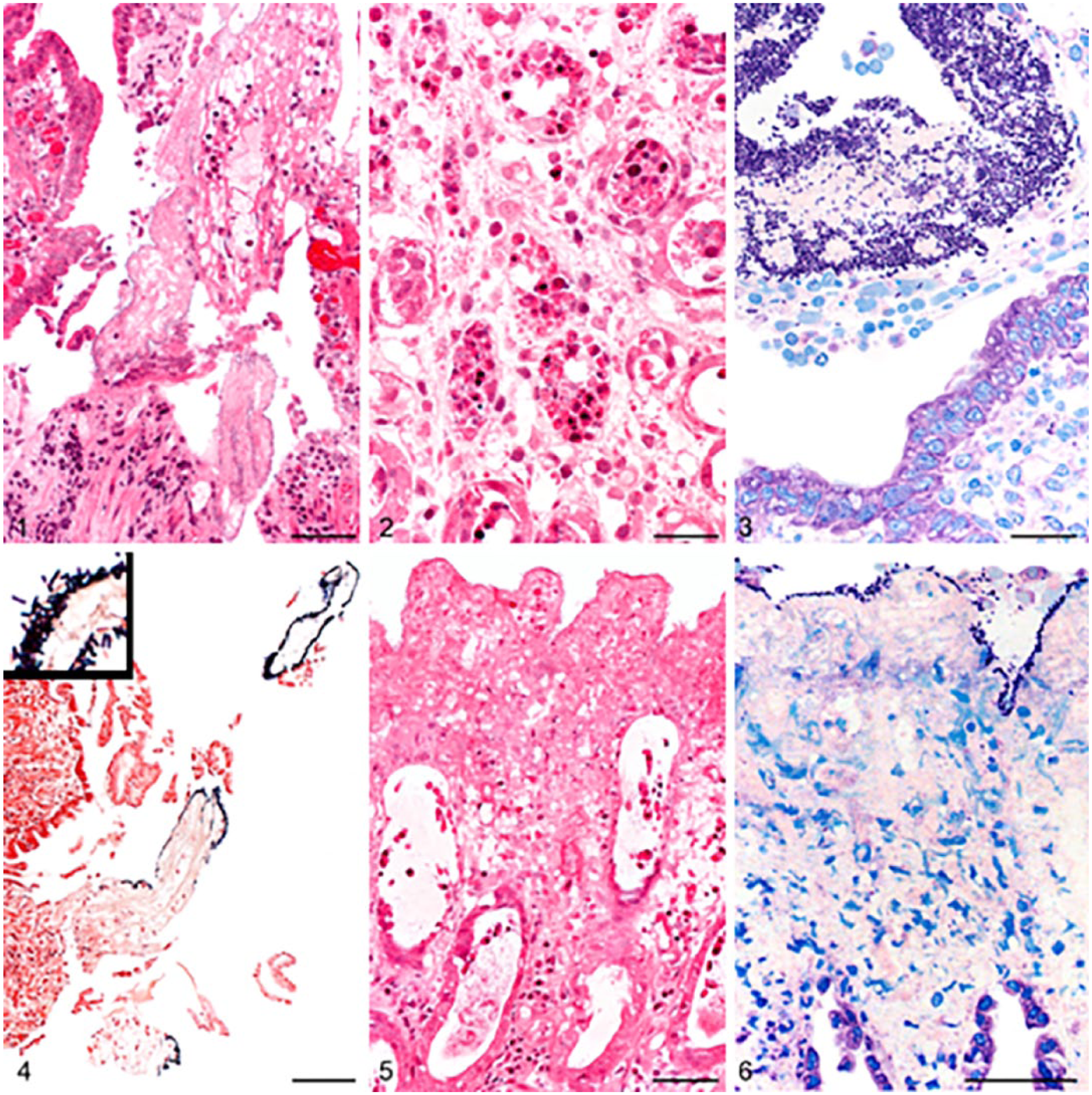
Intestinal biopsies of dogs with acute hemorrhagic diarrhea syndrome.
Ileum
Ileal biopsies were available from 8 dogs (2–9 biopsies per animal, n = 36). The lesions of the ileum resembled those of the duodenum. The severity of the ileal lesions was mild-to-moderate in 3 animals each and severe in 1 dog. Epithelial injury of villi and/or crypts was present in 6 of 8 dogs. Villi were blunted and fused in 7 dogs. Dilated crypts were found in 3 dogs. Edema of the lamina propria was obvious in 5 dogs; hyperemia (3 dogs) or focal hemorrhage (3 dogs) was also observed. Neutrophilic infiltration of the lamina propria was found in 7 dogs. Increased numbers of lymphocytes and plasma cells (n = 1), eosinophils (n = 2), as well as mast cells (n = 1) were occasionally present in the ileal mucosa. Rod-shaped, plump, gram-positive bacteria were adherent to the necrotic mucosal surface (3 of 8) and to necrotic debris and/or mucus in the gut lumen (2 of 8) of 5 dogs.
Colon
Biopsies of the colon were available from 9 dogs (2–10 biopsies per animal, n = 57). In all 9 animals, acute necrotizing lesions were found in the colon. The severity of the acute inflammatory and necrotizing changes was assigned as mild in 2 dogs, moderate in 3 dogs, and severe in 4 dogs. Lesions ranged from focal erosions of the surface epithelium to severe necrosis reaching into deeper parts of the lamina propria, including the crypt epithelium (Fig. 5). In 6 of 9 dogs, necrotic lesions were present in a moderate-to-severe degree. Furthermore, edema (4 of 9) and focal hemorrhages (5 of 9) of the lamina propria or erythrocytes and fibrin in the intestinal lumen (6 of 9) were found. Dilated crypts were obvious in 5 animals. Neutrophilic infiltrations were found in 9 animals and were located around necrotic lesions as well as diffusely in the intact lamina propria. The colonic mucosa was infiltrated by increased numbers of lymphocytes and plasma cells (n = 2), by eosinophils (n = 1), as well as by mast cells (n = 4). Increased numbers of intraepithelial lymphocytes were observed in 2 animals. Rod-shaped, plump, gram-positive bacteria adhering to the necrotic mucosal surface were found in 7 dogs (Fig. 6). Similarly shaped gram-positive bacteria could be found in the remaining 2 dogs, but only on luminal necrotic debris or mucus.
There was no significant difference in the necrosis scores of the duodenum, ileum, and colon with either per-protocol or intention-to-treat analysis (p > 0.05; Table 3). Histologic examination revealed heterogeneous results for the dogs of the control group (Table 2), which were not consistent with the histologic findings in dogs with AHDS.
Distribution of acute histologic lesions in the intestine of 10 dogs diagnosed with acute hemorrhagic diarrhea syndrome.
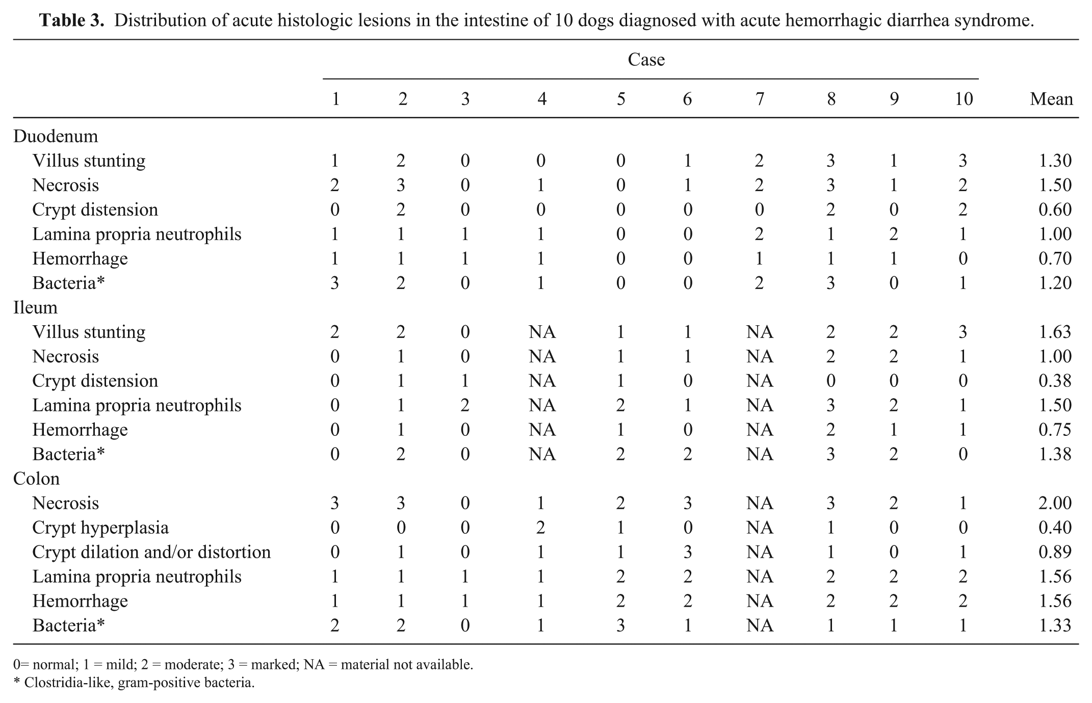
= normal; 1 = mild; 2 = moderate; 3 = marked; NA = material not available.
Clostridia-like, gram-positive bacteria.
Immunohistochemistry
Clostridium spp
Clostridium spp. antigen was detected in all 10 dogs with AHDS in at least 1 location (Table 4). Antigen was detected in the duodenum of 4 of 8 dogs examined, in the ileum of 5 of 6 dogs, and in the colon of all 9 dogs. A positive signal was located on the necrotic mucosal surface, at the surface of the necrotic debris, and within the mucus in the gut lumen, where bacteria were also detected histologically. Antigen detection was restricted to the necrotic debris and mucus in the gut lumen in the ileum and colon biopsies of 2 cases each. In no case was antigen detectable on the intact mucosal surface or in deeper parts of the lamina propria where the cryptal lesions occurred.
Results of immunohistochemistry (IHC) using antibodies against Clostridium spp. compared to histologic detection of bacteria.
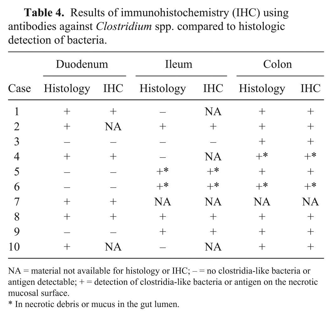
NA = material not available for histology or IHC; – = no clostridia-like bacteria or antigen detectable; + = detection of clostridia-like bacteria or antigen on the necrotic mucosal surface.
In necrotic debris or mucus in the gut lumen.
Parvovirus
Parvoviral antigen could not be demonstrated in the small intestine in any of the cases.
Bacterial culture, genotyping of C. perfringens isolates, and determination of genes encoding for toxin netF
Bacterial culture of duodenal biopsies revealed growth of bacteria in 8 of 9 dogs with AHDS and no bacterial growth in 1 dog with AHDS. MALDI-TOF typing of representative colonies resulted in C. perfringens (6 of 9 dogs), Escherichia coli (5 of 9), Stenotrophomonas maltophilia (2 of 9), Pasteurella spp. (2 of 9), α-hemolytic Streptococcus spp. (2 of 9), Pseudomonas aeruginosa (2 of 9), Corynebacterium sp. (1 of 9), Staphylococcus sp. (1 of 9), Neisseria sp. (1 of 9), Proteus mirabilis (1 of 9), and Enterococcus sp. (1 of 9 dogs). In 6 dogs, the quantification of C. perfringens was determined to be +++, +, and (+) in 2 cases each. In 5 dogs with AHDS, the clostridial isolate used for genotyping was classified as C. perfringens type A based on detection of the cpa gene and negative results for the other major toxins (Table 5). Additionally, both the cpb2 toxin gene and cpe gene were identified in 4 of 5 isolates and the netF toxin gene in 5 of 5 isolates.
PCR results for the toxin-encoding genes in clostridial strains isolated from 5 dogs with acute hemorrhagic diarrhea syndrome and 2 dogs from the control group (C).
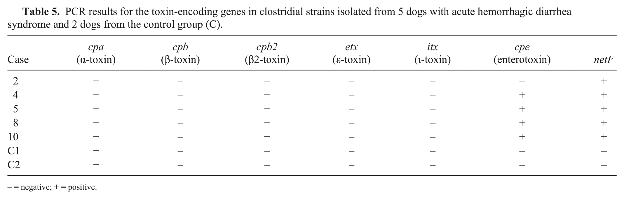
– = negative; + = positive.
Growth of bacteria was observed in 8 of 9 biopsies from control dogs. The following bacteria were identified: E. coli (5 of 9, hemolytic 1 of 5), P. aeruginosa (2 of 9), Bacillus spp. (2 of 9), S. maltophilia (1 of 9), Citrobacter sp. (1 of 9), Morganella morganii (1 of 9), Bergeyella zoohelcum (1 of 9), Neisseria sp. (1 of 9), Lactobacillus sp. (1 of 9), Acinetobacter baumannii (1 of 9), and Clostridium spp. (not C. perfringens; 2 of 9).
In 2 dogs, a 10-y-old Beagle, in which endoscopy was performed to remove a foreign body, and in a 4-y-old Havanese, in which lymphangiectasia was diagnosed, C. perfringens were isolated from duodenal biopsies and were used as control strains in order to assess the significance of the detection of pore-forming toxins in dogs with AHDS. Both strains were negative for the netF toxin gene.
Discussion
Although AHDS is common and a clinical entity well-known to veterinarians, little is known about the pathology and pathogenesis of the syndrome. 6 Although it has been reported that untreated dogs can die from hypovolemic and endotoxic shock soon after onset of clinical signs, 6 dogs usually recover completely after supportive treatment.27,32 Histologic studies of AHDS have described lesions in single cases.8,21,23,24 Moreover, in these studies, all dogs died because of the disease, which usually only occurs in the most severe case or results from additional complicating factors. The low mortality rate and rapid autolysis of the intestine after death has prevented acquisition of sufficient material for detailed histologic characterization of the lesions.6,11
Our study extends previously published histopathologic findings 31 based on rapidly processed endoscopic biopsies of a larger number of acutely diseased dogs with AHDS that had a typical course of disease with recovery and convalescence within a few days. The bacteria associated with the lesions in these dogs are also characterized.
The major histopathologic feature was necrotizing and neutrophilic inflammation of the mucosa of the small and large intestine. Lesions were detectable in the entire intestinal tract with variable individual distribution. The colonic mucosa was affected in each case, together with at least one part of the small intestine. In previous reports, distribution of lesions has been variable: small and large intestine except very proximal and distal portions 13 ; whole small intestine to colon 6 ; isolated segment of jejunum, terminal jejunum 6 ; ileum, cecum, and proximal colon 6 ; whole intestine with highest degree in the colon. 21 In our study, we detected no significant differences in the necrosis scores of the duodenum, ileum, and colon (Table 3). Because samples were obtained through endoscopy, it was not possible to obtain biopsies from the jejunum, but it is likely from our study that all sections of the small and large intestines are affected.
Interestingly, significant differences in lesion severity were observed between animals. This might be explained by the different time points of presentation after onset of clinical signs and by the different times required for individual dogs to be stabilized before anesthesia for endoscopy was performed. These differences in times together with the ability for rapid regeneration of the intestinal mucosa could be responsible for the observed difference in severity of necrosis and neutrophilic infiltration found in the histologic examination. In previous AHDS studies using postmortem specimens,21,24 the gastric mucosa was occasionally involved. In our study, only chronic lesions, likely not related to AHDS, were found. Therefore, the most commonly used name “hemorrhagic gastroenteritis (HGE)” in dogs is not an adequate term for the syndrome. The term “acute hemorrhagic diarrhea syndrome”8,31 seems to be more appropriate.
Morphologic findings in the intestine resemble previously reported histologic findings in postmortem examinations of dogs with AHDS.13,21,24 In previous reports, villus necrosis was described without evidence of inflammation 13 and cryptal involvement,13,23 whereas in our study at least a mild acute inflammatory infiltration was present in all cases, and the cryptal epithelium was affected in most cases including cryptal necrosis and regeneration.
As also found in our study, other case reports have described bacteria on the necrotic mucosal surface of dogs with AHDS.21,23,24 In previous reports, lesions were suggested to be associated with clostridial infection.21,23,24 Given the close association of bacteria and epithelial lesions, a toxic insult is considered to be the most likely explanation for the mucosal damage. In contrast to a previous study, 24 we did not detect clostridial organisms in deeper parts of the lamina propria. Clostridia-like, gram-positive bacilli, and clostridial antigen were only demonstrated on the necrotic mucosal surface and in necrotic debris in the intestinal lumen in our study population. This difference between the dogs presented in our study and the fatal case of the disease might be related to the severity of clostridial overgrowth with isolation of C. perfringens in a large number described in that earlier postmortem study. 24
The etiology of AHDS has remained unclear for decades. 27 Over time, the suspicion of involvement of clostridia in the pathogenesis of AHDS has grown.8,11,21,23,24 Currently, a pathogenic role of clostridia is assumed, given that the demonstration of clostridia-like bacteria histologically and the detection of C. perfringens by bacteriologic investigation were the only common findings in the investigated dogs. Moreover, the results of a quantitative PCR assay performed on feces from 13 dogs with AHDS confirmed a significant increase in C. perfringens compared to healthy dogs. 28 Similar histologic findings in the ileum and the large intestine have been described in dogs with suggested clostridial infections secondary to drug treatment or to parvoviral infections.22,30 Proliferation of clostridia naturally occurring in the large intestine, with upstream migration into the small intestine followed by production of toxin, could explain the extensive lesions, but a descending infection from the stomach is equally plausible. 14 Various physical stresses, depressed immune reaction, or sudden modifications in diet can decrease intestinal peristalsis or reduce normal anaerobic bacterial flora, and might predispose to overgrowth of C. perfringens in the small intestine.22,27 Our study suggests that netF-positive type A C. perfringens may play an important role in AHDS in dogs. This recently identified novel pore-forming toxin netF2,18 was found in each of the 5 cases in which C. perfringens was isolated, and PCR testing for the toxin gene was performed. It was not present in the C. perfringens isolated from the 2 dogs with unrelated gastrointestinal disorders. The netF toxin was originally identified in a C. perfringens strain isolated from a dog with fatal necrotizing enteritis, 21 and has also been isolated from neonatal foals with fatal necrotizing enteritis.17,18 NetF is a pore-forming toxin, which has cytotoxic activity and could be responsible for the typical necrotizing mucosal lesions in dogs with AHDS. 18 Further focus on this toxigenic strain in cases of AHDS will enhance understanding of its likely important role in AHDS.
Despite an extensive search, a common triggering event or predisposing factor could not be found in the 10 dogs examined. 31 As found in other studies, 18 most of the dogs were small breed (9 of 10 dogs ≤13 kg body weight) in our study. Previously, the high incidence of AHDS in dogs of small breed was associated with the common occurrence of pancreatitis in these breeds. 18 However, pancreatitis was not confirmed in the cases examined in our study.
Apart from Clostridium spp., E. coli was the only bacterial species isolated from these dogs that is considered an enteropathogen. E. coli, notably enterotoxigenic (ETEC), enteropathogenic (EPEC), and enterohemorrhagic (EHEC) strains, are associated with canine gastrointestinal diseases, particularly in young puppies.3,10,15 EHEC and EPEC strains produce typical attachment and effacement lesions in the intestine,4,10,15 whereas generally few or no mucosal lesions are seen in diarrhea produced by ETEC. 5
Given that specific virulence factors of the cultured E. coli strains were not investigated, a causative role of these bacterial isolates cannot be ruled out. However, the absence of lesions typical of pathogenic E. coli,4,10,15 coupled with detection of C. perfringens by culture together with the clostridia-like, gram-positive rods on the necrotic intestinal mucosa, makes involvement of clostridia more probable than E. coli in the dogs examined.
Some of the other bacteria (e.g., Pseudomonas, Stenotrophomonas, and Acinetobacter), grown in study and control animals, are neither enteropathogens nor normal intestinal commensals and are therefore considered to be contaminants, possibly from endoscopic investigation. 1
C. perfringens causes gastrointestinal infections in many mammalian species, and toxins produced in the intestine can cause mucosal destruction, which is similar to the lesions found in the dogs with AHDS. 26 Specifically, beta2 toxinogenic C. perfringens type A strains have been associated with intestinal disorders in dogs. 29 However, a review of the role of beta2 toxin in animals concluded that the evidence for its contribution to enteric disease is ambiguous. 33 Furthermore, a strong association between diagnosis of acute hemorrhagic diarrhea and detection of C. perfringens enterotoxin (CPE) was reported. 8 However, the results of a 2015 published study questions whether CPE plays a relevant role in the pathogenesis of AHDS in dogs. 7 Moreover, CPE can be detected in the feces of up to 14% of non-diarrheic, healthy dogs, which complicates the interpretation of a positive test result. 16
To identify the specific C. perfringens genotype involved in AHDS, a multiplex PCR was performed, and the results of this limited but unique study suggest that netF-positive C. perfringens type A is the most important genotype associated with the disease process. Recent work on netF-positive C. perfringens has shown that the cpe gene is consistently present, as usually is cpb2. 18 This earlier research has shown that netF is present on one large tcp-conjugative plasmid and that cpb2 and cpe are found on a second large tcp-conjugative plasmid. 17 The role, including possible interaction, of the cpb2, cpe, and netF genes in disease associated with netF-positive strains remains to be determined.
Footnotes
Declaration of conflicting interests
The authors declared no potential conflicts of interest with respect to the research, authorship, and/or publication of this article.
Funding
The authors received no financial support for the research, authorship, and/or publication of this article.