
Abstract
Measurement of serum trypsin-like immunoreactivity (TLI) is used to assess exocrine pancreatic function in dogs and cats. Ferrets (Mustela putorius furo) serve as valuable animal models for human diseases such as cystic fibrosis and other pulmonary diseases, and may be a useful model of other diseases including pancreatitis. We developed and analytically validated a competitive radioimmunoassay (RIA) for measurement of TLI in ferret serum by determination of analytical sensitivity, assay linearity, accuracy of spiking recovery, precision, and reproducibility. Analytical sensitivity of the assay was 0.55 μg/L. Observed-to-expected (O/E) ratio for dilutional parallelism was 90.2–127.9% (mean: 108.1 ± 11.9%). The O/E ratio for spiking recovery was 94.5–113.0% (mean: 103.9 ± 7.2%). The intra- and inter-assay coefficients of variation (CVs) were 2.7–5.7% and 3.5–8.2%, respectively. The reference interval (RI) for serum TLI derived from 31 healthy ferrets was 28–115 μg/L; the 90% confidence interval for the lower and upper limits of the RI were 10.0–32.1 μg/L and 103–126 μg/L, respectively. This TLI RIA is analytically sensitive, sufficiently linear, accurate, precise, and reproducible for the measurement of TLI in ferret serum samples.
Introduction
Ferrets (Mustela putorius furo) can serve as a valuable animal model for the study of many human diseases, including cystic fibrosis (CF), severe acute respiratory syndrome, and influenza.2,3,11,18 CF transmembrane conductance regulator (CFTR)-knockout ferrets exhibit disease characteristics similar to humans, 17 especially in the lungs 19 and the exocrine pancreas. 23 CF ferrets have been shown to exhibit exocrine pancreatic insufficiency, which mirrors that observed in human CF patients. 17 As CF progresses, pancreatic acinar cell destruction leads to decreased secretion of pancreatic enzymes and decreased serum trypsin-like immunoreactivity (TLI) concentrations, characteristic of pancreatic insufficiency. Human CF patients with pancreatic insufficiency require lifelong enzyme supplementation. 10 Therefore, a serum TLI radioimmunoassay (RIA) could have utility for the assessment of exocrine pancreatic function in CF ferrets.
Assays for TLI measure not only trypsin, but also its zymogen trypsinogen, and some trypsin molecules bound to protease inhibitors. Trypsinogen is only synthesized by acinar cells of the pancreas; therefore, measurement of this inactive precursor in serum serves as a minimally invasive tool for the assessment of exocrine pancreatic function. Serum concentrations of trypsin and trypsinogen, and therefore TLI, may increase in pancreatitis. 16 For this reason, a RIA for the measurement of TLI in ferret serum could potentially be useful for diagnosing pancreatitis in this species.
To date, there are 3 other species in addition to humans for which an immunoassay for the measurement of TLI in serum has been developed: dogs, cats, and horses.5,14,21 Despite ferrets being a well-known animal model for human CF and a potential animal model for human pancreatitis, to our knowledge, there is no immunoassay available for the measurement of serum TLI concentrations in ferret serum. 18 Therefore, we developed and analytically validated a RIA for the measurement of TLI in serum from ferrets.
Materials and methods
Purification and partial characterization of ferret trypsin from pancreatic tissue
Pancreata were collected from 12 ferrets euthanized for various unrelated research projects at the University of Iowa, Carver College of Medicine (Iowa City, IA) and were placed into a 15-mL centrifuge tube (2 pancreata per tube) and shipped overnight on dry ice to the Gastrointestinal Laboratory at Texas A&M University (College Station, TX). Ferret pancreata were received frozen and kept at −20°C until purification. Ferret trypsin was purified from pancreatic tissue using slightly modified protocols described previously.6,15,20,22 The frozen tissue was thawed at room temperature and sectioned into 1-cm 3 pieces, removing fatty tissue.
Proteins were extracted using a standard protocol that was modified to account for the low weight of the ferret pancreata. Briefly, 10 mL of 0.125 M sulfuric acid was added to a beaker containing 1 pancreas, regardless of weight. The beaker was quickly placed into a bucket of ice and covered. The solution was homogenized with a tissue grinder until the mixture was smooth and all of the tissue had been liquefied. The mixture was poured into a 50-mL centrifuge tube and spun in the centrifuge at 18,000 × g at 4°C for 45 min. The supernatant was carefully decanted and saved for further purification; the pellet was reserved for evaluation.
Ammonium sulfate precipitation was carried out in 2 steps using 0.8 M for step 1; 3.0 M ammonium sulfate was used for step 2. The supernatant was kept after step 1; the pellet was retained after step 2. All fractions were analyzed by a sodium dodecyl sulfate–polyacrylamide gel electrophoresis (SDS-PAGE) gel and screened for tryptic activity. Briefly, a 50-μL aliquot was taken from each fraction, and the pH was adjusted by the addition of 50 μL of buffer (1 M Tris-HCl, pH 8.0). Fifty μL of a porcine enteropeptidase solution (1 mg/mL in 1 M Tris-HCl buffer, pH 8.0) was added to activate any trypsinogen present. After incubation at 37°C, all samples plus controls were assayed for tryptic activity. 12
Gel filtration was performed on a column (Sephacryl S-100 26 × 60 HR, GE Healthcare, Uppsala, Sweden) with a mobile phase of 200 mM NaCl, 50 mM CaCl2, and 0.1 g/L NaN3, at pH 2.6. After the column was equilibrated with buffer, the pellets from step 2 of the ammonium sulfate precipitation were completely dissolved in 5 mL of the same buffer and then filtered through a 0.2-μM syringe filter. The solution was loaded onto the column with a flow rate of 1 mL/min, and 5-mL fractions were collected for analysis and further purification.
All active fractions were pooled, and the pH was adjusted to 8.0 by adding concentrated NaOH. A solution of 1 M Tris-HCl, pH 8.0, with 1 mg/mL of enteropeptidase was added to begin activation. Initially, ~3 mL of the activation solution was added and allowed to incubate. Every 2 h, 50 μL was taken from the pool and screened for tryptic activity. If the activity did not increase after 8–10 h, more of the enterokinase solution was added, and screening was continued every 2 h. After the activity had plateaued or slightly decreased, the solution was further purified by affinity chromatography using a benzamidine sepharose column (GE Healthcare) with a series of buffers to remove contaminants, elute the purified trypsin, and regenerate the column. All fractions were collected and pooled, then the pH was adjusted to pH 8.0 using concentrated NaOH. Immediately after pH 8.0 was reached, 2 mL of a 1 mg/mL Nα-p-tosyl-L-lysine chloromethyl ketone (TLCK) solution was added to inhibit trypsin activity. Tryptic activity was measured again, and, if activity persisted, more of the TLCK solution was added and the activity was further monitored. The solution of trypsin and TLCK was left to incubate at room temperature for 6 h. The solution was then dialyzed against 4 L of 1 mM HCl for 4 h, 3 times for a total of 12 h, after which the trypsin was evaluated to ensure complete inhibition of activity.
Partial characterization of the TLCK-inhibited ferret cationic trypsin (TLCK-mcT) consisted of SDS-PAGE gel analysis and sequencing the first 6 amino acids at the N-terminal. Gels were run according to manufacturer’s instructions, with the following parameters: 12% Bis-Tris gels (Invitrogen, Carlsbad, CA), 200 V constant voltage for 47 min, with molecular weight markers (SeeBlue Plus2, Mark 12, Invitrogen). After electrophoresis, the gels were stained (Imperial stain, Pierce Chemical, Rockford, IL) for 45 min, then destained with distilled water for 2 h. Gel pictures were taken (Gel Doc XR+ system, Bio-Rad Laboratories, Hercules, CA) to determine purity. The concentration was determined before final preparation and storage. The pure ferret trypsin was sent to the Protein Chemistry Laboratory at Texas A&M University (College Station, TX) for amino acid sequencing by automated Edman degradation and to determine sequence homology with the trypsin reported for other species. The N-terminal amino acid sequence for the first 6 amino acids was determined.
Production of anti-ferret trypsin antiserum in rabbits
Animal use was approved by the Institutional Animal Care and Use Committee of Lampire Biological Laboratories, Pipersville, PA. Two New Zealand White rabbits (Oryctolagus cuniculus) were given injections of pure TLCK-mcT at Lampire Biological Laboratories, in order to produce anti-ferret trypsin antiserum. Briefly, each rabbit was given a subcutaneous injection of 1 mL of a solution consisting of 300 µg of TLCK-mcT in 0.5 mL of phosphate-buffered saline (PBS) mixed with 0.5 mL of complete Freund adjuvant. Repeat inoculations with 300 µg of TLCK-mcT in 0.5 mL of PBS mixed with 0.5 mL of incomplete Freund adjuvant were also performed on days 7, 14, 28, 56, and 84 after the initial inoculation. Blood samples were collected by venipuncture of the ear vein after the first injection, and 2 wk after each of the last 3 booster injections. After a total of 6 injections, both rabbits were exsanguinated, and the collected serum was sent to the Gastrointestinal Laboratory at Texas A&M University for the development of a RIA.
Iodination of ferret trypsin
Titers of anti-ferret trypsin antibodies in the serum of the rabbits were assessed using TLCK-mcT labeled with 125I using the chloramine T method. 1 All buffers for this procedure were prepared fresh, and the iodination procedure was carried out under a fume hood. The bottle containing 125I was kept behind a lead shield within the fume hood. The chloramine T procedure was carried out as described previously by our laboratory.13,14
As 125I has a half-life of 60 d, this iodination procedure was performed monthly to ensure consistent radioactivity of the tracer. 1 Fraction 4 had the highest radioactivity of all fractions and was chosen for the production of the tracer. Tracer was produced as needed and was adjusted to ~30,000 counts per min/100 µL tracer by dilution with RIA buffer. Both the final tracer solution and the remainder of fraction 4 were kept at 4°C and discarded with other radioactive materials after 30 d.
Ferret trypsin-like immunoreactivity RIA procedure
Anti-ferret trypsin rabbit antiserum was diluted with RIA buffer to determine the best total binding. Antibody dilutions of 1:1,000, 1:2,000, 1:4,000, 1:8,000, 1:10,000, and 1:20,000 were assessed for optimal binding. Incubation times of 2, 24, and 48 h after addition of tracer to the sample tubes were evaluated. Standards of TLCK-mcT were made in concentrations of 0.78, 1.56, 3.13, 6.25, 12.5, 25.0, 50.0, 100, and 200 μg/L by serial dilution, and standards were kept at −80°C until they were used in a RIA.
Each RIA procedure was carried out in a radiation room at room temperature (~23°C). Briefly, each ferret serum sample was diluted 1:10 with RIA buffer. Standards were removed from the freezer, and 100 µL each were pipetted into 2 polypropylene tubes. Next, 100 µL of diluted ferret serum samples were aliquoted into 2 polypropylene tubes before tracer and antiserum were added to all standards and samples.
The 2 tubes labeled TC contained only 100 μL of tracer. The 2 tubes labeled NB contained 200 μL of RIA buffer and 100 μL of tracer. The 2 tubes labeled B0 contained 100 μL of RIA buffer, 100 μL of tracer, and 100 μL of anti-ferret trypsin antibodies. After 100 μL of tracer and 100 μL of diluted antiserum were added to all standards and ferret serum samples, all tubes were vortexed and allowed to incubate at room temperature for 24 h. After 24 h, 100 μL of rabbit carrier serum (Sigma Chemicals, St. Louis, MO; 1 mL of serum from a healthy rabbit in 99 mL of RIA buffer) and 1 mL of precipitating solution (MP Biomedicals, Santa Ana, CA) were added to every tube, except TC. Next, the tubes were vortexed and spun in a centrifuge at 3,355 × g for 30 min at 4°C. The polypropylene tubes were promptly removed from the centrifuge and transferred to foam racks. The samples were then decanted and read with a gamma counter (2470 Wizard2, PerkinElmer, Waltham, MA).
Analytical validation of the ferret trypsin-like immunoreactivity RIA
Excess ferret serum samples from unrelated projects at the Gastrointestinal Laboratory at Texas A&M University were used to analytically validate the assay by determination of dilutional parallelism (dilutional linearity), spiking recovery, intra-assay variability, and inter-assay variability, adhering to the excess material policy at Texas A&M University. Assay sensitivity was assessed by setting up 10 duplicates of B0 (100 μL of anti-ferret trypsin antiserum plus 100 μL of RIA buffer) and calculating the mean and standard deviation (SD) of the raw counts. The mean count minus 3 SD was calculated, and the value was estimated on the standard curve. 14 The calculated sensitivity was used as the limit of detection (LOD) of the assay.
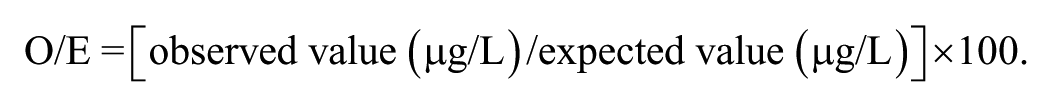
All ferret serum samples were kept undiluted at −80°C until use and then diluted to the standard 1:10 dilution using RIA buffer after thawing the samples at room temperature. These serum samples had remained frozen at −80°C since 2010. Standard dilutions were prepared using 1 part serum to 9 parts RIA buffer. Assay linearity was evaluated by assessing dilutional parallelism for 7 different serum samples at serial dilutions of 1:10, 1:20, 1:40, and 1:80 for each sample by taking the original 1:10 and diluting 1:2, 1:4, and 1:8. The accuracy of the assay was measured by spiking 6 different serum samples with equal volumes of solutions with known concentrations of ferret trypsin (0.39, 0.78, 1.56, 3.12, 6.25, 12.5, 25, 50 µg/L). The percentage of standard antigen recovery was calculated as the observed-to-expected (O/E) ratio:
To evaluate the precision of the RIA, 7 different serum samples were analyzed 10 times within the same assay run. The intra-assay coefficient of variation (CV) was calculated as:

Assay reproducibility was evaluated by assaying 9 different serum samples in 10 separate assay runs, followed by calculating the inter-assay CVs. All ferret serum validation samples were run in duplicate, and the same 1:8,000 antiserum dilution and 24-h incubation was used for each assay.
Ferret trypsin-like immunoreactivity RIA reference interval
Serum samples from 31 healthy ferrets from unrelated projects were used to establish a reference interval (RI). Ferrets were determined to be healthy by owner questionnaire. Ferrets were 1–7-y-old (n = 20, 18 males, 13 females) and had been evaluated by a veterinarian in Florida between 2008 and 2010. All samples were run at the standard 1:10 dilution and their concentrations recorded. The RI for the serum TLI RIA was determined using Microsoft Excel freeware add-in Reference Value Advisor 4 at 90% confidence interval (CI). The data were examined for outliers using the Tukey outlier detection method. The data were examined for normality using D’Agostino, Pearson, and Shapiro–Wilk tests. As well as visually observing the quantile-quantile plot, a robust Box–Cox was used, keeping in mind that the values cannot go lower or higher than the minimum and maximum RI sample values.
Results
Purification and partial characterization of ferret trypsin
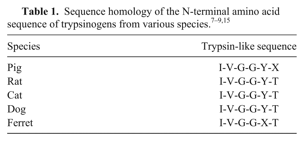
Trypsin was successfully purified from ferret pancreatic tissue. The molecular weight of ferret trypsin was estimated by SDS-PAGE to be 22–25 kDa. The amino acid sequence of the first 6 amino acids of the mature end of the trypsin was determined to be I-V-G-G-X-T (X is an unknown amino acid). The amino acid sequence was determined to be homologous to trypsin from several other species (Table 1) according to the sequence analysis report provided by the Protein Chemistry Laboratory at Texas A&M University.7–9,15
Production of anti-ferret trypsin antiserum in rabbits
Serum anti-ferret trypsin antibody titers were evaluated in both rabbits after inoculation. After the last inoculation, the antibody titers at different dilutions were determined by RIA. Titers from both rabbits were sufficient for RIA development (Fig. 1).
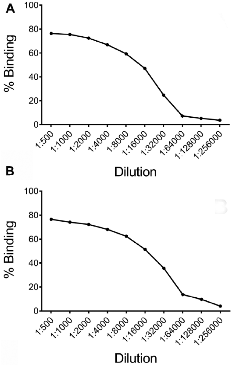
Serum anti-ferret trypsin antibody titers of rabbit 1
Development and analytical validation of a RIA for ferret trypsin-like immunoreactivity
The sensitivity of the TLI RIA was calculated to be 0.55 μg/L. The O/E ratios for dilutional parallelism for the 7 serum samples tested were 90.2–127.9% (mean ± SD; 108.1 ± 11.9%; Table 2). The O/E ratios for spiking recovery for the 6 serum samples assayed were 94.5–113.0% (103.9 ± 7.2%; Table 3). CVs for intra-assay variability for the 7 serum samples analyzed were 2.7–5.7% with a mean of 4.2% (Table 3). CVs for inter-assay variability for the 9 serum samples tested were 3.5–8.2% with a mean of 5.9% (Table 4). The RI for TLI established from 31 healthy ferrets was 28–115 μg/L, with a median of 65.3 μg/L, minimum of 28 μg/L, and maximum of 119 μg/L (Fig. 2); the 90% CIs for the lower and upper limit of the reference interval were 10.1–32.1 μg/L and 103–126 μg/L, respectively.
Dilutional parallelism of ferret serum samples in a trypsin-like immunoreactivity (TLI) radioimmunoassay.
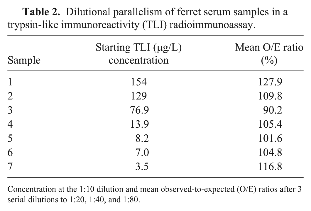
Concentration at the 1:10 dilution and mean observed-to-expected (O/E) ratios after 3 serial dilutions to 1:20, 1:40, and 1:80.
Observed-to-expected ratios for spiking recovery of ferret serum samples from a trypsin-like immunoreactivity (TLI) radioimmunoassay.
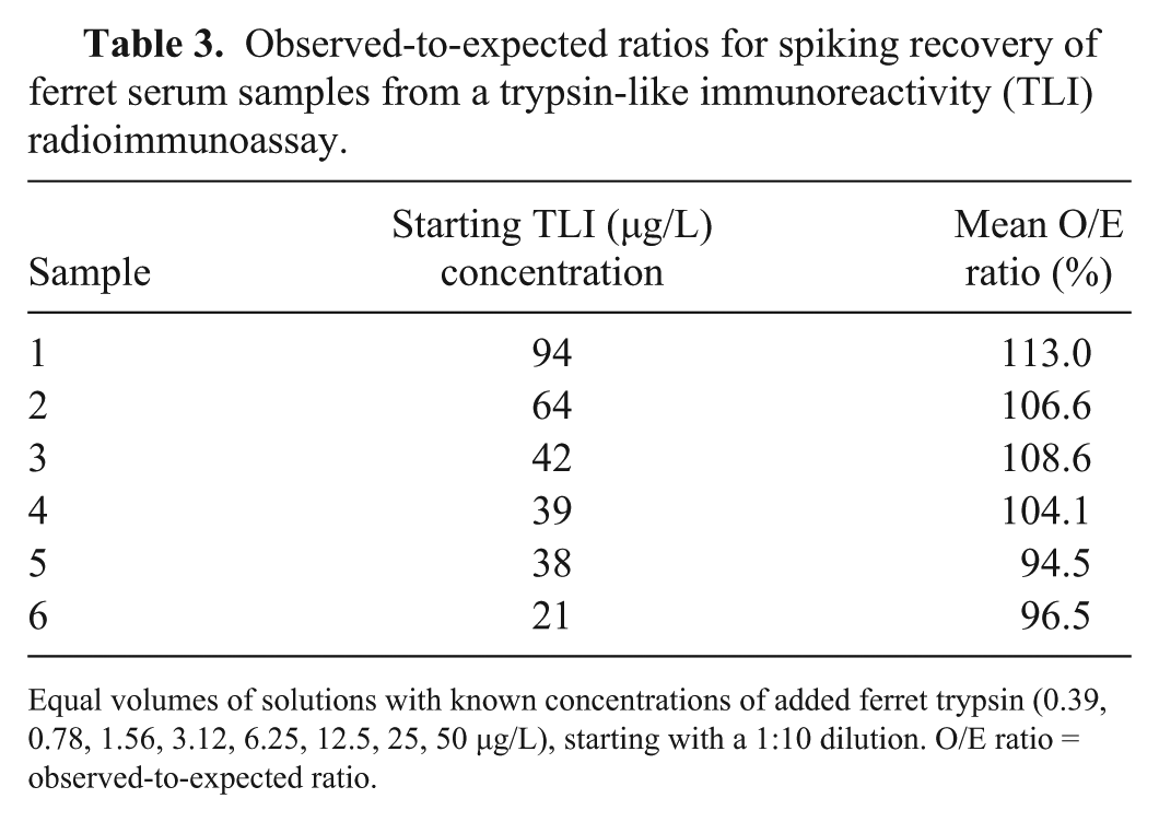
Equal volumes of solutions with known concentrations of added ferret trypsin (0.39, 0.78, 1.56, 3.12, 6.25, 12.5, 25, 50 µg/L), starting with a 1:10 dilution. O/E ratio = observed-to-expected ratio.
Precision and reproducibility of the trypsin-like immunoreactivity radioimmunoassay.
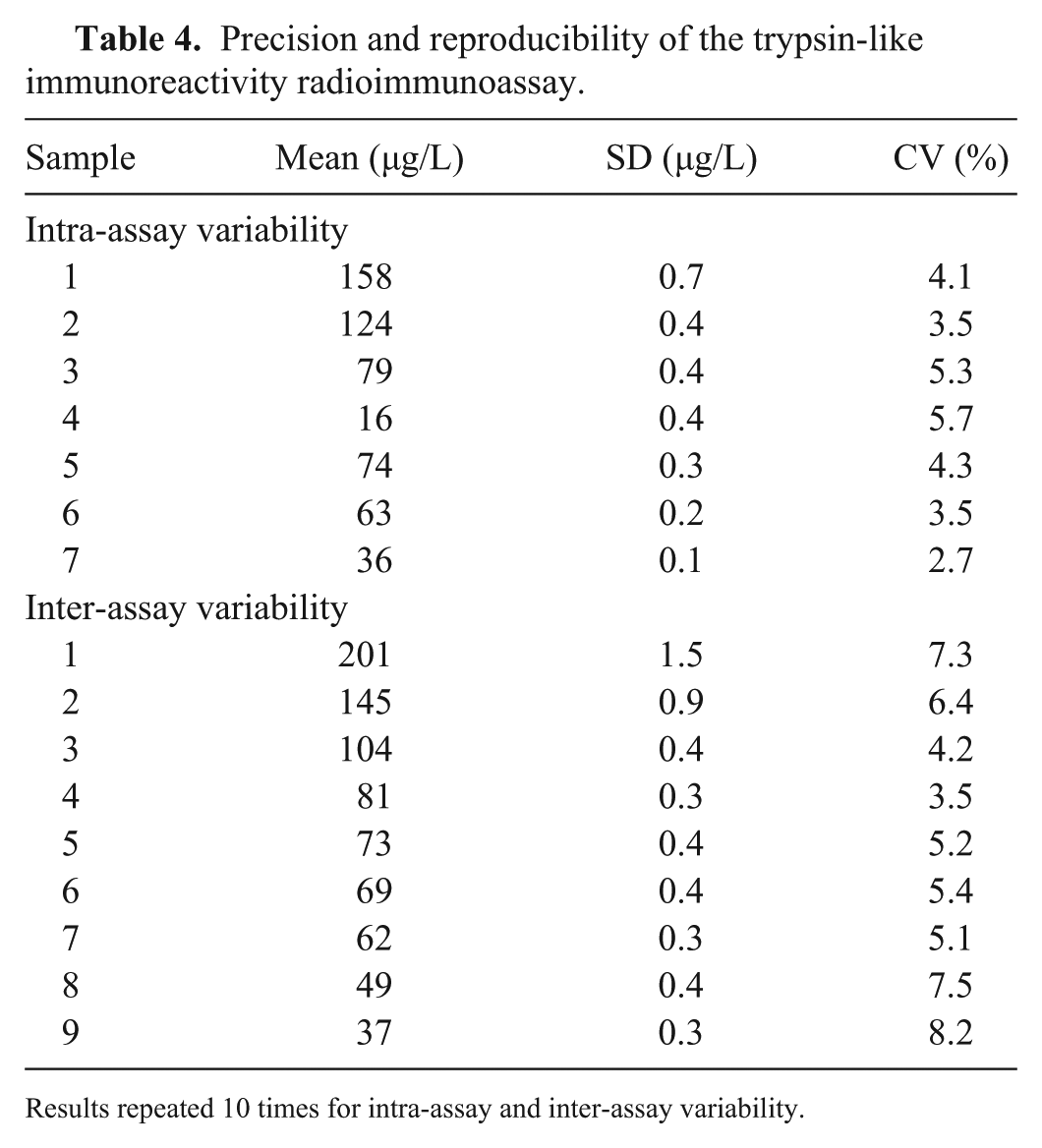
Results repeated 10 times for intra-assay and inter-assay variability.
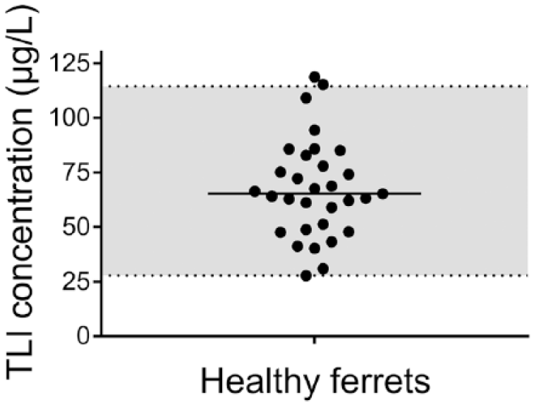
Scatter plot of serum trypsin-like immunoreactivity (TLI) concentrations from 31 healthy ferrets. Median (solid horizontal line) was 65.3 μg/L; the minimum and maximum concentrations were 28 μg/L and 119 μg/L, respectively. The calculated reference interval (RI; shaded area) was 28–115 μg/L (dotted lines) with 90% confidence interval for the lower and upper limit of the RI as 10.1–32.1 μg/L and 103–126 μg/L, respectively.
Discussion
Our purification of pancreatic tissue followed various other trypsin purification procedures reported previously for other species.7–9,15 After the final affinity chromatography purification step, the purified material was represented by a single band on SDS-PAGE gel, suggesting a pure protein. This fraction was also shown to have tryptic activity. The molecular weight of ferret trypsin was estimated to be 22–25 kDa, similar to trypsins and trypsinogens from other species. For example, canine cationic trypsin is reported to have a molecular weight of 23.8 kDa, and feline trypsinogen and feline trypsin are reported to have molecular weights of 22.6 kDa and 21.0 kDa, respectively.15,22 The N-terminal amino acid sequence of ferret trypsin was homologous to that of trypsinogen in other species.7–9,15 Ferret trypsin may have a modified amino acid in position 5 that cannot be identified by the standard procedures of Edman degradation. In the sequence of trypsinogen from other species, the amino acid in this position is a tyrosine. The protein was not further characterized because of cost constraints. Because of the similarities between our results and the results of purifications for TLI from other species, we concluded that the protein purified was pure trypsin and/or trypsinogen.15,22
We successfully established a TLI RIA for ferret serum. An antiserum dilution of 1:8,000 was used for each assay, after testing several dilutions. Also, an incubation time of 24 h was considered optimal after testing 2-, 24-, and 48-h incubation periods. Our assay has a working range of 0.55−200 μg/L, as defined by the LOD and the highest standard concentration. Samples that contain >200 μg/L can be diluted to fall within the working range of the assay, as demonstrated during assessment of dilutional parallelism. There are no standardized cutoff values for O/E ratios for dilutional parallelism or spiking recovery, or for CVs for intra-assay and inter-assay variability. However, generally acceptable O/E ratios are considered to be 80–120%, and acceptable CVs are <15%.13,14 The O/E ratios for serial dilution were 90.2–127.9%, with only one sample >120%, which suggests that the assay is sufficiently linear. The sample that showed the high O/E ratio was also the sample with the highest TLI concentration used for the validation, which may suggest limited linearity in the very upper region of the working range. However, the sample with the next highest TLI concentration had an O/E ratio of <110%. The O/E ratios for spiking recovery were 94.5–113.0%, which suggests that the assay is accurate. These samples only covered the middle and lower regions of the working range. The upper region may have limited accuracy, similar to the dilutional parallelism. CVs for intra-assay variability were all <6.0%, which suggest that the assay is precise. CVs for inter-assay variability were all <9.0%, which suggests that the assay is reproducible. The RI for TLI (n = 31) was 28–115 μg/L and is well within the assay working range. Therefore, unmeasurably low serum samples should be considered abnormal based on this preliminary RI. However, additional validation is necessary, especially in the upper range of the assay. In the future, the TLI assay may assist in assessing exocrine pancreatic function in CFTR-knockout ferrets.
Although the objective of developing and analytically validating a TLI RIA for ferret serum was accomplished, our study was not without limitations. First, the ferret serum samples used for RIA validation and construction of a RI in our study had been collected for unrelated projects conducted at the Gastrointestinal Laboratory at Texas A&M University between 2008 and 2010. At that time, ferret serum samples were collected and logged, accompanied by owner questionnaires that categorized the ferret as either healthy or diseased. Serum samples had remained frozen at −80°C since 2010. Therefore, degradation of serum sample quality could have affected our results, potentially resulting in a falsely altered RI. The sample size of 31 healthy ferret samples may be considered small, and these ferrets were only determined healthy based on owner questionnaires. However, this may be overcome by studies that utilize their own healthy controls, allowing direct comparison between healthy and diseased ferrets. Also, the current RI could be expanded by adding additional ferrets. Furthermore, an entirely new RI could be created from at least 120 healthy ferrets, adhering to ASVCP guidelines (https://www.urikapathology.com/publications/FriedrichsHarrRefInt2012.pdf). Although ferrets serve as an established animal model for CF, 18 we did not assay any serum samples from CF ferrets to determine if these ferrets exhibit exocrine pancreatic insufficiency. Further studies are underway involving ferrets with acute pancreatitis and CF.
Footnotes
Acknowledgements
We thank Robynne Gomez and Kim Green for their assistance with the iodination procedure, and Dr. Larry Dangott from the Protein Chemistry Laboratory at Texas A&M University for sequencing data. The material contained in this manuscript will be published as a dissertation at Texas A&M University in recognition of partial fulfillment of requirement of a MS degree for CS Bridges.
Declaration of conflicting interests
The Gastrointestinal Laboratory at Texas A&M University provides serum trypsin-like immunoreactivity (TLI) tests for dogs and cats on a fee-for-service basis.
Funding
The authors from Texas A&M University received no financial support for the research, authorship, and/or publication of this article. The University of Iowa component of this project was funded by NIH (P30 DK054759, R24 HL123482, and R24 DK096518) to JF Engelhardt.