
Abstract
Astroviruses are viral pathogens that have been associated with enteric and neurologic disease in a variety of species. The domestic cat is a prominent host, with reports of astroviral infection being both highly prevalent and widely distributed in the feline population. Despite the potential for inducing significant disease, especially within shelter environments, there is currently only one reliable method of detection: standard reverse-transcription PCR using pan-astrovirus degenerate primers (consensus RT-PCR) with product sequencing. Unfortunately, this process is relatively slow and costly. Quantitative real-time PCR (qPCR) represents an efficient, economical alternative, with the added benefit of viral load quantification. We developed a RT-qPCR assay using probe hybridization technique to detect conserved regions of mamastrovirus 2 extracted from fecal samples of domestic cats. Known positive and negative samples were tested, and results were compared with gold standard consensus RT-PCR and sequencing. A standard curve was employed to determine limits of detection. In order to assess analytic specificity, we tested several additional samples that had been collected from non-felid species and were known to contain non-target astroviruses. Discrepant results between consensus RT-PCR and RT-qPCR testing were further analyzed with a validation RT-PCR assay, using mamastrovirus 2–specific primers. Our probe hybridization RT-qPCR assay is reliable and effective for the detection of mamastrovirus 2. This assay will allow rapid, affordable detection and facilitate further research on astroviral infection within domestic cats.
Introduction
Astroviruses are round, nonenveloped, positive-stranded RNA viruses. The family Astroviridae was first discovered in humans in 1975 via electron microscopy. Since that time, astroviruses have been found in numerous other mammalian and avian species, thus prompting further classification into 2 genera: Avastrovirus and Mamastrovirus, found within avian and mammalian hosts, respectively. 6
The best known clinical manifestation of astroviral infection in mammals is diarrhea, which has been reported in a variety of species, including humans,7,16 dogs, 19 sea lions, 17 and cheetahs. 1 Astroviruses have also been indicated as causative agents of neurologic conditions in humans, 4 mink, 2 and cattle. 3 The domestic cat (Felis catus) is a relatively well-recognized host, with reports of infection documented on a multinational scale. 1 Astrovirus is not only cosmopolitan within this species, but also highly prevalent: studies11,20 have shown it to be one of the most common fecal viral isolates in feline shelter populations. Within the shelter cat population of Alachua County, Florida, consensus RT-PCR and sequencing of fecal samples revealed mamastrovirus 2 to be the most prevalent of the astroviral species (Lawler et al, in press). Previously known as feline astrovirus, species Mamastrovirus 2 is of particular significance because it has been shown to induce enteritis in experimentally infected specific pathogen–free cats. 8
Enteritis is an important clinical concern, especially within animal shelters. These facilities tend to be high-stress, close-contact environments, and they house high numbers of young animals particularly susceptible to enteric disease. The role of shelters in development of pathogenicity is best exemplified by feline calicivirus (FCV). Virulent strains have caused several fatal outbreaks in multi-cat populations and, in most of these outbreaks, viral virulence developed in a shelter. 9 Compared to FCV, mamastroviruses are similar in abundance and structure, with small genomes capable of rapid mutation and evolution. Thus, the concern that astroviruses may lead to fatal outbreaks in shelter populations is worth investigating.
Astroviruses are difficult to isolate in culture; they often require trypsin concentrations toxic to host cells. Astroviruses enhance epithelial permeability of the gut without inducing cell death. 13 Thus, histopathologic findings are usually absent and are an insensitive test for astroviral presence. Furthermore, astroviruses are morphologically similar to a number of other viruses, including caliciviruses, making definitive identification via electron microscopy very difficult. 14 To date, consensus RT-PCR and sequencing has been the only reliable method for identification of astroviral presence within fecal samples. Protocols for this methodology have been validated and applied to astroviral detection in a variety of species, including cheetahs 1 and cetaceans. 17 However, this approach can be expensive and laborious.
Alternatively, reverse-transcription quantitative PCR (RT-qPCR) employing probe hybridization could be a more rapid and informative means of astroviral detection. Quantitative PCR is not only more rapid than traditional gel-based PCR, but also tends to be a simpler and less expensive means of testing. Moreover, qPCR provides quantitative measurements, allowing for analyses of both viral presence and viral load, while often having the added benefit of greater sensitivity as compared to consensus PCR. Although RT-qPCR has been used to identify and quantify astrovirus in human fecal samples,5,10,18 it has yet to be applied to domestic cats. We developed and validated a probe hybridization RT-qPCR assay for detection of mamastrovirus 2 in domestic cats, which will provide a more efficient and economical test option. It will also provide quantitative information essential for further investigation of this virus and its influence on enteric disease within the species.
Materials and methods
Feline fecal samples (n = 120) were collected from litter boxes at Alachua County Animal Services (a county animal shelter) as well as the University of Florida College of Veterinary Medicine Community Outreach Program, a clinical program providing spay/neuter services to local animal rescues and shelters. Samples were collected between October 2012 and July 2013 and stored at −80°C prior to analysis. Six additional samples containing non-target astroviruses from other mammalian species were used for evaluation of analytic specificity. These included bottlenose dolphin astrovirus 1 (GenBank accession ADX97499), 2 (ADX97503), and 3 (ADX97507); minke whale astrovirus 1 (ADX97519) and 2 (ADX97522); and a squirrel astrovirus (unpublished data).
A maximum of 30 mg of feces from each sample was aliquoted for RNA extraction (Maxwell AS1280 LEV simply RNA tissue kit, Promega, Madison, WI). All extractions took place under a fume hood, with fecal samples on ice. Samples were not handled directly, and latex gloves were changed between samples. Negative control extraction samples were run in parallel with 26 of the 120 extracted RNA samples, and spectrophotometry (NanoDrop, Thermo Fisher Scientific, Waltham, MA) was used to measure total RNA concentrations within all samples. Standard RT-PCR reactions were then performed, following a nested protocol. The conserved region of astroviral open reading frame (ORF)1b (RNA-dependent RNA polymerase) was targeted and amplified using degenerate primers derived from conserved astroviral sequences, following previously published protocols1,17 for astrovirus detection by consensus RT-PCR (Table 1). Subsequent products underwent electrophoresis in 1% agarose gel. Bands of interest at 150 bp and 450 bp were cut from the gel and extracted (QIAquick, Qiagen, Valencia, CA). Direct sequencing was performed (Big-Dye terminator kit, Applied Biosystems, Thermo Fisher Scientific) using ABI automated sequencers at the University of Florida’s Interdisciplinary Center for Biotechnological Research.
Primers and associated sequences employed in PCR assays for mamastrovirus 2.
ORF = open reading frame; RT-qPCR = reverse-transcription quantitative PCR.
Prior to performance of the qPCR assay, extracted RNA samples were converted to complementary (c)DNA through the use of random hexamer primers (Advantage RT-for-PCR kit, Clontech Laboratories, Mountain View, CA). This protocol was used in lieu of a direct RT-qPCR because standard curve quality was optimized through this technique. To address the possibility of cross-contamination during reverse transcription, previously mentioned negative control extraction samples from RNA extractions were also converted to cDNA and subjected to qPCR. The probe hybridization RT-qPCR assay was targeted at a conserved region of RNA polymerase of mamastrovirus 2. The target was designed to be specific, excluding other astroviral strains. Primers and probes were established using MAFFT (https://goo.gl/RzBMn7) and Primer3 (https://goo.gl/SvFfD) analysis of mamastrovirus 2 sequences, followed by inspection for degeneracies and manual modification. Primers and probes were compared to all available astroviral sequences in GenBank in order to avoid nonspecific binding to other astroviruses. Based on this sequencing and analysis, the following primer and probe sequences were created for the RT-qPCR assay: forward primer FCAstqPCR-F (5′-CTNTTCAGGCATATYAAAGARCTCCG-3′), reverse primer FCAstqPCR-R (5′-GATGGRAGGAGRACRTRCCTGTT-3′), and probe FCAstqPCR-P (5′-ATGATCAACAARGARCAGCGKGAKAAGTATGCA-3′; Table 1). All reactions took place on a 96-well reaction plate (MicroAmp Fast optical reaction plates, Applied Biosystems). Each reaction well consisted of a 20-µL solution containing 1 µL each of forward primer, reverse primer, and probe (from solutions containing 18 µM of each primer and 5 µM of probe), 4 µL of sample (at 25 ng/µL cDNA), 10 µL of TaqMan master mix (Fast universal PCR master mix 2×, Life Technologies, Grand Island, NY), and 3 µL of nuclease-free water. Samples were run in duplicate, and a third well of each sample was run with Eukaryotic 18S rRNA endogenous control (VIC/MGB probe, Life Technologies) in order to confirm the presence of cDNA. Three wells per plate were designated as non-template controls and filled with nuclease-free water in lieu of sample. If cDNA material was detected within these controls, results were rejected and all samples on the plate were re-run.
In order to establish a standard curve for quantification, RNA from a known positive sample was amplified using the aforementioned consensus RT-PCR protocol, then extracted from the agarose gel and sequenced. RNA concentrations of the gel extractions were established via spectrophotometry. This information, along with amplification length, was used to calculate the number of molecular RNA copies present in the sample. Standard aliquots of 108 copies were prepared for single use and stored at −80°C; 10-fold serial dilutions of 101–108 were prepared and run on each plate, in triplicate. Slopes and R2 values were evaluated for each standard curve, in order to verify proper function of the primers and probe. Acceptable values were defined as efficiencies of 80–110% and R2 > 0.98. Reactions were amplified (7500 Fast real-time PCR system, Applied Biosystems) under the following conditions: denaturation at 95°C for 20 s, then 50 cycles of amplification, consisting of 95°C for 3 s followed by 60°C for 30 s. Given that standard curves were amplified effectively, no further optimization was pursued.
The diagnostic sensitivity and specificity of the RT-qPCR assay were assessed using the consensus RT-PCR and sequencing assay as the gold standard for comparison. Analytic sensitivity was evaluated based on the lowest standard curve concentration that consistently replicated above the limit of detection. Analytic specificity was determined by performing the assay on samples from other species, which were previously found to be positive for astroviral strains other than mamastrovirus 2 on consensus RT-PCR. To assess assay precision, RT-qPCR amplifications were performed on 10 replicates of a single known-positive sample on a single reaction plate at a consistent concentration.
In order to assess discrepancies between results in the RT-qPCR assay and gold standard consensus RT-PCR, a final validation RT-PCR protocol was run on the discordant samples. Following the same method for creation of RT-qPCR primers, a set of mamastrovirus 2–specific primers was developed for a final validation RT-PCR, with the following sequences: forward primer MamAst2F1 (5′-AGARCTCCGCTGGARRATGA-3′) and reverse primer MamAst2R (5′-RTCCATRGTGGTRGAGAACT-3′; Table 1). The validation RT-PCR assay was performed with the following conditions: 50°C for 30 min, then 95°C for 15 min, followed by 36 cycles of 94°C for 30 s, 54°C for 30 s, then 72°C for 1 min. Products underwent gel electrophoresis and sequencing, as described for initial consensus RT-PCR.
Results
Sequence analysis confirmed the presence of mamastrovirus 2 within 29 of the 120 samples. The remaining 91 samples were negative for mamastrovirus 2 on consensus RT-PCR and sequencing, although several samples were identified as positive for other astroviral strains, such as bat and fox astroviruses, based on sequence analysis.
All standard curves represented linear regressions (slopes of 3.37–3.80), with corresponding efficiency ranges of 83.3–98.1% and R2 values of 0.997–0.999 (Fig. 1). The range of threshold cycle values spanned from 16 cycles for 108 copies to 42 cycles for 10 copies, and samples were compared to the standard curve as an estimation of copy quantity. Although detection at 10 copies per well was observed fairly frequently, the reliable limit of detection was set at a concentration of 100 copies per well. This represents a satisfactory degree of analytic sensitivity for the assay, given that the average viral load of RT-qPCR–positive samples was much higher, at almost 400,000 copies.
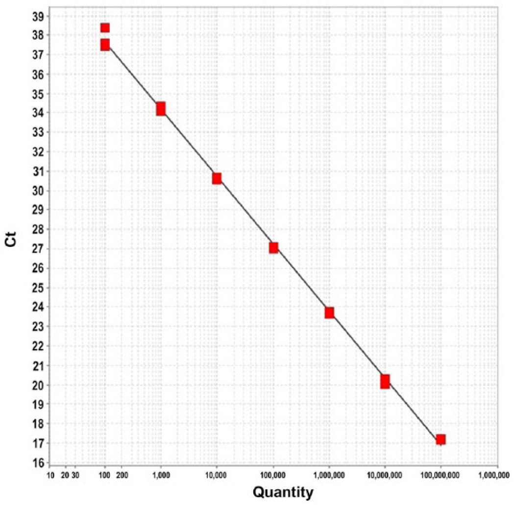
Example standard curve for reverse-transcription quantitative PCR (RT-qPCR) detection of mamastrovirus 2. Horizontal axis represents copy number on a logarithmic scale; vertical axis represents threshold cycle (Ct).
Six additional samples derived from non-feline mammalian species had previously been identified as non-mamastrovirus 2 astrovirus-positive. None of these 6 samples was detected positive by RT-qPCR assay, thus providing an excellent analytic specificity (100%; Table 2). No extraction negative controls were positive.
Comparative results of RT-qPCR for samples found not to contain mamastrovirus 2 by gold standard consensus RT-PCR and sequencing.
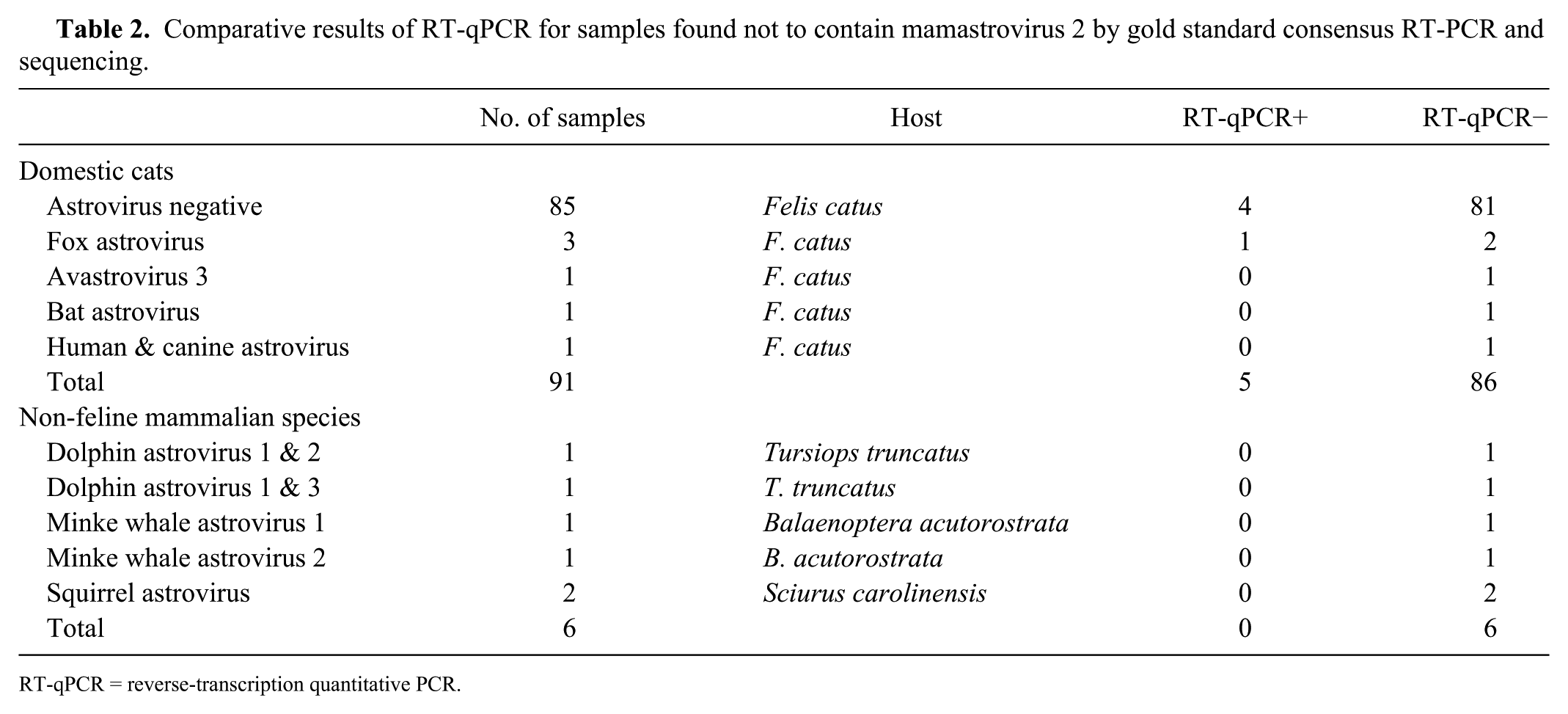
RT-qPCR = reverse-transcription quantitative PCR.
Of the 29 feline fecal samples that were identified as mamastrovirus 2–positive by consensus RT-PCR, 24 were confirmed positive by the probe hybridization RT-qPCR assay (Table 3). Thus, with consensus RT-PCR and sequencing used as the benchmark, RT-qPCR detected 82% of the positive samples (95% confidence interval [CI]: 63–93%). Ninety-one feline samples were used as negative controls based on their consensus RT-PCR results. Of those, 86 were determined to be negative by RT-qPCR and only 5 were positive, representing a diagnostic specificity of 94% (95% CI: 87–98%; Table 3).
Comparative results of probe hybridization RT-qPCR for mamastrovirus 2 in comparison to gold standard consensus RT-PCR and sequencing.
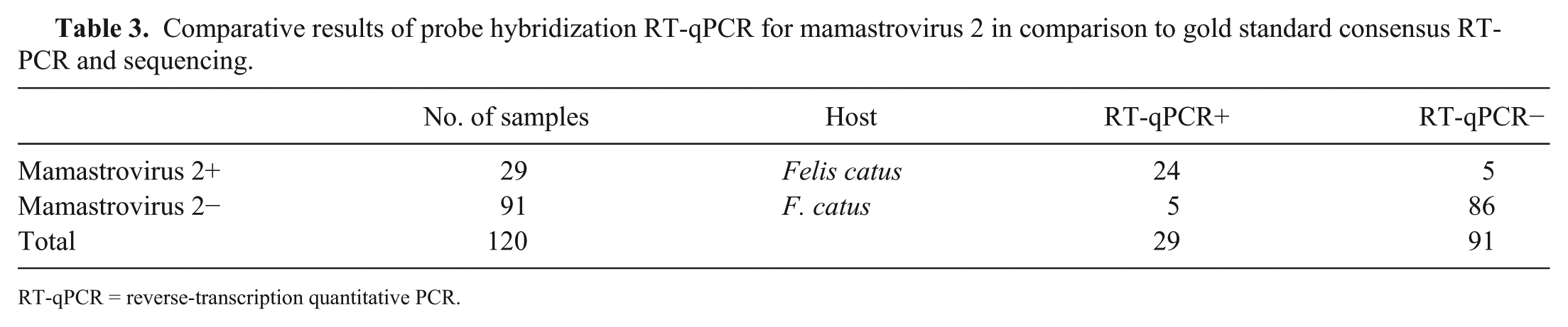
RT-qPCR = reverse-transcription quantitative PCR.
A known-positive sample was run in 10 replicates on a single plate in order to assess repeatability of results. For that sample, a mean of 1,240,770 amplified copies was detected, with a standard error of 37,819 copies and a 95% CI of ±85,553 copies. Given the high viral load within this sample—a fairly representative quantity for the population—the precision of the assay is adequate for clinical purposes. Reproducibility was not systematically tested; however, it should be noted that the sample used for repeatability analysis had a consistent copy quantification of approximately half its original detected quantity 2 y prior, consistent with degradation during storage.
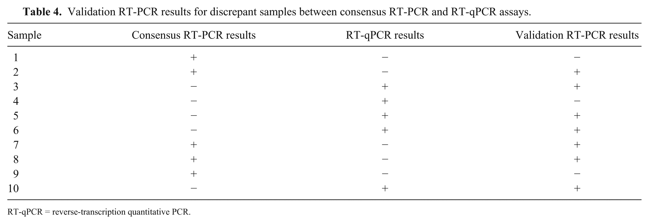
Based on initial findings, 10 samples had discrepant results between consensus RT-PCR and RT-qPCR. The validation RT-PCR test run on these samples revealed that, of the 10 samples, RT-qPCR correctly identified 4 positive and 2 negative samples that were misidentified by initial consensus RT-PCR (Table 4). Three of the remaining 4 samples (2, 7, 8) showed amplification curves by RT-qPCR but, because their concentrations fell below the established limit of detection, they must be deemed false negatives. The final discrepant sample (4) was negative for mamastrovirus 2 on both consensus and validation RT-PCR runs, with sequencing results consistent with fox astrovirus. This sample was incorrectly identified as positive with 281 copies by RT-qPCR, despite correct negative identification of 2 other fox-associated samples.
Validation RT-PCR results for discrepant samples between consensus RT-PCR and RT-qPCR assays.
RT-qPCR = reverse-transcription quantitative PCR.
Discussion
Based on these results, the RT-qPCR assay developed in our study is effective for the detection and quantification of mamastrovirus 2. It provides valuable, accurate results for the disease analysis of feline patients, while maintaining economic and timely advantages over the current gold standard.
The RT-qPCR assay produced positive results for 5 of the 91 consensus RT-PCR–negative samples. However, additional validation RT-PCR testing identified 4 of these samples to be true positives, proving RT-qPCR to be a more sensitive test for mamastrovirus 2 detection than its “gold standard” counterpart in this instance. Moreover, if not for a conservative determination of analytic sensitivity threshold, the RT-qPCR assay would have achieved 100% diagnostic sensitivity based on validation RT-PCR results. The use of a 2-step protocol, rather than a single-step RT-qPCR procedure, may introduce a potential for contamination; however, studies comparing 1-step versus 2-step RT-qPCR have not found significant differences in analytic sensitivity of the protocols. 15
RT-qPCR also obtained comparable or slightly better diagnostic specificity when considering tie-breaker test results: of the 3 negative samples identified by validation RT-PCR, 2 were correctly identified as negative by RT-qPCR but not initial consensus RT-PCR. The reason for false-positive analysis of the third sample by RT-qPCR is unknown. It is interesting to note, however, that this sample was found to contain fox astrovirus (AGK45543) upon sequencing. Although 2 other fox astrovirus–positive samples were correctly identified as mamastrovirus 2–negative by RT-qPCR, this sample may represent some degree of cross-reaction and a limitation in diagnostic specificity for the assay. We recommend future additional in vitro investigations of the in silico predicted analytic specificity of the assay.
The number of cDNA copies detected by RT-qPCR is not an exact quantification of viral load within feline feces. RNA loss during extraction cannot be accounted for. Moreover, the RT-qPCR assay detects cDNA sequences specific to the virus, rather than actual virions, and may additionally detect messenger RNA sequences not targeted for replication. As well, fecal samples commonly contain PCR inhibitors and nucleases, which may hinder amplification. 12 It is a limitation of our study that amplification controls beyond 18S were not used to evaluate the impacts of inhibitory substances. However, it should be noted that samples were fairly dilute (25 ng/µL), thus reducing the likelihood of strong inhibition. Reproducibility of the assay was not formally tested, despite changes in copy quantity being noted during repeatability testing. Though we surmise that the observed changes were secondary to multiple freezing and thawing processes, the possibility of poor reproducibility within the assay cannot be ruled out.
Footnotes
Acknowledgements
We thank the personnel at the Wildlife and Aquatic Veterinary Disease Laboratory for their guidance and assistance, as well as the staff of the Alachua County Animal Services and the University of Florida Veterinary Community Outreach Program for their support in data collection.
Declaration of conflicting interests
The authors declared no potential conflicts of interest with respect to the research, authorship, and/or publication of this article.
Funding
This work was funded by Morris Animal Foundation grant D13FE-009.